Letters to the Editor
Radiol Bras. 2015 Nov/Dez;48(6):399–403
401
http://dx.doi.org/10.1590/0100-3984.2015.0119 ing from sequelae, generally associated with tuberculosis. The
occurrence of lung cancer in cavities mimicking fungus ball or air crescent sign is quite rare(1,2,5). The tumor tends to infiltrate in the adjacent pulmonary parenchyma causing a paracicatricial effect, and may lead to emphysematous or cystic changes adja-cent to the neoplastic process(1).
In conclusion, lung cancer must be considered in the differ-ential diagnosis for patients who present with a fungus ball-like le-sion, particularly in cases where the nodule is fixed to the cavity wall.
REFERENCES
1. Wang LF, Chu H, Chen YM, et al. Adenocarcinoma of the lung present-ing as a mycetoma with an air crescent sign. Chest. 2007;131:1239–42. 2. Gazzoni FF, Severo LC, Marchiori E, et al. Pulmonary diseases with imaging findings mimicking aspergilloma. Lung. 2014;192:347–57.
Bruno Fernandes Cavalcante1, Gláucia Zanetti1, Edson Marchiori1
1. Department of Radiologiy – Universidade Federal do Rio de Janeiro (UFRJ), Rio de Janeiro, RJ, Brazil. Endereço para correspondência: Dr. Edson Marchiori. Rua Thomaz Cameron, 438, Valparaíso. Petrópolis, RJ, Brazil, 25685-120. E-mail: [email protected].
3. Truong MT, Ko JP, Rossi SE, et al. Update in the evaluation of the soli-tary pulmonary nodule. Radiographics. 2014;34:1658–79.
4. Watanabe H, Uruma T, Tsunoda T, et al. Lung metastasis of transi-tional cell cancer of the urothelium, with fungus ball-like shadows closely resembling aspergilloma: a case report and review of the literature. Oncol Lett. 2014;8:95–8.
5. Bandoh S, Fujita J, Fukunaga Y, et al. Cavitary lung cancer with an aspergilloma-like shadow. Lung Cancer. 1999;26:195–8.
Extramedullary plasmacytoma in the right pulmonary hilum
Plasmocitoma extramedular no hilo pulmonar direito
Dear Editor,
A 53-year-old black, asymptomatic man, driver, being as-sessed to be released for physical activity. The patient denied smoking as well as having comorbidities.
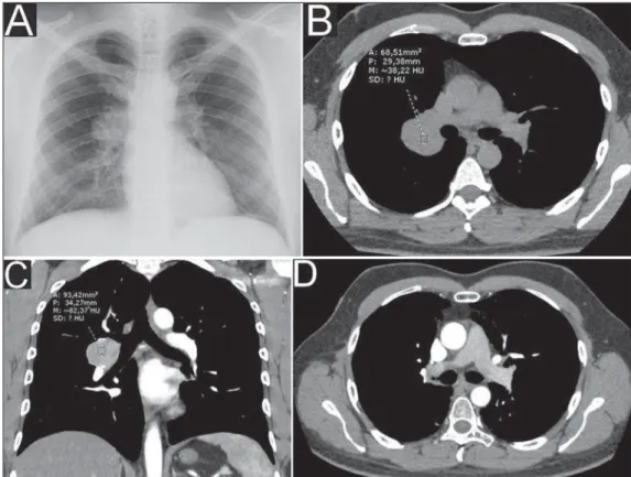
Chest radiography performed on February 1st, 2011 showed ovoid opacity in the right hilar region, with no other abnormality (Figure 1A). Chest computed tomography (CT) performed on March 13, 2011 identified circumscribed round opacity with soft parts attenuation in the right hilar region, presenting enhance-ment after intravenous contrast agent injection, adjacent to the ipsilateral main pulmonary artery and its branches. Absence of other findings (Figures 1B and 1C).
Lesion biopsy result: macro/microscopy – hypercellular light-brownish fragments showing well-differentiated plasmacytoid cells with small, eccentric and hyperchromatic nuclei;
immunohis-tochemical analysis – positive for CD138 and lambda antibodies; and negative for CD3, CD20, AE1/AE3 and kappa antibodies.
The investigation proceeded with abdominal CT (on May 16, 2011) that showed the presence of a liver cyst and signs of fat infiltration into the liver; normal blood count; negative Bence-Jones proteinuria; protein electrophoresis with no abnormalities; absence of noteworthy findings at bone scintigraphy and bone marrow aspiration.
Radiotherapy was the treatment of choice, with satisfactory response.
Chest CT performed on November 9, 2012 (Figure 1D) and other radiological studies with no suspect finding of disease re-currence/progression until May 20, 2015.
Diagnosis: extramedullary plasmacytoma (EMP) in the pul-monary hilum.
Plasmacytoma are primarily classified into solitary bone marrow/bone plasmacytoma (solitary myeloma), extramedullary plasmacytoma or one of multiple myeloma components(1,2). Such
Letters to the Editor
Radiol Bras. 2015 Nov/Dez;48(6):399–403
402
http://dx.doi.org/10.1590/0100-3984.2015.0074 tumors are constituted of plasmacytoid cells, presenting
malig-nant degeneration and producing a specific immunoglobulin mol-ecule(3–7).
The incidence of EMP is higher in men than in women, at a 3–4:1 ratio, most frequently occurring around the age of 50– 60(1,4,6,7). It is estimated that such tumor represents 2–4% of plas-macytoid neoplasms whose most relevant representative is the multiple myeloma(1,3–7), the latter representing up to 1% of all general malignancies(8).
Approximately 80–90% of EMP cases involve craniocervical structures (upper aerodigestive tract; larynx; nasopharynx; tonsilla; nasal and paranasal cavities)(1–8), but the number of cases does not reach 1% of all neoplastic head and neck lesions(5). Other sites such as gastrointestinal and urogenital tracts, central nervous system, thyroid, parathyroid glands, salivary glands, lymph nodes, skin, lungs, and breasts are uncommon(2,3,5,6). Lymph node in-volvement in pulmonary hila is extremely rare, with rates as low as less than 2% of cases(2).
Generally, they present as masses with nonspecific soft parts density(3). Histologically, such tumors do not originate directly from the bone marrow and cannot be distinguished from mul-tiple myelomas. Also the differentiation from plasmacytoid cell granulomas and other inflammatory reactions is difficult, essen-tially requiring immunophenotyping(1,4).
The diagnosis of EMP is made after rigorous investigation to rule out the presence of multiple myeloma, highlighting the histological confirmation by means of immunohistochemical analysis, biopsy/bone marrow puncture showing < 5% of plasma-cytoid atypia; to rule out the presence of osteolytic lesions, serum and urinary protein dosage and electrophoresis (to rule out the pres-ence of M and Bpres-ence-Jones proteins, respectively); and non-exist-ence of anemia(1–4,6,7).
EMP may be the initial manifestation of multiple myeloma, with progression in about 30% of cases(1,2,7).
Lenara Renó Arbex Coelho1, Gabriel Pinheiro Coelho1, Rodolfo Mendes Queiroz1, Marcus Vinicius Nascimento Valentin1
1. Documenta – Hospital São Francisco, Ribeirão Preto, SP, Brazil. Mailing Address: Dr. Rodolfo Mendes Queiroz. Documenta – Centro Avançado de Diagnóstico por Imagem. Rua Bernardino de Campos, 980, Centro. Ribeirão Preto, SP, Brazil, 14015-130. E-mail: rod_queiroz@ hotmail.com.
Treatments of choice include radiotherapy due the high ra-diosensitivity in 80–100% of cases, and surgery for localized le-sions(1,3–5,8). With such treatments, one observes recurrence and dissemination rates between 20% and 40%(1,,2,5–7), and ten-year survival in 70% of cases(1,5–7).
REFERENCES
1. Luh SP, Lai YS, Tsai CH, et al. Extramedullary plasmacytoma (EMP): report of a case manifested as a mediastinal mass and multiple pulmo-nary nodules and review of literature. World J Surg Oncol. 2007;5:123. 2. Nakayama K, Okada D, Koizumi K, et al. Excision of extramedullary plas-macytoma in a hilar lymph node. Japanese Journal of Lung Cancer. 2006; 46:723–6.
3. Ooi GC, Chim JC, Au WY, et al. Radiologic manifestations of primary solitary extramedullary and multiple solitary plasmacytomas. AJR Am J Roentgenol. 2006;186:821–7.
4. Bertolami A, Henriques AC, Penha FG, et al. Plasmocitoma extramedular. Arq Med ABC. 2005;30:58–60.
5. Ching ASC, Khoo JBK, Chong VFH. CT and MR imaging of solitary extramedullary plasmacytoma of the nasal tract. AJNR Am J Neuroradiol. 2002;23:1632–6.
6. Galieni P, Cavo M, Pulsoni A, et al. Clinical outcome of extramedullary plasmacytoma. Haematologica. 2000;85:47–51.
7. Lee SY, Kim JH, Shin JS, et al. A case of extramedullary plasmacytoma arising from the posterior mediastinum. Korean J Intern Med. 2005;20: 173–6.
8. Ferrari S, Tecchio C, Turri G, et al. Unusual case of solitary intraparen-chymal brain plasmacytoma. J Clin Oncol. 2012;30:e350–2.
PET/CT and brown fat in the evaluation of treatment response in Hodgkin lymphoma
PET/CT e gordura marrom na avaliação da resposta terapêutica no linfoma de Hodgkin
Dear Editor,
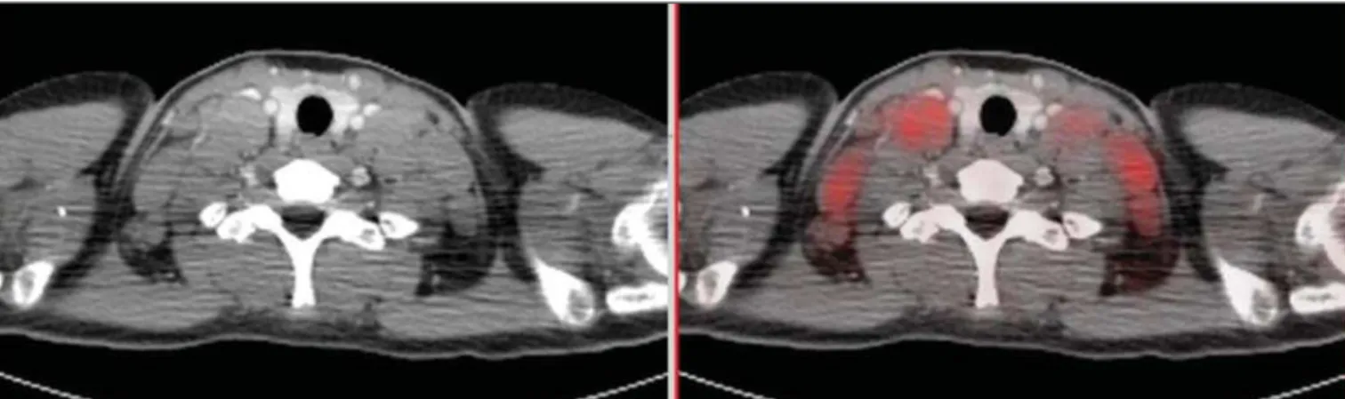
A female, 15-year-old patient presented with insidious onset of weight loss and low fever. Hodgkin’s lymphoma was diagnosed after biopsy of a palpable enlarged lymph node. 18
F-FDG PET/ CT was performed during the initial staging, demonstrating
hy-permetabolic mediastinal, axillary and cervical lymph node enlarge-ment (Figure 1). The findings were interpreted as lymphoma in activity in the mentioned sites. At basal PET/CT study, one could not observe metabolic activity in brown fat. Chemotherapy was initiated with adriblastine, bleomycine, vinblastine and dacar-bazine at days D1 and D15 for every 28-day cycles.
Six chemotherapy cycles were uneventfully performed. A new FDG PET/CT performed after about three months to evaluate the therapeutic response demonstrated complete regression of all the lesions interpreted as lymphoma in activity at the first study.