Letters to the Editor
Radiol Bras. 2015 Jul/Ago;48(4):263–270
265
Uncommon presentation of perforated Meckel’s diverticulum in preterm newborn
Divertículo de Meckel perfurado como causa incomum de pneumoperitônio em recém-nascido pré-termo
Dear Editor,
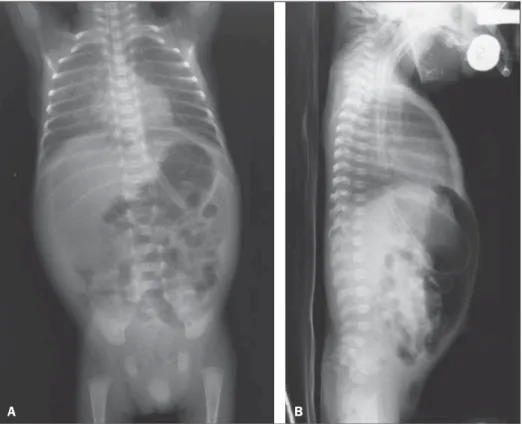
A male neonate with gestational age of 30 weeks, weighting 940 g at birth, with respiratory failure right after birth, and radio-logical signs compatible with hyaline membrane disease. At his tenth day of life, the patient presented vomiting and abdominal distention, presenting with radiological signs of pneumoperito-neum (Figure 1).
Initially, the neonate was submitted to peritoneal drainage, due to the lack of surgical conditions, and at the 19th day, after
gaining weight and present with hemodynamically stable condi-tions, was submitted to exploratory laparotomy. During the sur-gery, a Meckel’s diverticulum (MD) was found, with jejunal per-foration, hepatic blockage and obstruction distal to the blockage due to the development of adherence. Resection of about 6 cm of the jejunal loop including the perforated area was performed, with later termino-terminal anastomosis. The anatomopathological result was subacute diverticulitis with ulcer and severe peridiverti-culitis. The neonate presented a favorable evolution and was dis-charged at his 82nd day of life.
Meckel’s diverticulum represents the most common con-genital malformation of the digestive tube, and is asymptomatic in most cases(1–3). Symptomatic cases of MD are rarely found, affecting less than 20% of all pediatric cases(1). Bowel obstruction
http://dx.doi.org/10.1590/0100-3984.2014.0064
Ronaldo Garcia Rondina1, Ricardo Andrade Fernandes de Mello1, Gabriel Antônio de Oliveira1, Laís Bastos Pessanha1, Luiz Felipe Alves Guerra1, Diego Lima Nava Martins1
1. Universidade Federal do Espírito Santo, Vitória, ES, Brazil. Mailing Address: Dr. Ronaldo Garcia Rondina. Rua Júlio Cesar de Oliveira Serrano, 135, ap. 302, Bloco 03, Mata da Praia. Vitória, ES, Brazil, 29065-720. E-mail: [email protected].
2. Silveira RB, Lopes FAR, Reis ALB, et al. Dysplasia epiphysealis hemi-melica (Trevor-Fairbank disease): case report. Radiol Bras. 2013;46: 59–60.
3. Alves MPT, Fonseca COP, Granjeiro JM, et al. Carpal tunnel syndrome: comparative study between sonographic and surgical measurements of the median nerve in moderate and severe cases of disease. Radiol Bras. 2013;46:23–9.
4. Bayerl JS, Oliveira ARN, Peçanha PM, et al. Osteomyelitis of the wrist in a patient with disseminated paracoccidioidomycosis: a rare presenta-tion. Radiol Bras. 2012;45:238–40.
5. Arend CF. Tenosynovitis and synovitis of the first extensor compartment of the wrist: what sonographers should know. Radiol Bras. 2012;45:219– 24.
6. Tyring SK, Lupi O, Hengge UR. Tropical dermatology. 1st ed. George-town: Landes Bioscience; 2001.
7. Silva Lima JF. Ainhum. Molestia ainda não descripta, peculiar à raça ethiopica e affectando os dedos mínimos dos pés. Gaz Med Bahia. 1867;2:146–72.
8. Bertoli CL, Stassi J, Rifkin MD. Ainhum – an unusual presentation in-volving the second toe in a white male. Skeletal Radiol. 1984;11:133–5. 9. de Araujo DB, Lima SM, Giorgi RD, et al. Ainhum (dactylolysis sponta-nea): a case with hands and feet involvement. J Clin Rheumatol. 2013; 19:277–9.
10. Olivieri I, Piccirillo A, Scarano E, et al. Dactylolysis spontanea or ain-hum involving the big toe. J Rheumatol. 2005;32:2437–9.
Figure 1. A: Chest and abdominal radiography – Image acquired with the patients in supine position, with vertical x-rays, demonstrating hypertransparent abdominal cavity due to accumulation of free air. B: Chest and abdominal radiography – Image acquired with the patient in supine position with horizontal x-rays, demonstrating the free air collection located between the anterior abdominal wall and the bowel loops.
Letters to the Editor
Radiol Bras. 2015 Jul/Ago;48(4):263–270
266
Beatriz Regina Alvares1, Aline Satomi Yumioka1, Isabela Gusson Galdino dos Santos1
1. Faculdade de Ciências Médicas da Universidade Estadual de Campi-nas (FCM-Unicamp), CampiCampi-nas, SP, Brazil. Mailing Address: Dra. Beatriz Regina Alvares. Rua Alberto de Salvo, 238, Barão Geraldo. Campinas, SP, Brazil, 13084-759. E-mail: [email protected].
http://dx.doi.org/10.1590/0100-3984.2014.0134
is the most common symptom, usually occurring as a result from inflammation or ileal volvulus(1,4). Meckel’s diverticulum rupture is rarely found in neonates, occurring in less than 10% and mani-festing at radiography as pneumoperitoneum(1). In such situations, the differential diagnosis should be made with necrotizing entero-colitis, since this disease is responsible for 41% of cases of neona-tal pneumoperitoneum(4).
In the present case, there was a clinical suspicion of necrotiz-ing enterocolitis, but this hypothesis was ruled out as the pres-ence of a perforated MD was intraoperatively confirmed.
The causes of MD include inflammatory reaction, mucosal ulceration and defective muscular layer of the diverticulum(1,2). Rarely, MD perforation may occur as a result from umbilical cath-eterization by means of an umbilical vein connection with the MD via umbilical cord(6). In the present case, catheterization of umbili-cal vein and artery was performed with two hours of life; but the late symptoms onset and the exploratory laparotomy demonstrated that the catheterization was not related to the MD perforation.
Hirschsprung’s disease may also predispose to MD perfora-tion due to delayed passage of meconium, determining increased pressure upstream of the diverticulum(5). Such a condition occurs with typical symptoms of bowel obstruction, abdominal pain and bilious vomiting(5). In the present case, despite the symptoms of bowel obstruction and abdominal discomfort at palpation, bilious vomiting was not observed. Furthermore, the histopathological analysis of the surgical specimen ruled out the hypothesis of Hirschsprung’s disease.
Finally, MD should be included as a diagnostic hypothesis in the absence of other factors that might justify the presence of pneumoperitoneum in a neonate. Such a complication is confirmed by means of a surgical procedure.
REFERENCES
1. Aguayo P, Fraser JD, St Peter SD, et al. Perforated Meckel’s diverticulum in a micropremature infant and review of the literature. Pediatr Surg Int. 2009;25:539–41.
2. Chang YT, Lin JY, Huang YS. Spontaneous perforation of Meckel’s di-verticulum without peritonitis in a newborn: report of a case. Surg To-day. 2006;36:1114–7.
3. Gandy BJ, Byrne P, Lees G. Neotatal Meckel’s diverticular inflammation with perforation. J Pediatr Surg. 1997;32:750–1.
4. Oyachi N, Takano K, Hasuda N, et al. Perforation of Meckel’s diverticu-lum manifesting as aseptic peritonitis in a neonate: report of a case. Surg Today. 2007;37:881–3.
5. Skelly BL, Ervine E, Bisharat M, et al. Small bowel skip segment Hirschprung’s disease presenting with perforated Meckel’s diverticulum. Pediatr Surg Int. 2012;28:645–8.
6. Costa S, De Carolis MP, Savarese I, et al. An unusual complication of umbilical catheterization. Eur J Pediatr. 2008;167:1467–9.
Desmoplastic fibroma with perineural spread: conventional and diffusion-weighted magnetic resonance imaging findings
Fibroma desmoplásico com disseminação perineural: achados nas sequências convencionais de ressonância magnética e na difusão
Dear Editor,
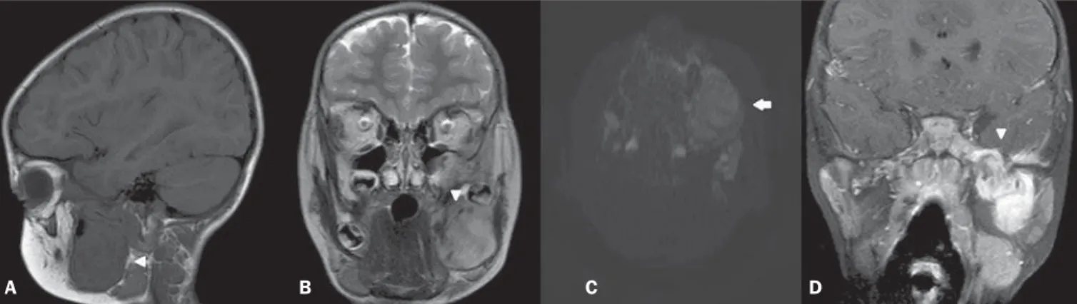
A male, three-year-old child with morphostructural alteration developed over the last year in the region of the mandible at left, presenting with recent onset of pain, with no other associated complaints. Laboratory tests did not demonstrate any alteration and magnetic resonance imaging (MRI) (Figure 1) showed a le-sion with predominant iso/hyposignal on T1-weighted image, hypersignal on T2-weighted image with subtle low signal inten-sity foci, absence of signal loss on susceptibility-weighted
se-quences and absence of diffusion restriction. After gadolinium injection, exuberant enhancement was observed in addition to perineural dissemination through the third division of the trigemi-nal nerve. Histopathological atrigemi-nalysis revealed spindle cells with-out atypias and pleomorphism, besides areas with acellular fibrous connective tissue, with immunohistochemical negative for S100, and positivity for vimentin and SMA, with Ki-67 < 5%. Such find-ings are compatible with desmoplastic fibromas. The patient was submitted to incomplete surgical excision supplemented with radiotherapy.
Desmoplastic fibroma is an extremely rare, benign bone tu-mor with aggressive and usually insidious behavior, representing 0.1% of all primary bone tumors(1–5). The mandible is the most affected site, particularly in its posterior portion, corresponding to 22% of cases(1,2,4), followed by the metaphyseal region of long
Figure 1. A: Sagittal, T1-weighted image showing lesion with hyposignal affecting the mandible (arrowhead). B: Coronal, T2-weighted sequence showing heteroge-neous lesion with subtle hypersignal intermingled with foci of low signal intensity (arrowhead). C: Axial, functional diffusion-weighted sequence does not demonstrate diffusion restriction (arrow). D: Contrast-enhanced coronal, T1-weighted sequence with fat suppression demonstrating exuberant gadolinium enhancement and notice-able perineural dissemination in the third division of the trigeminal nerve (arrowhead).