ABSTRACT
http://dx.doi.org/10.1590/1678-775720150065
Analysis of radiopacity, pH and cytotoxicity of a
new bioceramic material
Letícia Chaves de SOUZA1,2, Mamatha YADLAPATI2, Samuel O. DORN2, Renato SILVA2, Ariadne LETRA2
1- Department of Materials Science, Military Institute of Engineering, Rio de Janeiro, RJ, Brazil.
2- Department of Endodontics, School of Dentistry, University of Texas Health Science Center at Houston, Houston, Texas, USA.
Corresponding address: Ariadne Letra - Department of Endodontics, School of Dentistry - University of Texas Health Science Center at Houston - 7500 Cambridge St. - Ste. 6413 - Houston, TX 77054 - 713-486-4228 - e-mail: [email protected]
6XEPLWWHG)HEUXDU\0RGL¿FDWLRQ-XQH$FFHSWHG-XQH
O
bjective: RetroMTA® is a new hydraulic bioceramic indicated for pulp capping,this study was to compare the radiopacity, pH variation and cytotoxicity of this material to ProRoot® MTA. Material and Methods: Mixed cements were exposed to a digital x-ray
incubation period of 3, 24, 48, 72 and 168 hours. The cytotoxicity of each cement was tested was performed using ANOVA and Tukey’s post hoc in GraphPad Prism. Results: ProRoot®
MTA had higher radiopacity than RetroMTA®!"""#$%
&""'$ pH levels of both materials reduced over time. Both ProRoot® MTA and RetroMTA® allowed
!"""#$% statistical difference was observed between ProRoot® MTA and RetroMTA® cytotoxicity level
in all test parameters, except for the ProRoot® MTA 48-hour extract media in the NR assay
!""'$( ) and biological properties of Retro® MTA concerning radiopacity, pH and cytotoxic effects
* +/0® meets the
radiopacity requirements standardized by ANSI/ADA number 572, and similar pH values
and biocompatibility to ProRoot® MTA. Further studies should be performed to evaluate
additional properties of this new material.
Keywords: Dental materials. Endodontics. Physical and chemical properties. Cytotoxicity.
INTRODUCTION
Mineral trioxide aggregate (MTA) is considered a gold standard material for several endodontic applications such as pulp capping, perforations surgery9. The main elemental components of MTA
are Portland cement, bismuth oxide and gypsita23,24.
MTA is commercially available as ProRoot® MTA
(Dentsply Tulsa Dental, Tulsa, OK, USA) and MTA-Angelus® (Angelus, Londrina, PR, Brazil) in white
and gray colors. However, studies have reported on
/0 18,
delayed setting time (around 150 min)14, elevated
cost15,18 and tooth discoloration19.
The possibility of tooth discoloration associated
with the use of MTA has been attributed to the presence of bismuth oxide (radiopacifying agent) in its composition, and raises a major concern in clinical practice as it may negatively impact the patients’ anterior esthetics. Marciano, et al.19 (2014)
analyzed the color change in the tooth structures induced by bismuth oxide and white MTA-Angelus®,
as well as the interaction of the radiopacifying agent (bismuth oxide) with collagen, the main constituent of the dentin. Spectrophotometer was used for the color assessment of the tooth structure, and visual observation was used for the color assessment of the chemical interaction between bismuth oxide and collagen. The authors concluded that the white MTA-Angelus® caused discoloration and dentin
with bismuth oxide, resulting in a grayish color, and therefore suggested the use of an alternative
/0 19.
RetroMTA® (BioMTA, Seoul, Korea) has recently
been introduced in the market as a new hydraulic bioceramic material proposed for use in similar endodontic applications as MTA (pulp capping, and apical surgery). However, unlike MTA, this material does not contain Portland cement, and hydraulic calcium zirconia is included as a radiopacifying agent. According to the manufacturer, RetroMTA® is ideal for aesthetic repair, since it has
no discoloration and has a fast setting (initial setting #'" $ considering the moist environment of the oral cavity.
There are only two studies in literature reporting on some of the physicochemical and biological properties of RetroMTA®. Kang, et al.15 (2015)
compared the discoloration of ProRoot® MTA with
MTA-Angelus®, ENDOCEM Zr® (Maruchi, Wonju,
Korea) and RetroMTA®. Test samples of the four
materials were analyzed regarding changes in color after being irradiated with light for 15 and 30 minutes. In vitro tooth discoloration was also materials and measured for a 16-week period. From their results, these authors concluded that RetroMTA® and ENDOCEM Zr® showed less
discoloration than ProRoot® MTA and MTA-Angelus®
in both experiments15. Ghorbanzadeh, et al.11 (2015)
evaluated the marginal adaptation of ProRoot® MTA,
OrthoMTA® (BioMTA, Seoul, Korea) and RetroMTA®
? @ J+Q /0 OrthoMTA® or RetroMTA® and stored in phosphate
buffer saline for one week or for two months until evaluation of the marginal adaptation of each test material to dentin under scanning electron microscope. Results showed that all of the tested materials presented similar marginal adaptation for both time periods11.
The composition of RetroMTA® seems to be
promising in several aspects, such as fast setting
time and no discoloration, hence it could be a possible substitute to MTA. Therefore, the aim of this study was to evaluate the radiopacity, pH variation and cytotoxicity of RetroMTA® in
comparison to ProRoot® MTA.
MATERIAL AND METHODS
Material
The chemical composition of ProRoot® MTA and
RetroMTA® are presented in Figure 1. Both materials
were mixed according to the manufacturer’s * XY for RetroMTA® manipulation cite mixing 0.3 g of
powder with 3 drops of the liquid for 20 seconds with the use of a plastic spatula.
Radiopacity assays
Mixed samples (n=3 per group) were placed into stainless steel rings (10 mm in diameter and 1 mm in height) and incubated at 37±1°C and 95% relative humidity for 24 hours. The samples were placed onto an occlusal phosphor plate along with an aluminum stepwedge with 1 mm of increments (1 to 9 mm). Radiographic images were taken with FocusTM X-ray (Instrumentarium Dental, Tuusula,
Finland) at 70 kVp and 7 mA, and a 30 cm ^_ 0 ` Rel. 4.6 Software (Zeiss, Jena, Germany). The gray pixel values of three points from each sample image and the aluminum step from the stepwedge were measured, and the averages calculated. The average pixel values were converted into millimeters of Aluminum (mm Al), as previously described6.
Then, a graph of the radiographic density versus the thickness of the aluminium stepwedge was plotted to all obtained radiographs, and a calibration curve was generated using logarithmic regression. The obtained equation was used to calculate the radiopacity of the materials in mm of Al6.
pH analysis assays
Mixed samples (n=5 per group) were placed into plastic tubes (1.0 mm of internal diameter and #$ ?
Material Composition* Manufacturer
ProRoot MTA Portland cement 75%
Calcium sulfate dehydrate 5% Bismuth oxide 20%
Dentsply Tulsa Dental, Tulsa, OK, USA
RetroMTA Calcium Carbonate 60%-80%
Silicon dioxide 5%-15% Aluminium oxide 5%-10% Calcium zirconia complex 20%-30%
BioMTA, Seoul, Korea
*According to the manufacturer’s material safety datasheet
were immersed in a glass vial with 10 ml of distilled water and incubated for 3, 24, 48, 72 hours and 7 days. After each experimental period, the pH was evaluated with a pH meter (Accumet basic AB 15, | ? J J0}?0$ were placed in a new vial with fresh water.
Multi-parametric cytotoxicity assays
Preparation of cement elutes (extract media)
Cement elutes were prepared as previously described29. In short, cements were mixed inside a
X #"""~ tips (VWR, Radnor, PA, USA) that were cut at 2.2 cm was attached to the lid of a microcentrifuge tube using an o-ring (5 mm in diameter and 2 mm in height). Upon closing of the lid, the tip containing the cement was immersed in the tube containing 0.5 ml of Dulbecco’s Modified Eagle Medium (DMEM) (ATCC, Manassas, VA, USA) supplemented with 10% (v/v) heat-inactivated fetal bovine serum (FBS) (Gibco, Grand Island, NY), 100 U/ mL penicillin (PEN) and 100 mg/mL streptomycin (STREP) (Sigma-Aldrich, Saint Louis, MO, USA). Each cement sample was incubated at 37°C, 95% humidity and 5% CO2 for 24 and 48 hours. After each incubation period, the samples were removed X vortexed, transferred to a new microcentrifuge tube and stored at -20oC until further use.
Cell culture
J$ were cultured in DMEM medium supplemented with 10% FBS, 100 U/mL PEN and 100 mg/mL STREP and incubated at 37°C, 95% humidity and 5% CO2.
Cell viability assays
J 2x10⁴ cells/well in a 96-well plate and incubated
%""~ test material was added to each well and incubated for an additional 24 hours. Fresh culture media was used as a negative control, and 0.1% sodium dodecyl sulfate (SDS) (Bio-Rad, Hercules, CA, USA) as a positive control. All experiments were performed in triplicate and in three independent reactions.
The cytotoxicity of the test materials was evaluated using a multi-parametric assay kit (In Cytotox XTT-NR-CVDE, Xenometrix, Allschwill, ? _$) @ @ methoxy-4-nitro-5-sulfopheny)-2H-tetrazolium-5-carboxyanilide inner salt] assay measures the ability of viable cells to convert XTT, a tetrazolium salt, into formazan by the succinate dehydrogenase, which belongs to the mitochondrial respiratory chain
second, the Neutral Red assay (NR) measures the ability of viable cells to incorporate and bind NR ( ` Dye Elution (CVDE) assay stains viable DNA and provides quantitative information about the cell density8. The results were analyzed using an ELISA
plate reader (Dynex Tecnologies, Chantilly, VA, USA). Cell viability was calculated in function of the density (OD) of test sample × 100 / mean OD of negative control]. According to the ISO standard number 10993-513, a reduction in cell viability by
more than 30% is considered to have a cytotoxic effect.
Statistical analysis
Statistical analysis was performed using one-way analysis of variance (ANOVA) followed by Tukey’s multiple comparison test as implemented in GraphPad Prism 6.0 (GraphPad Software Inc., (0 }?0$ '""'$
RESULTS
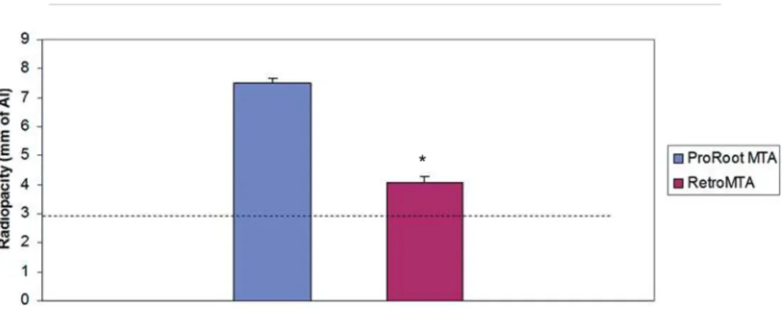
Figure 2 shows the mean values and standard deviations of the radiopacity levels for ProRoot® MTA
and RetroMTA® in mm of Al. While both materials
achieved the minimum required radiopacity value of 3 mm of Al, as recommended by ANSI/
ADA2, ProRoot® /0
radiopacity values (7.52±0.15 mm of Al) when compared to RetroMTA® (4.07±0.20 mm of Al)
!"""#$
levels of ProRoot® MTA and RetroMTA® throughout
&""'$ ProRoot® MTA varied from 9.93 to 8, while the
values for RetroMTA® varied from 9.93 to 7.9. It is
worth noting that the pH of both materials tended to decrease over time (Figure 3).
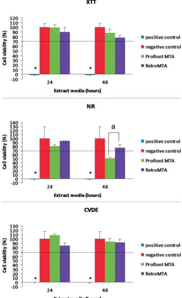
Both ProRoot® MTA and RetroMTA® allowed for
!"""#$ the XTT, NR and CVDE values for ProRoot® MTA
and RetroMTA® among the experimental periods,
the 48-hour extract media in the NR assay, in
which ProRoot®/0
DISCUSSION
In this study, we evaluated the radiopacity, pH variation and cytotoxicity of RetroMTA® and
ProRoot® MTA, and provided novel information on
some physicochemical and biological properties of this new material.
0%?^00 '2 recommends
that an endodontic material must have a radiopacity value higher than 3 mm of Al. Both ProRoot®
MTA and RetroMTA® met the established criteria,
however ProRoot®/0
radiopacity when compared to RetroMTA®. Our
results for the radiopacity of ProRoot® MTA are
in agreement with previous reports using similar
methodology4,5,140
on the radiopacity of RetroMTA®
literature, our values observed for radiopacity are similar to the values presented by the manufacturer (5 mm of Al).
Moreover, according to the manufacturers, the pH of ProRoot® MTA is ~12, and the pH of RetroMTA®
is initially 12.5, decreasing to pH 8 after four weeks. To evaluate the pH of the materials at different time periods, distilled water was changed after X increase over that period27. In the present study,
the pH of the tested materials was alkaline, varying from 9.93 to 8 for ProRoot® MTA and from 9.93 to
7.9 for RetroMTA®. The lower pH levels observed
here when compared to the manufacturers values may be explained by the different methodological approaches used. In this study, to simulate the surface area of the material exposed during clinical use, the cements were placed in plastic tubes for the analysis, thus decreasing the surface area of the material in contact with the liquid20,27, which may
have contributed to the lower pH values observed. similar sample sizes in which lower pH values for the ProRoot® MTA were observed when compared
Y 10,22,26.
It is worth noting that both materials showed a decrease in the pH over time. The cements were immersed in distilled water while fresh, allowing the material surface to be exposed before setting of the material. Further, this allows the release of hydroxyl ions, thus increasing the pH. Over time, the pH decreases, probably because the setting is complete27.
We also evaluated the cytotoxicity of ProRoot®
MTA and RetroMTA® using a multiparametric
cytotoxicity assay which allows the simultaneous evaluation of the toxic effects of tested materials Figure 3- Results of pH analysis over time
Figure 2- Radiopacity of ProRoot® MTA and RetroMTA® in mm of Al. Dashed line represents the radiopacity of 3 mm of Al
as recommended by ANSI/ADA No. 572.
in the same sample through three different parameters: mitochondrial metabolism and respiratory toxicity (XTT), lysossomal integrity and membrane permeability (NR), and cell proliferation and presence of DNA (CVDE). Our results showed that both ProRoot® MTA and RetroMTA® promoted
% differences were observed when comparing XTT, NR and CVDE values for ProRoot® MTA and RetroMTA®,
except for the 48-hour period in the NR assay. At this experimental period in the neutral red assay, the cell viability decreased in the ProRoot® MTA
group, although no major decrease in the XTT and CVDE values were observed, corroborating the
8 (2009). The observed
lower viability in the neutral red assay may indicate some adverse effect of ProRoot® MTA on membrane
integrity that could contribute to possible cell toxicity in vitro. Nonetheless, the effect alone may not be enough to provoke damage to normal cell function, as mitochondrial activity values were within normal limits and no effects of DNA damage were suspected in the XTT and CVDE assays. Moreover, the excellent in vivo biocompatibility of ProRoot®
has been reported on several studies17,21,25,30. The
main advantage of this multiparametric assay in comparison to other commonly used cytotoxicity / @'@ _@@ yl)-2,5-diphenyltetrazolium bromide)] is that it mechanisms through which the materials may be cytotoxic8,9. Although the MTT assay measures the
mitochondrial metabolic activity rate as an estimate of cell viability, an observed excess of metabolic activity may also represent a response to increased cellular stress due to toxicity, thus caution is recommended with the use of MTT as its estimation of cell viability may be misleading28,29. Moreover,
the MTT requires the cells to be killed, making it impossible to follow up on the cells in culture1, and
yet requires an extended incubation time with lower sensitivity than the XTT3. Our experimental model
designed to obtain extract media for the cell viability assay and the use of human periodontal ligament cells resembles a clinical scenario, simulating the amount and surface area of the material that usually comes in contact with surrounding tissues in most clinical applications8,9,12,29. Further, the use
of the extract media also simulates the scenario in which toxic elements released from the materials could be leaching into the periodontal ligament16.
Recently, Chung, et al.7 (2015) also investigated
the cytotoxicity of calcium-silicate cements including ProRoot® MTA and RetroMTA® on human
derived cells using a XTT assay. Human pulp-derived cells were grown in direct contact with the material, Dycal, or no cement for seven days.
Initial cell attachment, viability, calcium release, and the levels of vascular endothelial growth factor `|$ factor (FGF-2) were evaluated. The cell viability was tested with freshly mixed and set materials after three and seven days. These authors reported that the overall biocompatibility of RetroMTA® was
similar to those of the control and ProRoot® MTA,
CONCLUSION
The current study provides new and important data about the physicochemical and biological properties of RetroMTA® concerning radiopacity,
pH and cytotoxic effects on human periodontal
* +/0®
meets the radiopacity requirements stipulated by ANSI/ADA2, and presents similar pH values
and biocompatibility to ProRoot® MTA. Further
studies should be performed to evaluate additional properties of this new material.
ACKNOWLEDGEMENTS
from CAPES - Coordination of Higher Education and Graduate Training (BEX: 1099/12-4) to Leticia Souza. This study was supported by UTSD - University of Texas Health Science Center at Houston School of Dentistry startup funds to Ariadne Letra. HPDL cells were kindly provided by Dr. Isabel Gay, Department of Periodontics, University of Texas Health Science Center at Houston School of Dentistry. Thanks to Min Zhao for technical assistance.
X to this study.
REFERENCES
1- Al-Nasiry S, Geusens N, Hanssens M, Luyten C, Pijnenborg R. The use of alamar blue assay for quantitative analysis of viability, migration and invasion of choriocarcinoma cells. Hum Reprod. "")# "@
2- American National Standards Institute, American Dental 0 0%?^00 ? % ') ( )0%?^00"""
3- Atay A, Bozok Cetintas V, Cal E, Kosova B, Kesercioglu A, Guneri P. Cytotoxicity of hard and soft denture lining materials. Dent Mat "# #)#"@
4- Camilleri J. Evaluation of the physical properties of an J @ ^"#" ) #@" '@( / ^ "#" )#@ "
7- Chung CJ, Kim E, Song M, Park JW, Shin SJ. Effects of two fast-setting calcium-silicate cements on cell viability and angiogenic factor release in human pulp-derived cells. Odontology. 2015. Epub ahead of print.
8- De-Deus G, Canabarro A, Alves G, Linhares A, Senne MI, Granjeiro JM. Optimal cytocompatibility of a bioceramic nanoparticulate cement in primary human mesenchymal cells. J "" ')# @"
9- De-Deus G, Canabarro A, Alves GG, Marins JR, Linhares AB, Granjeiro JM. Cytocompatibility of the ready-to-use bioceramic putty repair cement iRoot BP Plus with primary human osteoblasts. ^"#')'"@#
10- Duarte MA, Demarchi AC, Yamashita JC, Kuga MC, Fraga SC. @ ?/J+ ""#")"@#' 11- Ghorbanzadeh A, Shokouhinejad N, Fathi B, Raoof M, Khoshkhounejad M. An in vitro comparison of marginal adaptation of MTA and MTA-like materials in the presence of PBS at one-week @ $"###)'"@ 12- Gomes Cornélio AL, Salles LP, Paz MC, Cirelli JA, Guerreiro-Tanomaru JM, Guerreiro-Tanomaru Filho M. Cytotoxicity of Portland cement with different radiopacifying agents: a cell death study. J Endod. "## )" @#"
13- International Organization for Standardization. ISO 10993-5:2009. Biological evaluation of medical devices – Part 5: Tests for in vitro )^?""
14- Islam I, Chng HK, Yap AUJ. Comparison of the physical and mechanical properties of MTA and Portland cement. J Endod. "" )# @
15- Kang SH, Shin YS, Lee HS, Kim SO, Shin Y, Jung IY, et al. Color changes of teeth after treatment with various mineral trioxide aggregate-based materials: an ex vivo study. J Endod. "#'#) @#
16- Keiser K, Johnson CC, Tipton DA. Cytotoxicity of mineral """)@#
17- Koçak S, Erten H, Baris E, Türk S, Alaçam T. Evaluation of the biocompatibility of experimentally manufactured portland cement: ( "#)#@#
18- Komabayashi T, Spångberg LS. Comparative analysis of the particle size and shape of commercially available mineral trioxide J ) X _"" )@
19- Marciano MA, Costa RM, Camilleri J, Mondelli RFL, Guimarães BM, Duarte MA. Assessment of color stability of white mineral trioxide aggregate Angelus and bismuth oxide in contact with "#")# '@"
20- Massi S, Tanomaru-Filho M, Silva GF, Duarte MA, Grizzo LT, Buzalaf MA, et al. pH, calcium ion release, and setting time of an experimental mineral trioxide aggregate-based root canal sealer. "## )@
21- Menezes R, Bramante CM, Letra A, Carvalho VG, Garcia RB. Histologic evaluation of pulpotomies in dog using two types of mineral trioxide aggregate and regular and white Portland cement as wound dressings. Oral Surg Oral Med Oral Med Oral Pathol Oral + "") @
@ ? /0 0 0 / ? Garcia-Godoy F, et al. Effect of particle size on calcium release and elevation of pH of endodontic cements. Dent Traumatol. "#' #)#@"#
23- Song JS, Mante FK, Romanow WJ, Kim S. Chemical analysis of powder and set forms of Portland cement, gray ProRoot MTA, white ProRootMTA, and gray MTA-Angelus. Oral Surg Oral Med J+ ""#")"@#'
24- Storm B, Eichmiller FC, Tordik PA, Goodell GG. Setting expansion of gray and white mineral trioxide aggregate and J"" )"@
25- Tawil PZ, Trope M, Curran AE, Caplan DJ, Kirakozova A, Duggan DJ, et al. Periapical microsurgery: an in vivo evaluation of @ "" ') '@ 26- Vasconcelos BC, Bernardes RA, Cruz SM, Duarte MA, Padilha PM, Bernardineli N, et al. Evaluation of pH and calcium íon release @ ?/J + ""#")# '@
27- Vivan RR, Zapata RO, Zeferino MA, Bramante CM, Bernardineli N, Garcia RB, et al. Evaluation of the physical and chemical properties of two commercial and three experimental root-end ? / / J + "#"##")'"@
28- Wang P, Henning SM, Heber D. Limitations of MTT and MTS-based assays for measurement of antiproliferative activity of green J?%"#"')#""
29- Yadlapati M, Souza LC, Dorn S, Garlet GP, Letra A, Silva RM. Deleterious effect of triple antibiotic paste on human periodontal ^"#)@'