• Nenhum resultado encontrado

Arq. Bras. Cardiol. vol.89 número2 en a16v89n2

N/A
N/A
Protected

Academic year: 2018

Share "Arq. Bras. Cardiol. vol.89 número2 en a16v89n2"

Copied!
1
0
0

Texto

(1)

Image

Poor Left Ventricle Compaction - Diagnosis Optimized by Real-time

Three-Dimensional Echocardiography

Gabriel Antonio Stanisci Miguel and Henry Abensur

Real e Benemérita Associação Portuguesa de Beneficência de São Paulo - São Paulo, SP - Brazil

Mailing address: Gabriel Antonio Stanisci Miguel •

Rua Santa Madalena, 290/131 - 01322-020 - São Paulo, SP - Brazil E-mail: gasmiguel@cardiol.br

Manuscript received November 13, 2006; revised manuscript received February 16, 2007; accepted March 13, 2007.

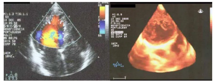
We treated a 37-year old male patient with diagnosis of poor left ventricle compaction. The two-dimensional echocardiography demonstrated extensive trabeculae associated with sinusoidal formation inside the left ventricle shown by color flow mapping. A real-time three-dimensional echocardiography confirmed these findings, and showed the

presence of several excessively prominent trabeculae and deep intertrabecular recesses, particularly in the apical region.

In cases of limited acoustic window, the three-dimensional imaging could provide more details through visualization of the cardiac structures by means of multiple observational plans, thus enhancing morphological and functional information (fig. 1).

Key words

Echocardiography; heart ventricles/anatomy & histology.

Fig. 1 -On the left, transthoracic two-dimensional echocardiography (in apical four chamber view), demonstrating sinusoids shown by color flow mapping. On the right, real-time transthoracic three-dimensional echocardiography (apical view), showing several trabeculae associated with intertrabecular recesses (trabecular sinusoids) inside the left ventricle.
Imagem

Fig. 1 - On the left, transthoracic two-dimensional echocardiography (in apical four chamber view), demonstrating sinusoids shown by color flow mapping

Referências

Documentos relacionados

Table 1 – Corrected coefficient (CC), amplitude (A), cyclic variation delay index (I) and pattern (P) of IBS curve for myocardial segments under study.. Values for continuous

Nowicki et al 20 , in their study on independent risk factors for aortic valve replacement surgery reported the following variables: age over 70 years, small body surface,

O objetivo do presente relato é descrever a caracterização anatômica e funcional do aneurisma apical em pacientes portadores de cardiopatia chagásica crônica, mediante

Together, these morphological and functional characteristics enable an accurate distinction to be made between the apical aneurysm of chronic Chagas’ heart disease and that

Os elementos clínicos sugerem diagnóstico de canal arterial, em decorrência da posição alta na borda esternal esquerda do sopro contínuo, raramente encontrada em outros tipos de

122.. 1 - Chest x-ray showing evidence of ductus arteriosus, as shown by the enlargement of the aorta and pulmonary arteries, in addition to left ventricular dilation and

Seus ensinamentos serão sempre um guia para nós todos, para a prática da medicina e o magistério sob os nobres princípios de dignidade e humanismo, que ele sempre defendeu.

No sumário, o artigo: “Modificação do padrão respiratório melhora o controle cardiovascular na hipertensão essencial” , considerar correto o nome do 1º autor: Carlos Hermano