Image
Saddle Shape of Mitral Valve Annulus: Three-Dimensional
Transthoracic Echocardiography
Marcelo Luiz Campos Vieira*, Prasad Maddukuri **, Natesa G. Pandian**, Wilson Mathias Jr.*, José Antônio F.
Ramires*
*Instituto do Coração do Hospital das Clínicas – FMUSP e ** Tufts University – New England Medical Center, São Paulo, SP, Brazil - Boston, MA, USA
Mailing Address: Marcelo luiz Campos Vieira •
Rua Cardoso de Melo, 463/21 - 04548-002 – São Paulo, SP, Brazil E-mail: [email protected]
Manuscript received October 11, 2005; revised manuscript received October 17, 2005; accepted October 17, 2005.
We are describing the case of volunteer, 28 years
old, male, submitted to transdimensional transthoracic
echocardiographic investigation (3D echo). Cardiac anatomy
showed to be normal. 3D echo analysis allowed the
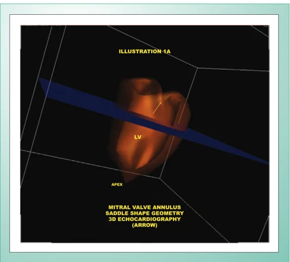
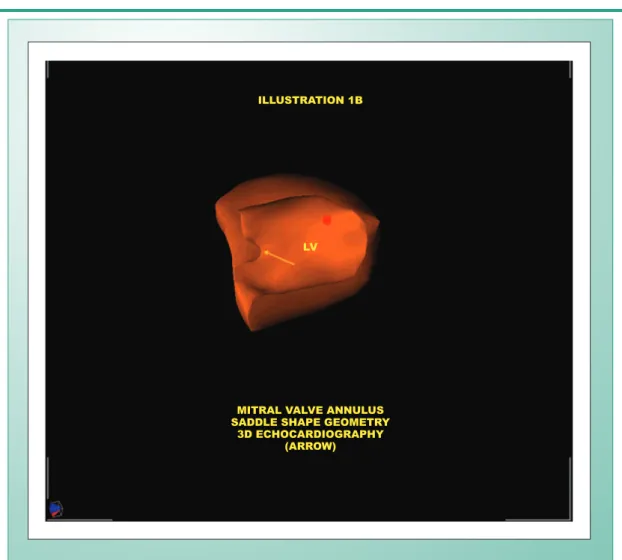
identification of saddle shape of mitral valve annulus (Fig. 1A
Key words
Echocardiography, three-dimensional echocardiography,
mitral valve, anatomy.
and Fig. 1B), which had not been identified by bidimensional
echocardiography. Three-dimensional echocardiography is
an imaging investigation method that leads to advancement
towards anatomic and diagnostic analysis
1,2.
This study was supported by CAPES, Brasilia, DF, Brazil.
Fig. 1A - Three-dimensional transthoracic echocardiogram (3D) (longitudinal, apical projection), showing saddle shape of mitral valve annulus (arrow). LV- left ventricle.
IllustratIon 1a
MItral ValVe annulus saddle shape GeoMetry
3d echocardIoGraphy (arroW)
lV
apex
Image
References
1. De Castro S, Salandin V, Cartoni D, et al. Qualitative and quantitative evaluation of mitral valve morphology by intraoperative volume rendered three-dimensional echocardiography. J Heart Valve Dis 2002;11(2):173-80.
2. Kwan J, Shiota T, Agler DA, et al. Geometric differences of the mitral apparatus between ischemic and dilated cardyomyopathy with significant mitral regurgitation: real-time three-dimensional echocardiography study. Circulation 2003;107(8):135-40.
Fig. 1B - Three-dimensional transthoracic echocardiogram (3D) (paraesternal, apical projection), showing saddle shape of mitral valve annulus (arrow). LV- left ventricle.
IllustratIon 1b
lV
MItral ValVe annulus saddle shape GeoMetry
3d echocardIoGraphy (arroW)