Original Article
2 2 2 Arq Bras Oftalmol. 2014;77(4):222-4 http://dx.doi.org/10.5935/0004-2749.20140057
INTRODUCTION
Posterior capsule opacification (PCO) is one of the most common postoperative complications following cataract surgery(1,2). It usually
leads to a decrease in visual acuity (VA), a loss of contrast sensitivity, and glare disability. It also precludes examination of the posterior segment(3). Patients who have visual function reduction in response
to PCO may be treated with Neodymium (Nd):YAG laser capsulotomy to restore VA, but this treatment is not free of complications(1,4).
Researchers are trying to identify the key factors in reducing the incidence of PCO(5,6). It has already been demonstrated that
sharp-edged intraocular lens (IOL) optics are preferable to round-edged IOL optics(7). Modification of the IOL surface, which can inhibit cell
and protein adhesion, has been suggested for reducing the incidence PCO(4). Furthermore, modifications of IOL design, maintaining an
open capsular bag, enhancing the aqueous endocapsular inflow, and even the use of a bag-in-the-lens technique, all appear to prevent capsular bag opacification(8,9).
The causes of opacification include cell proliferation and migra-tion, epithelial-to-mesenchymal cell transitions, collagen deposimigra-tion, and lens fiber regeneration of lens epithelial cells(10). The main factors
relating to PCO development are either patient related (e.g., the age and type of ocular disease), surgery related (e.g., the capsulorrhexis size, and whether irrigation/aspiration of the capsule, a hydrodis-section-enhanced cortical clean-up, sealed capsule irrigation, and in-the-bag IOL fixation, are performed), and IOL related (material and design of the implanted IOL)(8,10-12). The Ioflex (Mediphacos) is a 1-piece
foldable acrylic hydrophilic IOL for posterior chamber implantation, designed with square edges for PCO prevention. It has a low cost and
Incidence of posterior capsule opacification following the implantation of a foldable
hydrophilic acrylic intraocular lens: a 4 year follow-up study
Incidência da opaciicação da cápsula posterior após o implante de uma lente intraocular acrílica
hidrofílica dobrável: seguimento de 4 anos
Priscillade almeida Jorge1, delano Jorge2, camila Vieira Ventura3, Bruna Vieira Ventura3, Wagner lira3, marcelo carValho Ventura3, marcony rodrigues santhiago1, neWton Kara-Junior1
Submitted for publication: March 18, 2014 Accepted for publication: May 26, 2014
Study conducted at Altino Ventura Foundation (FAV) and in University of São Paulo (USP), Brazil. 1 Universidade de São Paulo (USP), São Paulo, SP, Brazil.
2 Fundação Leiria de Andrade (FLA), Fortaleza, CE, Brazil. 3 Fundação Altino Ventura (FAV), Recife, PE, Brazil.
Funding: No specific financial support was available for this study.
Disclosure of potential conflicts of interest: None of the authors have any potential conflicts of interest to disclose.
Corresponding author: Priscilla de Almeida Jorge. Rua Osvaldo Cruz, 201/401 - Fortaleza (CE) - 60125-150 - Brazil - E-mail: [email protected]
Aproved by Altino Ventura Foundation Ethics Committee: CEP: 054/2011.
ABSTRACT
Purpose: To evaluate the incidence of posterior capsule opacification (PCO) four years after the implantation of a hydrophilic acrylic intraocular lens (IOL). Methods: Fifty-eight randomly selected eyes from 58 patients were analyzed four years after phacoemulsification and Ioflex IOL implantation. The patients underwent an ophthalmic examination to detect PCO and a detailed medical history was obtained. The patients’ charts were reviewed for their corrected distance visual acuity prior to the IOL implantation, as well as for one month postoperatively. The Student’s t-test was used for statistical analysis.
Results: The mean age of patients without PCO was 74.6 ± 9.5 years, compared to 70.3 ± 15 years in patients with PCO. Four years after surgery, 39 of the 58 eyes (67%) had detectable PCO and 24 eyes (41.3%) had decreased visual acuity (VA) due to PCO. These patients were referred for Nd:YAG laser capsulotomy. Three pa-tients (5.1%) had decreased VA due to glaucoma, IOL opacification, or age-related macular degeneration. Twelve eyes (20.7%) presented mild PCO with unchanged VA. Systemic arterial hypertension was reported by 45% of the patients, and in 3.5% of these cases this was associated with diabetes mellitus.
Conclusion: This study found the incidence of PCO to be 67% four years after phacoemulsification and Ioflex IOL implantation.
Keywords: Posterior capsule of the lens; Capsule opacification; Postoperative complications; Cataract/epidemiology; Visual acuity
RESUMO
Objetivo: Avaliar a incidência da opacificação da cápsula posterior com o implante de uma lente intraocular acrílica hidrofílica.
Métodos: Cinquenta e oito olhos, de 58 pacientes, selecionados de forma aleatória, foram examinados 4 anos após a cirurgia de facoemulsificação com implante da lente intraocular Ioflex em uma campanha comunitária para pessoas carentes. Os pacientes foram submetidos ao exame oftalmológico a fim de detectar opacificação da cápsula posterior. Foi obtido histórico médico detalhado. A acuidade visual corrigida antes e 1 mês após a cirurgia foi obtida através de revisão em prontuário médico. O teste tde student foi utilizado para a análise estatística.
Resultados: A idade média dos pacientes sem opacificação da cápsula posterior foi 74,6 ± 9,5 anos, e 70,3 ± 15 anos nos pacientes com opacificação da cápsula posterior. Após 4 anos da cirurgia, 39 olhos (67%) foram diagnosticados com opacificação da cápsula posterior, e 24 olhos (41,3%) tiveram redução da acuidade visual causada pela opacificação da cápsula posterior, sendo encaminhados para realização de capsulotomia com Nd:YAG laser. Três olhos (5,1%) tiveram redução da acuidade visual causada por glaucoma, opacificação da lente intraocular e degeneração macular rela-cionada à idade. Em outros 12 olhos (20,7%) que apresentaram opacificação da cápsula posterior, a acuidade visual ficou mantida. Dentre as doenças sistêmicas, a hipertensão arterial foi relatada por 45% da amostra avaliada e 3,5% referiram diabetes mellitus. Conclusão: O estudo encontrou incidência de 67% de opacificação da cápsula pos-terior na lente intraocular Ioflex 4 anos após a cirurgia.
JorgePA, et al.
2 2 3 Arq Bras Oftalmol. 2014;77(4):222-4 has been marketed for over ten years in several European, Asian, and
Latin American countries. Its postoperative performance has not yet been studied. Thus, this study aimed to evaluate the incidence of PCO four years after Ioflex IOL implantation.
METHODS
A sample of 150 eyes was randomly selected. This sample size calculation was based on the PCO incidence reported in the litera-ture. The patients were from a population that had undergone cataract surgery and Ioflex IOL implantation in 2007, as part of a community campaign for underprivileged people. A total of 50 eyes (31 patients) could not be located, either due to communication difficulties relating to geographical distance, or to patient death. Of the 100 eyes located, 13 were not presented for examination. Thus, 87 eyes corresponding to 58 patients, were available for examination. However, to avoid bias, in patients who underwent cataract surgery in both eyes, only the right eye was included in the study. Therefore, a final total of 58 eyes from 58 patients were submitted to a detailed ophthalmic examination.
The inclusion criteria comprised being subjected to phacoemul-sification and Ioflex IOL implantation between January and June 2007. The exclusion criteria comprised intraoperative complications, such as posterior capsule rupture, and non-Ioflex IOL implantation. The same surgeon performed all surgical procedures using a Uni-versal II Phacoemulsifier (Alcon). In each case a 2.8 mm incision was made under peribulbar anesthesia, using balanced salt solution (BSS®, Alcon) and 2% methylcellulose viscoelastic (Ophthalmos). Identical continuous curvilinear capsulorrhexis and cortical clean- up procedures were used in all cases. The ophthalmic examination in -clu ded measurements of corrected distance visual acuity (CDVA) and biomicroscopy slit lamp evaluations after pupil dilation with Mydriacyl (Alcon). Patients with a VA decreased in response to PCO were referred to Nd:YAG laser treatment. A detailed medical history for each patient was also obtained. The patients’ charts were reviewed for their CDVA prior to IOL implantation as well as for one month pos -toperatively.
This study was approved by an Ethics Committee (CEP: 054/2011) and followed the Declaration of Helsinki principles. The variables were expressed as the mean and standard error of the mean. We used the Student’s t-test for independent samples to check for possi-ble differences in VA, and the Student’s t-test for paired samples to analyze the mean VA of patients with and without PCO during the evaluation periods. A p value of <0.05 was adopted for rejection of the null hypothesis.
RESULTS
Of the 58 patients, 27 were male and 31 female. The mean age of patients without PCO in the four year follow-up period was 74.6 ± 9.5 years, and the mean age of patients with PCO over the same period was 70.3 ± 15.1. There was no statistically significant difference between the mean age of the patients with or without PCO (p=0.182). All surge-ries were uneventful both intra and postoperatively.
There was no statistically significant difference between the mean VA of the patients with or without PCO during the evaluation period. Preoperative CDVA ranged from hand movement to 0.30 logMAR. One month after cataract surgery with Ioflex IOL implantation, impro-vement of VA was observed in all but two patients. These two patients either had glaucoma or an age-related macular degeneration. On sys-temic disease evaluation, 45% (26 patients) reported syssys-temic arterial hypertension and 3.5% (2 patients) had diabetes mellitus.
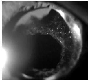
Four years after surgery, 39 out of 58 patients (67%) had PCO as detected by a slit lamp examination. Of the total, 24 eyes (41.3%) had decreased VA due to PCO and were referred for Nd:YAG laser capsulotomy (Figure 1). Three eyes (5.1%) had decreased VA due to glau coma, IOL opacification, and age-related macular degeneration,
respectively. In a further 12 eyes (20.7%) with mild PCO, VA remained unchanged.
DISCUSSION
This study aimed to evaluate the PCO rates in Ioflex IOLs four years after surgery. We found that 67% of patients suffered from this postoperative complication. Clinical studies have shown varied incidences of PCO for other hydrophilic IOLs, ranging from 4.6% to
50%(1,13-15). Notably, all of these incidence rates are less than the one
we have found. To our knowledge, there are no other published stu-dies that have reported the incidence of PCO in the Ioflex IOL. As the eradication of PCO is a major goal of researchers working to improve IOL materials and design, as well as surgical techniques(6,10,16), it is
important that additional research is performed.
The IOL material and design is able to influence the incidence of PCO(5,7,8). It has been well documented that a hydrophobic lens
ma terial causes less PCO than a hydrophilic material, even though
the latter is more uveal biocompatible(17,18). However, PCO may
pro-gresses significantly with time and is more extensive following poly-methylmetacrylate IOL implantation, while its progression rate after silicone and acrylic IOL implantation is low(19).
Previous reports have shown that hydrophilic IOLs lead to at least twice the number of Nd:YAG laser treatments when compared with hydrophobic lenses(1,13,17,18). Although Nd:YAG laser capsulotomy is
the standard treatment for PCO and is generally found to be safe and effective, it is also expensive and not free of complications(1,11,20),
inclu-ding a significant rate of morbidity from postoperative complications. The most common complication is intraocular pressure elevation, while other complications include cystoid macular edema, retinal hemorrhage or detachment, iritis, vitreous prolapse, corneal damage, vitritis, iris damage, pupillary blockage, hyphema, IOL subluxation, and localized exacerbation of endophthalmitis(21,22). PCO is also
asso-ciated with socioeconomic factors, since an outpatient examination is required to diagnose it and, depending on local policies, an addi-tional hospital visit may be required for Nd:YAG laser treatment(23).
All of these factors emphasize the importance of efforts aimed at minimizing Nd:YAG laser capsulotomy necessity.
Following technological advances, IOL design has been modified from round-edged to square-edged, and this has resulted in a reduction in the incidence of PCO(24). However, studies suggest that square-edged
IOLs should completely encompass the 360 degrees around the IOL optic in order to provide an effective barrier(25). Furthermore, another
study has shown differences between brands of square-edged
Incidence of posterior capsule opacification following the implantation of a foldable hydrophilic acrylic intraocular lens: a 4 year follow-up study
224 Arq Bras Oftalmol. 2014;77(4):222-4
ses, and suggested that variations in their edge profiles may account for clinical differences in postoperative PCO rates(26). In addition, it has
been reported that acrylic IOLs appear to lose their PCO preventive effect, despite their sharp optic edges(27).
Although the Ioflex is a 1-piece lens, this not considered to be a factor in the development of PCO, since both 1- and 3-piece IOLs have been studied to evaluate their influence on PCO formation and no statistically significant differences between them have been found(28,29).
PCO is also age-dependent, being more frequently observed as a postoperative complication in young people and children(7,30). This
study evaluated an adult population and thus did not find a statis-tically significant difference on the independent analysis of age between patients with and without PCO.
Decreased VA induced by PCO was noted in 41% of patients. This value is supported by data in the published literature, and confirms that it represents the most frequent cause of vision reduction follo-wing cataract surgery(7,15). Only one patient presented IOL
opacifica-tion, but we had previously reported, in another study with the same IOL, an incidence as high as 7% of long term IOL opacification, with no correlation to PCO(16).
CONCLUSION
This study found a PCO incidence of 67% four years after Ioflex IOL surgery. This rate is higher than that reported for other hydro-philic lenses described in the literature. A better understanding of the pathogenic mechanisms of PCO is highly desirable as a basis for improving the outcome of cataract surgery and for eradicating this serious postsurgical complication.
REFERENCES
1. Johansson B. Clinical consequences of acrylic intraocular lens material and design: Nd:YAG-laser capsulotomy rates in 3 x 300 eyes 5 years after phacoemulsification. Br J Ophthalmol. 2010;94(4):450-5.
2. Auffarth GU, Brezin A, Caporossi A, Lafuma A, Mendicute J, Berdeaux G, Smith AF; Eu ropean PCOStudy Group. Comparison of Nd:YAG capsulotomy rates following pha-coemulsification with implantation of PMMA, silicone, or acrylic intraocular lenses in four European countries. Ophthalmic Epidemiol. 2004;11(4):319-29.
3. Senne FM, Temporini ER, Arieta CL, Pacheco KD. Perception of difficulties with vision-related activities of daily living among patients undergoing unilateral posterior capsulotomy. Clinics. 2010;65(5):459-68.
4. Apple DJ, Peng Q, Visessook N, Werner L, Pandey SK, Escobar-Gomez M, et al. Eradi-cation of posterior capsule opacifiEradi-cation: documentation. of a marked decrease in Nd:YAG laser posterior capsulotomy rates noted in an analysis of 5416 pseudophakic human eyes obtained postmortem. Ophthalmology. 2001;108(3):505-18. Comment in: Ophthalmology. 2002;109(4):625; author reply 625-6; Ophthalmology. 2002;109(4):626; author reply 626-7.
5. Hazra S, Palui H, Vemuganti GK. Comparison of design of intraocular lens versus the material for PCO prevention. Int JOphthalmol. 2012;5(1):59-63.
6. Kavoussi SC, Werner L, Fuller SR, Hill M, Burrow MK, McIntyre JS, et al. Prevention of capsular bag opacification with a new hydrophilic acrylic disk-shaped intraocular lens. J Cataract Refract Surg. 2011;37(12):2194-200. Comment in: J Cataract refract Surg. 2012;38(5):924-5; author reply 925.
7. Buehl W, Findl O. Effect of intraocular lens design on posterior capsule opacification. J Cataract Refract Surg. 2008;34(11):1976-85.
8. Leishman L, Werner L, Bodnar Z, Ollerton A, Michelson J, Schmutz M, et al. Prevention
of capsular bag opacification with a modified hydrophilic acrylic disk-shaped intrao-cular lens. J Cataract Refract Surg. 2012;38(9):1664-70.
9. Tassignon MJ, Gobin L, Mathysen D, Van Looveren J, De Groot V. Clinical outcomes of cataract surgery after bag-in-the-lens intraocular lens implantation following ISO standard 11979-7:2006. J Cataract Refract Surg. 2011;37(12):2120-9.
10. Awasthi N, Guo S, Wagner B. Posterior capsular opacification: a problem reduced but not yet eradicated. Arch Ophthalmol. 2009:127(4):555-62.
11. Apple DJ, Werner L. Complications of cataract and refractive surgery: a clinicopatho-logical documentation. Trans Am Ophthalmol Soc. 2001;99:95-107; discussion 107-9. 12. Apple DJ. Influence of intraocular lens material and design on postoperative
intracap-sular cellular reactivity. Trans Am Ophthalmol Soc. 2000;98:257-83.
13. Gauthier L, Lafuma A, Laurendeau C, Berdeaux G. Neodymium:YAG laser rates after bilateral implantation of hydrophobic or hydrophilic multifocal intraocular lenses: twenty-four month retrospective comparative study. J Cataract Refract Surg. 2010; 36(7):1195-200.
14. Medeiros HA, Avila M, Santos PM. [Incidence of posterior capsule opacification in patients submitted to phacoemulsification and expandable acrylic intraocular lens implantation]. Arq Bras Oftalmol. 2006;69(4):371-5. Portuguese.
15. De Senne FM, Cardillo JA, Rocha EM, Kara-Jose N. Long-terns visual outcomes in the Cataract-Free Zone Project in Brazil. Acta Ophthalmol Scand. 2002;80(3):262-6. 16. Jorge PD, Jorge D, Ventura CV, Ventura BV, Lira W, Ventura MC, et al. Late opacification
in hydrophilic acrylic intraocular lenses: Analysis of 87 eyes in a random sample of 102 patients. J Cataract Refract Surg. 2013;39(3):403-7.
17. Vasavada AR, Raj SM, Shah A, Shah G, Vasavada V, Vasavada V. Comparison of posterior capsule opacification with hydrophobic acrylic and hydrophilic acrylic intraocular lenses. J Cataract Refract Surg. 2011;37(6):1050-9.
18. Kugelberg M, Wejde G, Jayaram H, Zetterstroem C. Two-year follow-up of posterior capsule opacification after implantation of a hydrophilic or hydrophobic acrylic in -traocular lens. Acta Ophthalmol. 2008;86(5):533-6.
19. Hayashi K, Hayashi H, Nakao F, Hayashi F. Changes in posterior capsule opacification after poly(methyl methacrylate), silicone, and acrylic intraocular lens implantation. J Cataract Refract Surg. 2001;27(6):817-24.
20. Karahan E, Tuncer I, Zengin MO. The Effect of ND:YAG laser posterior capsulotomy size on refraction, intraocular pressure, and macular thickness. J Ophthalmol. 2014;2014: 846385. Epub 2014 Mar 3.
21. Burq MA, Taqui AM. Frequency of retinal detachment and other complications after neodymium:Yag laser capsulotomy. J Pak Med Assoc. 2008;58(10):550-2.
22. Billotte C, Berdeaux G. Adverse clinical consequences of neodymium:YAG laser treatment of posterior capsule opacification. J Cataract Refract Surg. 2004;30(10): 2064-71.
23. Chang A, Behndig A, Ronbeck M, Kugelberg M. Comparison of posterior capsule opacification and glistenings with 2 hydrophobic acrylic intraocular lenses: 5-to 7-year follow-up. J Cataract Refract Surg. 2013;39(5):694-8.
24. Maddula S, Werner L, Ness PJ, Davis D, Zaugg B, Stringham J, et al. Pathology of 157 human cadaver eyes with round-edged or modern square-edged silicone intraocular lenses: analyses of capsule bag opacification. J Cataract Refract Surg. 2011;37(4):740-8. 25. Werner L, Mamalis N, Pandey SK, Izak AM, Nilson CD, Davis BL, et al. Posterior capsule opacification in rabbit eyes implanted with hydrophilic acrylic intraocular lenses with enhanced square edge. J Cataract Refract Surg. 2004;30(11):2403-9.
26. Werner L, Tetz M, Feldmann I, Bucker M. Evaluating and defining the sharpness of intraocular lenses: microedge structure of commercially available square-edged hy-drophilic intraocular lenses. J Cataract Refract Surg. 2009;35(3):556-66.
27. Vock L, Menapace R, Stifter E, Georgopoulos M, Sacu S, Buehl W. Posterior capsule opacification and neodymium:YAG laser capsulotomy rates with a round-edged sili-cone and a sharp-edged hydrophobic acrylic intraocular lens 10 years after surgery. J Cataract Refract Surg. 2009;35(3):459-65.
28. Pandey SK, Apple DJ, Werner L, Maloof AJ, Milverton EJ. Posterior capsule opacifica-tion: a review of the aetiopathogenesis, experimental and clinical studies and factors for prevention. Indian J Ophthalmol. 2004;52(2):99-112.
29. Ness PJ, Werner L, Maddula S, Davis D, Zaugg B, Stringham J, et al. Pathology of 219 human cadaver eyes with 1-piece or 3-piece hydrophobic acrylic intraocular lenses: Capsular bag opacification and sites of square-edged barrier breach. J Cataract Refract Surg. 2011;37(5):923-30.