INTRODUCTION
Chemical exposure to the eye can cause trauma ranging from mild irritation to the severe damage of ocular surfaces such as the conjunctiva, limbus, and anterior segment. Such damage has the potential to result in permanent vision loss. Chemical burns of the cornea cause supericial and deep neovascularization(1-3) which can lead to signiicant vision loss because of scar formation and lipid deposition(4). Regarding the impact of strong alkalis, ocular tissues have a limited ability to protect against such burns, which denature
proteins and saponify lipids. Overall, chemical burns are responsible for an ocular trauma prevalence of between 7.7% and 18%.
Neovascularization is a poorly understood pathologic response of the cornea against chronic inlammation that is usually due to infection, sterile corneal ulceration, chemical or thermal injuries, or the immune rejection of corneal grafts. Following chemical burns, inlammatory cells such as polymorphonuclear leukocytes, and me-senchymal cells such as myoibroblasts, activated keratocytes, ma-crophages, and neovascularization factors are activated. Some of the
Comparison of the effects of subconjunctival and topical anti-VEGF therapy
(bevacizumab) on experimental corneal neovascularization
Comparação do efeito da terapia anti-VEGF (bevacizumab) subconjuntival e tópica na
neovascularização corneana experimental
Ozdemir Ozdemir1,Ozgul AltintAs2, levent AltintAs3, BernA OzkAn2, Cigdem AkdAg2, NurşeN Yüksel2
Submitted for publication: February 26, 2014 Accepted for publication: May 7, 2014 Study conducted at Kocaeli University.
1 Ophthalmology Department, Zekai Tahir Burak Women’s Health Education and Research Hospital, Ankara, Turkey.
2 Ophthalmology Department, Faculty of Medicine, Kocaeli University, Kocaeli, Turkey. 3 School of Occupational Health and Safety, Kocaeli University, Kocaeli, Turkey.
Funding: No specific financial support was available for this study.
Disclosure of potential conflicts of interest: None of the authors have any potential conflicts of interest to disclose.
Corresponding author: Ozdemir Ozdemir. Göz Hastalıkları Polikliniğis - Zekai Tahir Burak Kadın Sağlığı Eğitim Ve Araştırma Hastanesi - Talatpaşa Bulvarı, Altındağ, Ankara, Turkey
E-mail: [email protected] Project number: KOÜ HADYEK ¼-2010.
ABSTRACT
Purpose: To compare the effects of bevacizumab applied either subconjunctivally or topically, in a rat model of corneal neovascularization induced by alkali burn. Methods: The right corneas of 24 Wistar-Albino rats were cauterized with silver nitrate sticks. The rats were divided randomly and equally into three groups: no treatment control (n=8), subconjunctival bevacizumab treatment (n=8), and topical bevacizumab treatment (n=8). Immediately following cauterization, the subconjunctival group was treated with a 0.05 ml (1.25 mg) bevacizumab subcon-junctival injection. The topical group was treated with 10 mg/ml bevacizumab twice daily, and the control group received subconjunctival saline injections twice daily. The burn stimulus and neovascularization scores were evaluated using a technique previously described by Mahoney and Waterbury. Digital photographs were obtained before the eyes were enucleated and corneal sections were then analyzed by histopathology.
Results: The mean burn stimulus score was 1.86 ± 0.6 and there was no statistical difference between the groups (p=0.730). The mean neovascularization scores in the subconjunctival and topical bevacizumab groups were statistically lower than the control group (p<0.05). The mean percentage area of corneal neovascularization was 82.5 ± 22.1 in the control group, 42.7 ± 15.0 in the subconjunctival group, and 55.8 ± 18.2 in the topical group. The differences between the control and treatment groups were statistically significant (p<0.05). Histopathology showed that the treatment groups presented less neovascularization, inflammation, and fibroblast activity than the control group (p<0.05).
Conclusions: This study demonstrates that both subconjunctival and topical administrations of bevacizumab inhibit corneal neovascularization and decrease inflammation and fibroblast activity in a rat model of corneal neovascularization induced by alkali burn.
Keywords: Corneal neovascularization/chemically induced; Vascular endothelial growth factor A; Angiogenesis inhibitors/administration and dosage; Injections; Disease models, animal; Corneal diseases; Animals; Rats
RESUMO
Objetivo: Comparar o efeito de bevacizumab aplicado subconjuntival e topicamente em um modelo de neovascularização de córnea de ratos induzida por queimadura alcalina.
Métodos: Córneas direitas de 24 ratos Wistar-Albino foram cauterizados por nitrato de prata. Os indivíduos foram divididos aleatoriamente e igualmente em três grupos: controle (n=8), o bevacizumab subconjuntival (n=8), o bevacizumab tópico (n=8). Imediatamente após a cauterização, 0,05 ml (1,25 mg) de bevacizumab foi injetado no grupo subconjuntival. Grupo tópico foi inculcado com 10 mg/ml de bevacizumab duas vezes por dia. O grupo controle recebeu solução salina normal, topicamente, duas vezes ao dia. A graduação do estímulo da queimadura e a graduação da neovas-cularização foram avaliados utilizando a técnica descrita por Mahoney e Waterbury. Fotografias digitais foram obtidas dos olhos serem enucleados. Seções da córnea foram analisadas por histopatologia.
Resultados: A média da graduação do estímulo da queimadura foi de 1,86 ± 0,6 e não houve diferença estatisticamente entre os grupos (p=0,730). As médias das graduações da neovascularização no grupo bevacizumab subconjuntival e no grupo bevacizumab tópico foram estatisticamente menores do que o grupo controle (p<0,05). A percentagem média de área de neovascularização da córnea foi de 82,5 ± 22,1 no grupo controle, 42,7 ± 15,0 no grupo subconjuntival e 55,8 ± 18,2 no grupo tópico. As diferenças entre os grupos de tratamento e grupo de controlo foram estatisticamente significativos (p<0,05). A histopatologia mostrou que os grupos de tratamento apre-sentavam menos neovascularização, inflamação e atividade de fibroblastos do que o grupo controle (p<0,05).
Conclusões: Este estudo demonstra que a administração tanto subconjuntival quanto tópica de bevacizumab inibe a neovascularização da córnea, e diminui a inflamação e atividade de fibroblastos em córneas de ratos submetidas a queimaduras alcalinas.
factors responsible for the inducement of inlammation include vas-cular endothelial growth factor (VEGF), transforming growth factors, platelet-activating factor, basic ibroblast growth factor, and tumor necrosis factor-α(5-7). It has previously been reported that VEGF is upre-gulated in corneas with inlammation and vascularisation and that it is a signiicant angiogenic factor in corneal neovascularization(8,9).
Bevacizumab is a monoclonal antibody that binds to VEGF-A and all its isoforms. Its amino acid sequence comprises both human IgG (93%) and murine antibody (7%). It has been approved as an antian-giogenic pharmaceutical for the treatment of certain cancers and it has also been utilized to treat ocular neovascularization(10). In several studies it has been reported that topically or subconjunctivally applied bevacizumab results in the inhibition or regression of corneal neovascularization(11-13).
In this study we aimed to observe the efects of bevacizumab on experimentally produced corneal neovascularization in a rat model, and to compare the efects of this therapy when subconjunctivally or topically applied.
METHODS
Twenty-four male rats (Wistar-Albino) weighing between 250 g and 300 g and with two healthy eyes, were used in the study. All re-search obeyed the statement of the Association for Rere-search in Vision and Ophthalmology (ARVO) for the Use of Animals in Ophthalmic and Vision Research. It was approved by the Local Ethics Committee for Animal Experiments of Kocaeli University. All rats were kept in indivi-dual cages and were managed under identical conditions.
Xylazine hydrochloride (5 mg/kg) (Rompun®, Bayer, Turkey) and ketamine hydrochloride (50 mg/kg) (Ketalar®, Eczacibasi, Turkey) were administered intraperitoneally for anesthesia and analgesia. For inducing corneal neovascularization, a previously described cau-terization technique using silver nitrate was used(14). The right corneas of the rats were cauterized with a chemical applicator stick of 2 mm diameter, consisting of 75% silver nitrate and 25% potassium nitrate (Hemo Stop®, Istanbul, Turkey). This was touched onto the central corneas for eight seconds under an operating microscope. After cauterization, corneas and fornices were irrigated with 10 ml of normal saline in order to remove any residual silver nitrate. Burn stimulus res-ponses were evaluated using the technique described by Mahoney and Waterbury(14). The burn stimulus response was scored as grade 0 (no blister), + 1 (small blister), + 2 (medium blister), or + 3 (large blister), according to lesions detected on the corneal surface (Figures 1, 2). Following cauterization, the burn stimulus scores were immediately calculated and treatment was begun.
The rats were categorized randomly into three groups of eight. The rats in the subconjunctival bevacizumab group were treated with a subconjunctival injection of 0.05 ml (1.25 mg) bevacizumab (Altu-zan® 400 mg/16 ml, F. Hofmann-La Roche Ltd., Basel, Switzerland). The rats in the topical group were treated topically with bevacizumab solution at a concentration of 10 mg/ml, and the rats in the control group received subconjunctival saline injections twice daily. The sub conjunctival injections were performed with a 30 gauge needle under an operating microscope. Needles were inserted 2 mm pos-terior from the limbus at the superior temporal bulbar conjunctiva. Bevacizumab solution was prepared by dilution with normal saline under sterile situations and was kept at 4°C. The solution was applied twice per day (with the initial application performed immediately after cauterization) for seven days. All procedures were performed by the same investigator (O.O.).
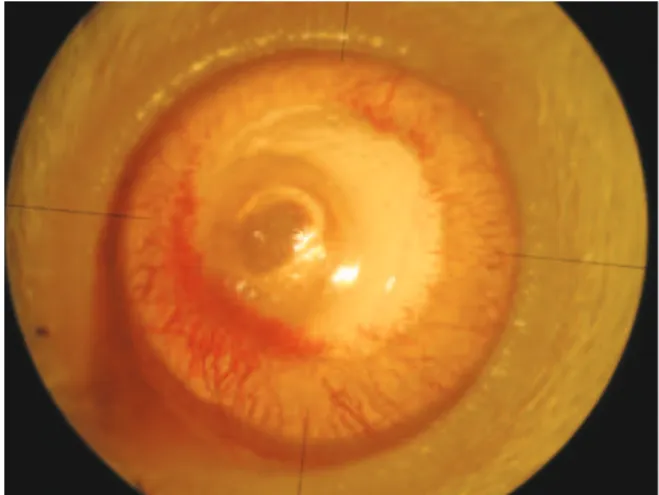
On the eighth day, a slit-lamp microscopy examination was per-formed and digital photographs were taken under anesthesia and analgesia (Figure 3). For each eye, the neovascularization response was evaluated toward the centrally placed burn from the entire cor-neal circumference, as follows: Grade 0, no visible vessels in cornea; Grade 1.5, 1/4 distance to burn; Grade 2, 1/3 distance to burn; Grade 3,
1/2 distance to burn; Grade 4, 2/3 distance to burn; Grade 4.5, 3/4 dis-tance to burn; and Grade 6, vessels reach burn(14). The area of corneal neovascularization was analyzed using a software program (Matlab™
Figure 2. Image of a cornea following cauterization.
Figure 1. Baseline image of a non-cauterized cornea.
R2007b version 7.5, Math Works, Natick, Massachusetts, USA). An investi-gator who was blinded to the study groups initially drew blood vessel borders and cornea circumferences on the photographs. The pro-gram then calculated the total cornea area and the neovascularized area. It then estimated the percentage of corneal neovascularization (neovascularized area/total cornea area × 100).
The rats were sacriiced on the eighth day using a high dose of pentothal sodium (Pentothal®, Abbott, Italy). The globes were then enucleated and ixed in 10% bufered formalin for 24 h. Corneas were then excised from the limbus and 5 µm thick sections were prepared. Sections were sliced from both the central region of the burn area and the intensive neovascularization area. They were then stained with haematoxylin-eosin and were analyzed using light microscopy. Sections were evaluated according to the intensity of neovasculari-zation, the intensity of inlammation, and the ibroblast activity. Light microscopic inspections were performed on sections by an examiner (D.K.Y.) who was blinded to the study groups. The following scaling system, previously described in a study on corneal neovasculariza-tion, was used (Figure 4)(15).
Intensity of neovascularization: + 1: minimal or close to negative vascularization. + 2: limited or focal vascularization in the subepithelial and prestromal areas. + 3: cases intermediate to groups 2 and 4. + 4: difuse and intense vascularization.
Intensity of inflammation: + 1: minimal or close to negative inlamma -tion. + 2: focal, low count of mixed inlammatory cell types such as lymphocytes, neutrophil leukocytes, and eosinophil leukocytes). + 3: cases intermediate to groups 2 and 4. + 4: intense, difuse, mixed inlammatory cell types.
Fibroblast activity: + 1: minimal or close to negative ibroblast acti -vity. + 2: focal ibroblast acti-vity. + 3: cases intermediate to groups 2 and 4. + 4: difuse and intense ibroblast activity.
S
TATISTICALANALYSISStatistical analyses were performed using SPSS 16.0 (SPSS™ Inc., Chi cago, IL, USA). Data were evaluated as the mean ± standard deviation (SD). The Kruskal-Wallis test was performed for multiple com parisons of groups. When a statistically signiicant diference was found, a pair-wise comparison of groups was performed using the Mann-Whitney U test. Signiicance was deined as a p value <0.05.
RESULTS
The mean burn stimulus score was 2.0 ± 0.7 for the control group, 1.8 ± 0.6 for the subconjunctival bevacizumab group, and 1.7 ± 0.4 for the topical bevacizumab group. No statistical diferences were found between the groups (p=0.730).
The mean neovascularization score was 5.5 ± 0.8 for the control group, 2.8 ± 1.7 for the subconjunctival bevacizumab group, and 3.5 ± 1.6 for the topical bevacizumab group. The mean neovascularization scores of the subconjunctival and topical bevacizumab groups were statistically lower than the control group (p=0.015 andp=0.035, res-pectively). In a comparison between the subconjunctival and topical bevacizumab groups, the mean neovascularization score was lower in the subconjunctival group. However, the diference was not statis-tically signiicant (p=0.512).
The mean percentage of corneal neovascularization area is shown in igure 5. It was found to difer signiicantly between groups (p=0.001). Both the subconjunctival and topical bevacizumab treat ments inhi-bited corneal neovascularization. However, there was no statistically signiicant diference between these two treatments (p=0.142).
In the histopathological evaluation, both treatment groups dis-played less neovascularization, inlammation, and ibroblast activity than the control group (p<0.05) (Table 1). The diference between the subconjunctival and topical bevacizumab groups was not statistically signiicant for the following evaluations: intensity of neovasculariza-tion (p=0.298), intensity of inlammation (p=0.960), and ibroblast acti vity (p=0.225).
DISCUSSION
Corneal neovascularization originates from venules and capillaries of the pericorneal (limbal) plexus. Types of corneal neovascularization include vascular pannus, supericial vascularization, and stromal vas-cularization. Many medical and surgical treatments have been repor-ted for the management of corneal neovascularization. Traditionally, topical steroids are recommended, primarily due to their suppression of actively growing neovascularization. Medical treatments may also include non-steroidal anti-inlammatory agents such as rapamycin, cyclosporine A, gene therapy approaches, methotrexate, and anti-VEGF agents. Laser and surgical treatments include the use of argon lasers, yellow lasers, Nd:YAG lasers, supericial keratecto my, diathermy, cau-terization, and photodynamic therapy(16).
VEGF is a family of proteins comprising VEGF-A, -B, -C, and -D, the viral VEGF homologue VEGF-E, and placental growth factor(10). VEGF-A is one of the most important mediators of angiogenesis. It is upregulated under conditions of neovascularization and it plays an important role in the development of pathological angiogenesis in inlammatory, neoplastic, and vascular diseases of the eye. Nume-rous studies have shown that anti-VEGF agents, used either alone or in combination with steroids, verteporin, or anti-tumor necrosis factorα microantibody, are efective in the treatment of corneal neo -vascularization(17). Anti-VEGF agents reported for the treatment of corneal neovascularization include bevacizumab, pegaptanib, and
Figure 4. Histopathological preparation of a cornea. Inlammatory cells (thin arrow), ibroblasts (black thick arrow), blood vessels, and stained erythrocytes within the blood vessels (white thick arrow) can be seen. The corneal specimens were stained with hematoxylin-eosin and examined using light microscopy (magniication × 400).
ranibizumab. It has been reported that bevacizumab, ranibizumab, pegaptanib, and trastuzumab all have the ability to inhibit corneal neo-vascularization, and that bevacizumab is the most efective treatment in an experimental rat model(18). Various experimental and clinical studies have reported that bevacizumab inhibits corneal neovascu-larization when used at diferent doses both topically and subcon-junctivally(11-13).
In our comparison of topical and subconjunctival applications of bevacizumab, there were no statistical diferences regarding the neovascularization score, the corneal neovascularization area, or the histopathological evaluation. Compared to the control group, we found that corneal neovascularization, inlammation, and ibroblast activity decreased in both the topical and subconjunctival bevacizu-mab treatment groups. These results indicate that both treatment methods inhibit the development of corneal neovascularization. Furthermore, we have demonstrated that both treatments produce meaningful anti-inlammatory efects and reduce ibroblast activity. However, upon careful examination of the data we propose that the subconjunctival application appears to be more efective, although we accept that the diference is not supported statistically. Firstly, this diference in efect may be related to the limited penetration of topical bevacizumab through the corneal epithelium and/or its rapid clearance by tears(19,20). Secondly, the twice daily application of topical bevacizumab could be an insuicient treatment dose in this study design.
There have been several previously published studies comparing topical and subconjunctival bevacizumab treatments(21-23). Using rat corneas cauterized with silver nitrate sticks, Öner et al.(21) injected 0.05 ml (1.25 mg) of bevacizumab subconjunctivally on the irst, fourth, and seventh days, or (in a second group) applied 4 or 12.5 mg/ml beva-cizumab topically twice daily. They reported that both treatments were safe and efective in controlling corneal neovascularization. This result is supported by another inding demonstrating that both subconjunctival (1, 5, and 25 mg/ml) and topical (25 mg/ml) beva-cizumab treatments prevent rat corneas from neovascularization(22). However, Dastjerdi et al.(23), in a model of vascularized corneal trans-plantation, investigated corneal graft survival rates following their treatment with either topical or subconjunctival bevacizumab. They showed that the regression of corneal neovascularization was more profound when subconjunctival treatment was applied. Furthermore, Ahmed et al.(19) compared topical (12.5 mg⁄ml, three times daily) and subconjunctival (5 mg and 10 mg) bevacizumab treatments in an ex-perimental rabbit model of corneal neovascularization, and found a signiicant decrease in the amount of neovascularization, particularly in the subconjunctival group. They reported that the subconjunctival treatment was superior because of the rapid clearance of the topi-cally applied drug by tears and its subsequent low concentration on the ocular surface. It was suggested that topically applied bevacizu-mab has a limited capacity to access corneas with an intact epithe-lium and that, contrastingly, subconjunctivally injected bevacizumab penetrates the corneal stroma in eyes with an intact corneal epithe-lium(19). Conversely, Kim et al. reported that topical administration of bevacizumab produced a longer lasting anti-angiogenic efect than subconjunctival injection in corneal neovascularization following chemical injury in rats. They suggested that the half-life of
bevaci-zumab after subconjunctival injection was probably not suiciently long to demonstrate a continuous efect(24).
In this investigation we replicated the methods of Mahoney and Waterbury(14), by touching an applicator stick made from silver nitrate onto the cornea in order to induce neovascularization, and by conducting the study over seven days duration. In experimental studies of corneal neovascularization there are some diferences of opinion regarding the timing of the anti-antigenic treatment and the duration of the investigation. The duration of previously reported studies has ranged from one to three weeks. For example, McCulley(25) grouped the clinical progress of chemical injuries into four distinct phases: immediate, acute (0-7 days), early reparative (7-21 days), and late reparative (>21 days). Regarding treatment, the injured eye or eyes are immediately irrigated until the pH returns to neutral, after which medical treatment is rapidly begun in order to promote epi-thelial wound healing, minimize inlammation, minimize ulceration, and control intraocular pressure(26). In the study of Mahoney and Waterbury(14), neovascularization appeared on the third day, reached a maximum level on the ifth post cauterization day, and decreased after 7-10 days. Other studies have shown that bevacizumab signii-cantly inhibits neovascularization when injected simultaneously with the chemical cauterization or when administered early after in jury(12,27). This is probably the result of the drugs easy penetration into the scar area and of it reaching a maximum concentration during the second phase of stromal wound healing(27). To summarize, the antiangiogenic efects of bevacizumab appear to preferentially afect new, rather than established vessels(16).
Despite the fact that bevacizumab does not have direct anti-in-lammatory efects and that its action during acute inlammation is not fully understood, we have conirmed that it is able to negatively afect ibroblast activity and decrease the number of inlammatory cells. Two high-ainity VEGF tyrosine kinase receptors have been described, kinase domain receptor (KDR) and fms-like tyrosine kinase (Flt-1). VEGF stimulates inlammation by modulating Flt-1 signaling(28). Recently, it has been shown that VEGF stimulates the proliferation of Tenon’s ibroblasts and inhibits the proliferation of both human and rabbit ibroblasts with diferent concentrations of bevacizumab(29). The recruitment of monocytes and macrophages by VEGF-A plays a crucial role in inducing inlammatory neovascularisation(30). Anti-VEGF antibodies can bind VEGFR1 resulting in macrophage inhibition and neutrophil chemotaxis. Saravia et al.(31) reported that subconjunctival administration of bevacizumab abolished the inlammatory respon-se, induced involution of new vessels, and resulted in the return of corneal function in herpes simplex virus type 1-infected rabbits. Fur-thermore, they demonstrated that a single subconjunctival injection of 10 μl of bevacizumab (25 μg/μl) leads to a signiicant decrease in neutrophils and blocks the inward low of inlammatory cells into the stroma. This was reported to result from a VEGF-induced chemotaxis response by neutrophils and macrophages via the VEGFR1 receptor. It is interesting to observe that the bevacizumab-induced inhibi-tion of corneal neovascularizainhibi-tion can occur within seven days. This suggests that the observed efects of bevacizumab might be increased by extending the duration of the study. Indeed, the main shortcomings of our experiment are the limited experiment time, along with the small sample size. Another limitation was the staining
Table 1. The mean (± SD) results for the histopathological evaluations of the three groups
Groups Intensity of neovascularization Intensity of inlammation Fibroblast activity
Control 3.5 ± 0.5* 3.5 ± 0.5* 3.3 ± 0.7*
Subconjunctival bevacizumab 2.4 ± 0.5* 2.2 ± 0.7* 2.1 ± 0.6*
Topical bevacizumab 2.7 ± 0.4* 2.8 ± 0.3* 2.5 ± 0.5*
P a 0.07* 0.014* 0.015*
of sections using haematoxylin-eosin alone. In future studies, speciic cell populations located in the cornea following cauterization can be detected using speciic markers. For example, a good immunohisto-chemical marker for ibroblast activity is increased expression of the alpha smooth muscle actin protein.
CONCLUSION
Subconjunctival and topical administrations of bevacizumab both efectively inhibit corneal neovascularization, and decrease inlamma-tion and ibroblast activity in a rat model of corneal neovasculariza-tion induced by alkali burn. Although the therapeutic concentraneovasculariza-tion of bevacizumab that penetrated the cornea was not determined, we have demonstrated that a single subconjunctival dose of 0.05 ml (1.25 mg), or a twice daily topically applied bevacizumab solution of 10 mg/ml, is efective. Additional research is required to determine the optimal treatment schedules, dosage, intervals of application, and possible side-efects.
REFERENCES
1. Rhee S. Goldstein MH. Acid and alkali burns. In: Yanof M, Duker JS, editors. Ophthal-mology. New York: Mosby; 2009. p.348-50.
2. Cameron JD. Surgical and nonsurgical trauma. In: Tasman W, Jaeger EA, editors. Duane’s Ophthalmology [CD-ROM]. Philadelphia: Lippincott Williams & Wilkins; 2007. Chapter 6.
3. Wagoner MD. Chemical injuries of the eye: current concepts in pathophysiology and therapy. Surv Ophthalmol. 1997;41(4):275-313.
4. Epstein RJ, Stulting RD, Hendricks RL, Harris DM. Corneal neovascularization: pathoge-nesis and inhibition. Cornea. 1987;6(4):250-7.
5. Heiligenhaus A, Heinz C, Schmitz K, Tappeiner C, Bauer D, Meller D. Amniotic mem-brane transplantation for the treatment of corneal ulceration in infectious keratitis. In: Reinhard T, Larkin F, editors. Cornea and external eye disease. Berlin: Springer-Verlag; 2008. p.15-31.
6. Chang JH, Gabison EE, Kato T, Azar DT. Corneal neovascularization. Curr Opin Ophthalmol. 2001;12(4):242-9.
7. Wagoner D, Kenyon KR. Chemical injuries: clinical course and management. In: Kuhn F, Pieramici DJ, editors. Ocular trauma: principles and practice. New York: Thieme; 2002. p.335-49.
8. Philipp W, Speicher L, Humpe C. Expression of vascular endothelial growth factor and its receptors in inlamed and vascularized human corneas. Invest Ophthalmol Vis Sci. 2000;41(9):2514-22.
9. Cursiefen C, Rummelt C, Küchle M. Immunohistochemical localization of vascular en-dothelial growth factor, transforming growth factor alpha, and transforming growth factor beta 1 in human corneas with neovascularization. Cornea. 2000;19(4):526-33. 10. Rodrigues EB, Farah ME, Maia M, Penha FM, Regatieri C, Melo GB. Therapeutic
mono-clonal antibodies in ophthalmology. Prog Retin Eye Res. 2009;28(2):117-44. 11. Bock F, Koenig Y, Kruse F, Baier M, Cursiefen C. Bevacizumab (Avastin) eye drops inhibit
corneal neovascularization. Graefes Arch Clin Exp Ophthalmol. 2008;246(2):281-4. 12. Hurmeric V, Mumcuoglu T, Erdurman C, Kurt B, Dagli O, Durukan AH. Efect of
subcon-junctival bevacizumab (avastin) on experimental corneal neovascularization in Guinea pigs. Cornea. 2008;27(3):357-62.
13. Erdurmus M, Totan Y. Subconjunctival bevacizumab for corneal neovascularization. Graefes Arch Clin Exp Ophthalmol. 2007;245(10):1577-9.
14. Mahoney JM, Waterbury LD. Drug efects on the neovascularization response to silver nitrate cauterization of the rat cornea. Curr Eye Res. 1985;4(5):531-5.
15. Ozdemir O, Altintas O, Altintas L, Yildiz DK, Sener E, Caglar Y. Efects of subconjuncti-vally injected bevacizumab, etanercept and the combination of both drugs on ex perimental corneal neovascularization. Can J Ophthalmol. 2013;48(2):115-20. 16. Gupta D, Illingworth C. Treatments for corneal neovascularization: a review. Cornea.
2011;30(8):927-38.
17. Özdemir Ö, Altıntaş Ö, Altıntaş L, Yildiz DK, Sener E, Yüksel N. The comparison of eicacy of topical bevacizumab, etanercept and the combination of both drugs on experi-mental corneal neovascularization. Turkiye Klinikleri J Ophthalmol. 2012;21(4):211-9. 18. Sener E, Yuksel N, Yildiz DK, Yilmaz B, Ozdemir O, Caglar Y, et al. The impact of sub conjuctivally
injected EGF and VEGF inhibitors on experimental corneal neovascularization in rat model. Curr Eye Res. 2011;36(11):1005-13.
19. Ahmed A, Berati H, Nalan A, Aylin S. Efect of bevacizumab on corneal neovasculari-zation in experimental rabbit model. Clin Experiment Ophthalmol. 2009;37(7):730-6. 20. Dastjerdi MH, Sadrai Z, Saban DR, Zhang Q, Dana R. Corneal penetration of topical and subconjunctival bevacizumab. Invest Ophthalmol Vis Sci. 2011;7;52(12):8718-23. 21. Öner V, Küçükerdönmez C, Akova YA, Çolak A, Karalezli A. Topical and subconjunctival bevacizumab for corneal neovascularization in an experimental rat model. Ophthal-mic Res. 2012;48(3):118-23.
22. Hashemian MN, Z-Mehrjardi H, Moghimi S, Tahvildari M, Mojazi-Amiri H. Prevention of corneal neovascularization: comparison of diferent doses of subconjunctival bevaci-zumab with its topical form in experimental rats. Ophthalmic Res. 2011;46(1):50-4. 23. Dastjerdi MH, Saban DR, Okanobo A, Nallasamy N, Sadrai Z, Chauhan SK, et al. Efects
of topical and subconjunctival bevacizumab in high-risk corneal transplant survival. Invest Ophthalmol Vis Sci. 2010;51(5):2411-7.
24. Kim J, Kim D, Kim ES, Kim MJ, Tchah H. Topically administered bevacizumab had longer standing anti-angiogenic efect than subconjunctivally injected bevacizumab in rat corneal neovacularization. Int J Ophthalmol. 2013;18;6(5):588-91.
25. McCulley JP. Chemical injuries. In: Smolin G, Thoft, RA, editors. The cornea. Boston: Little, Brown; 1987. p.527-42.
26. Brodovsky SC, McCarty CA, Snibson G, Loughnan M, Sullivan L, Daniell M, et al. Management of alkali burns: an 11 year retrospective review. Ophthalmology. 2000; 107(10):1829-35.
27. Papathanassiou M, Theodossiadis PG, Liarakos VS, Rouvas A, Giamarellos-Bourboulis EJ, Vergados IA. Inhibition of corneal neovascularization by subconjunctival bevaci-zumab in an animal model. Am J Ophthalmol. 2008;145(3):424-31.
28. Murakami M, Iwai S, Hiratsuka S, Yamauchi M, Nakamura K, Iwakura Y, et al. Signa ling of vascular endothelial growth factor receptor-1 tyrosine kinase promotes rheumatoid arthritis through activation of monocytes/macrophages. Blood. 2006;108(6):1849-56. 29. Li Z, Van Bergen T, Van de Veire S, Van de Vel I, Moreau H, Dewerchin M, et al. Inhibition of vascular endothelial growth factor reduces scar formation after glaucoma iltration surgery. Invest Ophthalmol Vis Sci. 2009;50(11):5217-25.
30. Cursiefen C, Chen L, Borges LP, Jackson D, Cao J, Radziejewski C, et al. VEGF-A stimu-lates lymphangiogenesis and hemangiogenesis in inlammatory neovascularization via macrophage recruitment. J Clin Invest. 2004;113(7):1040-50.