Case Report
2 5 6 Arq Bras Oftalmol. 2014;77(4):256-8 http://dx.doi.org/10.5935/0004-2749.20140064
Managing the consequences of aggressive conservative treatment for refractory
retinoblastoma with vitreous seeding
Lidando com as consequências do tratamento agressivo para retinoblastoma refratário
com semeadura vítrea
Aubrey brink1, ZéliA MAriA CorreA1,2, JAMes Geller1,2, Todd AbruZZo1,2, JAMes J. AuGsburGer1,2
Submitted for publication: August 12, 2013 Accepted for publication: February 10, 2014
Study conducted at University of Cincinnati Medical School and Cincinnati Children’s Hospital Medical Center.
1 University of Cincinnati Medical School, University of Cincinnati Medical Center, Cincinnati, OH,
USA.
2 Cincinnati Children’s Hospital Medical Center, Cincinnati, OH, USA.
Funding: This work was partially supported by an unrestricted grant from Research to Prevent Blindness to James J. Augsburger, MD, Chairman, Department of Ophthalmology, University of Cincinnati College of Medicine.
Disclosure of potential conflicts of interest: None of the authors have any potential conflicts of interest to disclose.
Corresponding author: Zélia M. Corrêa. 260 - Stetson Street, Suite 5300 - Cincinnati - OH - 45267 - EUA - E-mail: [email protected]
INTRODUCTION
Retinoblastoma remains the most common pediatric intraocular tumor(1,2).While current therapies have signiicantly limited patient mortality, the focus has recently shifted towards eye-preserving treatments. These new eye-preserving therapies are particularly relevant to 40% of the RB cases that are bilateral(3).Periocular chemo-therapy, selective ophthalmic artery infusion chemotherapy (SOAIC), and intravitreal (IVit) chemotherapy are relatively recent therapeutic options that have shown potential for eye-preservation(4-6).The ultimate goal of these therapies is to deliver a locally toxic dose of chemotherapeutic agent while avoiding the systemic side efects seen in intravenous chemotherapy(7).
Periocular carboplatin has been used to deliver a high, stable con centration of chemotherapeutic agents by sub-Tenon injec-tion(8).Reports indicated a favorable clinical response with tumor regression, but long-term fol low-ups have shown tumor recurrence and disease progression in the eyes treated with periocular
carbo-ABSTRACT
A 4 year-old girl with bilateral, non-familial retinoblastoma (RB) was referred to our care after primary enucleation OS and active tumor OD refractory to multiple therapies (intravenous chemotherapy, laser/cryotherapy, and I-125 plaque radiotherapy). Vitreous seeding OD, initially controlled by several sessions of Ophthalmic Artery Infusion Chemotherapy (OAIC) and periocular chemotherapy, recurred shortly thereafter. The patient underwent intravitreal (IVit) Melphalan injections achieving tumor control despite the concurrent development of keratopathy, pupillary synechiae, cataract, and necrosis of the inferior fornix and the adjacent orbital fat, all secondary to the treatments administered. Repeated amniotic membrane implants and tarsorrhaphy were performed to alleviate the symptoms. Despite being tumor free for 6 months, a poor fundus view and treatment-related complications prompted us to consider enucleation, but pa-rents declined. Following recent negative magnetic resonance imaging (MRI), her cataract was removed. She was then found to have tumor recurrence. Her eye was enucleated 12 months ago and she recovered well from the surgery. As ocular oncology embarks in eye-preserving treatments for retinoblastoma, it is important to address the cumulative effects and associated impact of such treatments and the possibility of failure.
Keywords: Retinoblastoma/drug therapy; Retinoblastoma/surgery; Melphalan/ administration & dosage; Vitreous body/pathology; Intravitreal injections; Retinal neoplasms/surgery; Eye enucleation; Humans; Female; Child; Case reports
RESUMO
Uma menina de 4 anos com retinoblastoma (RB) bilateral, não-familiar foi encaminhada após enucleação OE e tumor ativo OD refratário a múltiplas terapias (quimioterapia endovenosa, laser/crioterapia e braquiterapia com I-125). Semeadura vitrea OD, inicial-mente controlada por inúmeras sessões de Quimioterapia Intra-Arterial Oftálmica (QIAO) e quimioterapia periocular, recorreu em seguida. Paciente recebeu injeções intravítreas de Melphalan obtendo controle tumoral apesar do desenvolvimento con-comitante de ceratopatia, sinéquias pupilares, catarata, necrose do fór nice inferior e gordura periorbitária adjacente, todos secundários aos tratamentos usados. Implantes repetidos de membrana amniótica e tarsorrafias foram realizadas para melhora sinto-matológica. Apesar de estar livre de tumor por 6 meses, a baixa visibilidade do fundo e complicações terapêuticas nos levaram a considerar enucleação que foi descartada pelos pais. Após recente ressonância magnética nuclear (RMN) negativa, a catarata foi removida. Foi então detectada recorrência tumoral. O olho foi enucleado há 12 meses e ela se recuperou bem da cirurgia. Enquanto a oncologia ocular embarca em tratamentos para preservar em retinoblastoma, é importante considerar os efeitos cumulativos e impacto associado desses tratamentos, e a possibilidade de fracasso.
Descritores: Retinoblastoma/quimioterapia; Retinoblastoma/cirurgia; Melfalan/ administração & dosagem; Corpo vítreo/patologia; Injeções intravítreas; Neoplasias da retina/cirurgia; Enucleação ocular; Humanos; Feminino; Criança; Relatos de casos
platin(4).Side effects of this treatment include orbital swelling, orbi-tal fat atrophy, acute vision loss, and optic nerve atrophy/pallor(4,8). SOAIC was introduced to deliver chemotherapeutic agents di rectly into the ocular vasculature in eyes that were thought to be other wise unsalvageable. SOAIC has been shown to be efective, particularly in Group C and D eyes(9). Its side efects include orbital inlammation and difuse chorioretinal atrophy, among others(9).
The IVit chemotherapy is the most recent modality developed to deliver the chemotherapeutic agent directly into the vitreous(6,10). Studies have shown clearing of the vitreous seeding and avoidance of enucleation in the majority of cases(6).Complications included transient, localized vitreous hemorrhage.
Brinka, et al.
257 Arq Bras Oftalmol. 2014;77(4):256-8
Our case report aims to discuss this contemporary dilemma in the multi-modality eye-preserving treatment of retinoblastoma.
CASE REPORT
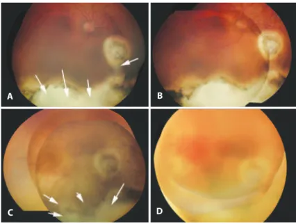
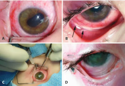
A 4 year-old girl with nonfamilial bilateral retinoblastoma was referred to our care, with refractory disease in her remaining eye. She had been treated elsewhere by primary enucleation, intravenous (IV) chemotherapy, laser/cryotherapy, and I-125 brachytherapy OD. Vitreous seeding and chorioretinal scarring with an inferior partial rhegmatogenous retinal detachment OD prompted referral to our center. No discrete tumor was visible, but vitreous seeding was con-irmed (Figure 1 A). The patient underwent 6 sessions of singledrug Ophthalmic Artery Infusion Chemotherapy (OAIC) (topotecan) and 2 sessions of OAIC topotecan with subtenon carboplatin. After a sub-optimal response, 2 additional sessions of single-drug OAIC (melphalan) were given. Following the treatment, the vitreous see-ding resolved (Figure 1 B), but the patient was noted to have difuse ke ratopathy, posterior synechiae, cataract, difuse conjunctival hype-remia, and localized inferior fornix necrosis (Figures 2 A, B). Her kera-topathy was managed with the help of a therapeutic contact lens.
Unfortunately, her vitreous seeding relapsed 6 months later (Figure 1 C). She then underwent IVit chemotherapy (melphalan 20 μg/0.1 mL) as previously reported(9). The vitreous seeding resolved following the irst IVit injection, but she complained of foreign body pain. She recei-ved one subsequent injection (Figure 1 D). Examination throughout the 6 months that followed showed continued vitreous clearing.
Regardless of 3 sequential placements of Prokera®
(Biotissue, Mia-mi, FL) and 2 tarsorrhaphies, her chronic keratopathy progressed (Fi-gure 2 C). Eventually, the keratopathy improved and her inferior fornix healed (Figure 2 D), but her cataract worsened, precluding a view of the fundus. Magnetic resonance imaging (MRI) showed the vitreous to be clear, prompting a subsequent uneventful cataract surgery. Fundus exam performed after the cataract removal revealed massive tumor recurrence. Therefore, the eye was enucleated. Histopathology showed the optic nerve margin to be tumor free. However she had
massive invasion of the choroid inferiorly. She has been tumor free for 12 months after secondary enucleation.
DISCUSSION
Retinoblastoma presents a unique narrative on the need for efective communication between physicians and families in order to establish realistic therapeutic expectations. This discussion is of pa ramount importance to children with only one remaining eye. In modern times, physicians and families have looked to new, eyepre -serving therapies to avoid enucleation. As these therapeutic options begin to be widely used, one must address the cumulative efects and associated impact of such treatments.
Our patient’s clinical course relects these concerns. On presenta-tion, she had already undergone primary enucleation OS and intense treatment OD that included IV chemotherapy, followed by sequen-tial aggressive local therapy (SALT) and I-125 plaque brachytherapy. Although the toxicity of these combined treatments has not been documented, it has been reported that IV chemotherapy increases the risk for radiation retinopathy following I-125 plaque therapy(11). Although salvage of the eyes with difuse vitreous or extensive su-bretinal seeding can be achieved with such treatments in a minority of cases(11), these treatments may have contributed to retinal scarring and de tachment, which, in addition to the vitreous seeding, threa-tened the vision in our patient’s only eye.
Our decision to use OAIC with additional periocular carboplatin as salvage therapy was based on the premise that this combination efectively delivers high-dose chemotherapy to the eye while sparing the side efects of systemic treatment. OAIC is currently used as a single or multi-drug treatment(5). At the time we irst saw this patient, our institution had initiated a protocol with topotecan (dose based on child’s age) as a single agent in an attempt to minimize the side efects of Melphalan, a toxic alkylating agent. After several cycles of treatment and limited response, she was switched to Melphalan. Currently, Melphalan is being used as a single agent in some treatment- naïve patients, using doses in the range of 3 to 5 mg, depending on
Figure 1. RetCam® photographs of the patient’s right eye at various time points. A) First exam at presentation. Note the extensive chorioretinal scarring inferiorly and poorly diferentiated matte of preretinal acellular vitreous seeds. B) Following single-drug ophthalmic artery infusion chemo-therapy (OAIC) using topotecan (8 sessions), and single-drug OAIC using melphalan (2 sessions), periocular injection carboplatin (2 sessions). Note the absence of preretinal vitreous seeds on exam. C) Recurrence of vitreous seeds OD 6 months after the completion of the previous treatment. D) Follo-wing the completion of intravitreal melphalan. Note the resolution of vitreous seeding.
A
C
B
Managing the consequences of aggressive conservative treatment for refractory retinoblastoma with vitreous seeding
258 Arq Bras Oftalmol. 2014;77(4):256-8
the child’s age and weight. In cases that prove to be refractory to OAIC treatment or in case of a late tumor relapse, some centers use a 3-drug regimen (melphalan 3-5 mg, Topotecan 0.3 to 0.5 mg, carbo-platin 20-30 mg)(5). Although our patient’s vitreous seeding resolved, its recurrence demanded a new discussion with the family regarding further management options. The family’s decision to preserve her eye prompted the use of Melphalan intravitreously.
Although the eye was tumor free, the patient faced complica-tions resulting from her many treatments. She developed a chronic keratopathy, which caused severe, persistent pain and loss of vision. Amniotic membrane graft and limbal stem cell transplant were dis-cussed. Her prior periocular chemotherapy had resulted in necrosis of the inferior fornix, prohibiting the suturing of an amniotic mem-brane graft. Although the complications of periocular carboplatin administration have been reported,these complications tend to be variable and dose-dependent(12).The use of periocular carboplatin was thoroughly discussed and was concluded to be a good option based on the reported synergy with topotecan(13). Limbal stem cell transplant from the patient’s mother was deemed unsuitable. There-fore, the focus shifted toward the placement of Prokera®
, a double amniotic membrane graft with implantation similar to a contact lens. Sequen tial placements of Prokera®
healed the previously necrotic inferior fornix. Unfortunately, the patient’s cataract and small pupil, which severely limited her vision, precluded fundus examination and our ability to monitor any tumor recurrence. Knowing the risks of cataract surgery, the patient’s parents chose to proceed with an MRI. After having put much efort to eradicate her RB, the inal decision to enucleate her remaining eye was very diicult. However, the tumor recurrence left us with no other choice.
Modern management of retinoblastoma requires that physicians be aware of the beneits and limitations of new eye-preserving the-rapies. These novel treatment modalities require a multi-disciplinary, open approach with families. As seen with our patient’s chronic kera-topathy and secondary cataract, the cumulative side efects of multiple therapies are still unknown and have limited our management options. This is of particular concern when complications preclude active assess-ment of tumor recurrence. Although tumor suppression is the main
treatment goal, it is imperative that physicians consider the long-term consequences of these progressive therapies and their impact on the lives of these children. Finally, it is important to realize that, at times, even extensive eforts may fail to save the child’s eye. In doing so, col-lectively, we will be more mindful of the treatment-associated morbi-dity and will anticipate the potential side efects and complications to possibly achieve better outcomes and improve patients’ quality of life.
REFERENCES
1. Abramson DH. Retinoblastoma incidence in the United States. Arch Ophthalmol. 1990; 108(1):1514. Comment on: Arch Ophthalmol. 1990;108(1):128-32.
2. Tamboli A, Podgor MJ, Horm JW. The incidence of retinoblastoma in the United States: 1974 through 1985. Arch Ophthalmol. 1990;108(1):128-32. Comment in: Arch Ophthal-mol. 1990;108(11):1514.
3. Shields JA, Shields CL. Ocular tumors. A text and atlas. Philadelphia, PA: Saunders; 1992. 4. Marr BP, Dunkel IJ, Linker A, Abramson DH. Periocular carboplatin for retinoblastoma: long-term report (12 years) on eicacy and toxicity. Br J Ophthalmol. 2012;96(6):881-3. 5. Abramson DH, Marr BP, Dunkel IJ, Brodie S, Zabor EC, Driscoll SJ, et al. Intra-arterial
chemotherapy for retinoblastoma in eyes with vitreous and/or subretinal seeding: 2-year results. Br J Ophthalmol. 2012;96(4):499-502.
6. Munier FL, Gaillard MC, Balmer A, Soliman S, Podilsky G, Moulin AP, et al. Intravitreal chemotherapy for vitreous disease in retinoblastoma revisited: from prohibition to conditional indications. Br J Ophthalmol. 2012;96(8):1078-83.
7. Rizzuti AE, Dunkel IJ, Abramson DH. The adverse events of chemotherapy for retino-blastoma: what are they? Do we know? Arch Ophthalmol. 2008;126(2):862-5. 8. Abramson DH, Frank CM, Dunkel IJ. A Phase I/II study of subconjunctival carboplatin
for intraocular retinoblastoma. Ophthalmology. 1999;106(10):1947-50.
9. Suzuki S, Yamane T, Mohri M, Kaneko A. Selective ophthalmic arterial injection the-rapy for intraocular retinoblastoma: the long-term prognosis. Ophthalmology. 2011; 118(10):2081-7.
10. Kaneko A, Suzuki S. Eye-preservation treatment of retinoblastoma with vitreous seeding. Jpn J Clin Oncol. 2003;33(12):601-7.
11. Murphree AL, Villablanca JG, Deegan WF 3rd, Sato JK, Malogolowkin M, Fisher A, et al. Chemotherapy plus local treatment in the management of intraocular retinoblas-toma. Arch Ophthalmol 1996;114(11):1348-56.
12. Schmack I, Hubbard GB, Kang SJ, Aaberg TM, Grossniklaus HE. Ischemic necrosis and atrophy of the optic nerve after periocular carboplatin injection for intraocular retinoblastoma. Am J Ophthalmol. 2006;142(2):310-5.
13. Nemeth, KM, Federico S, Carcaboso, AM, Shen Y, Schaiquevich P, Zhang J, et al. Sub-conjunctival carboplatin and systemic topotecan treatment in preclinical models of retinoblastoma. Cancer. 2011;117(2):421-34.
A
C
B
D