INTRODUCTİON
The tilted optic disc is a congenital, nonhereditary condition in which the optic nerve enters the eye at an oblique angle. Its preva-lence has been reported to be 1%-2% in the general population(1). In clinical practice, the diagnosis of tilted disc is based on ophthalmos-copic appearance. Clinically, tilted discs appear as an exaggerated oval or D-shaped optic nerve head with one hemisphere of the disc more elevated than the contralateral half(2). The elevated margin of the disc is usually adjacent to an intact neurosensory retina, pigment epithelium, and choroid and the depressed portion of the disc is commonly associated with a thinned choroid and thinned retinal pigment epithelium (RPE) that may be visible as fundus depigmen-tation on ophthalmoscopic evaluation(3-5).
The most common form of optic disc tilt consists of superotemporal rim elevation, often with inferonasal depression(5). Subretinal neovas-cularization and chorioretinal degenerative changes in the tilted disc syndrome have been reported(6-8).
ABSTRACT
Purpose: This study was performed to evaluate the retinal nerve fiber layer (RNFL) and peripapillary choroidal thickness in eyes with tilted optic disc in order to identify characteristic RNFL and peripapillary choroid patterns verified by optical coherence tomography (OCT ).
Method: Twenty-nine eyes of 29 patients with tilted optic discs were studied with spectral-domain (SD)-OCT and compared with age and sex-matched control subjects in a prospective design. The imaging of RNFL was performed using circular scans of a diameter of 3.4 mm around the optic disc using OCT. For measurements of peripapillary choroidal thickness, the standar d protocol for RNFL assessment was performed.
Results: SD-OCT indicated significantly lower superotemporal (p<0.001), supe-ronasal (p=0.001), and global (p=0.005) RNFL thicknesses in the tilted disc group than those of the control group. Peripapillary choroid was significantly thicker at the site of the elevated rim of eyes with tilted disc (p<0.001).
Conclusions: This study demonstrated a clinical characterization of the main tilted disc morphologies that may be helpful in differentiating a tilted disc from other altered disc morphologies. Further studies are recommended to study the comparison between glaucoma and tilted disc groups.
Keywords: Choroid/pathology; Nerve ibers; Tomography, optical coherence; Optic disc/abnormalities
RESUMO
Objetivo: Avaliar camada de fibras nervosas da retina (RNFL) e a espessura da coroide peripapilar em olhos com disco óptico inclinado a fim de identificar as características da RNFL e os padrões de coroide peripapilar verificados pela tomografia de coerência óptica (OCT ).
Método: Vinte e nove olhos de 29 pacientes com discos ópticos inclinadas foram estudados prospectivamente com OCT de domínio espectral (SD) e comparados com controles pareados por sexo e idade. As imagens da RNFL foi obtidas por meio de varreduras circulares com um diâmetro de 3,4 mm em torno do disco óptico usando OCT. Para as medições de espessura da coroide peripapilar, o protocolo padrão para avaliação da RNFL foi realizado.
Resultados: O OCT SD indicou diminuição das espessuras significativas da RNFL superotemporal (p<0,001), superonasal (p=0,001), e global (p=0,005) no grupo disco inclinado em relação aos do grupo controle. A coroide peripapilar foi significativamente mais espessa no local da borda elevada dos olhos com disco inclinado (p<0,001).
Conclusões: Este estudo demonstrou que a caracterização clínica das principais morfologias disco inclinado pode ser útil na diferenciação entre um disco inclinado de outras alterações morfológicas de disco. Seria importante a comparação entre grupos com glaucoma e com discos inclinados, estudos futuros.
Descritores: Coroide/patologia; Fibras nervosas; Tomografia de coerência óptica; Disco óptico/anormalidades
In most of the cases, RNFL thickness was found to be thinned on the sectors close to the elevated disc rim. Many studies have discus-sed altered RNFL patterns in cases of tilted disc, but the results did not relect the same results. Law et al.(9) found signiicant lower RNFL in eyes with tilted optic disc, whereas Rauscher et al.(10) found no as-sociation between optic disc tilt and RNFL. Controversially, the study by Hwang et al.(11) using a Cirrus HD OCT found a signiicantly thicker RNFL in eyes with tilted disc.
The characteristics of tilted optic disc make the diagnosis and mo-nitoring of glaucoma diicult due to the similarities with glaucoma-tous discs. Moreover, tilted disc is associated with visual ield defects, which also leads to diagnostic diiculties.
With the advent of OCT, all retinal layers can now be measured objectively, and the technique is widely used to assess the status of the RNFL in patients with glaucoma and other optic neuropathies(12). Spai-de et al. recently Spai-described a technique (enhanced Spai-depth imaging) in which SD-OCT was used to measure choroidal thickness(13).
Evaluation of peripapillary choroidal and retinal nerve fiber layer thickness in eyes
with tilted optic disc
Avaliação espessura da camada de ibras nervosas peripapilares da retina e coroide em
olhos com disco óptico inclinado
MuaMMer OzcıMen1, Yasar sakarYa1, sertan GOktas1, rabıa sakarYa1, ısMaıl s. ıvacık1, Halıl ı. Yener2, erkan erdOGan1
Submitted for publication: April 10, 2014 Accepted for publication: September 18, 2014
Study conducted at Konya Training and Research Hospital, Konya, Turkey.
1 Department of Ophthalmology, Konya Training and Research Hospital, Konya, Turkey. 2 Konya Eye Clinic, MD, Konya, Turkey.
Funding: No specific financial support was available for this study.
Disclosure of potential conflicts of interest: None of the authors have any potential conflicts of interest to disclose.
Corresponding author: Muammer Ozcımen. Konya Egitim ve Arastirma Hastanesi - Goz Klinigi -
Meram/Konya 42090 - Turkey - E-mail: [email protected]
Our aim was to study RNFL and peripapillary choroidal thickness in individuals with tilted disc and compare the values obtained from the control group in order to identify characteristic RNFL and choroi-dal thickness patterns.
METHODS
This prospective study included 35 eyes of 35 patients with tilted disc syndrome (14 men and 21 women). Records from the Ophthal-mology Department at Konya Training and Research Hospital were scanned. Patients who were diagnosed with tilted optic disc were invited to participate in the study from December 2013 to February 2014. The control group included 29 eyes of 29 age- and sex-matched control subjects. All patients underwent a complete ophthalmic exa-mination including slit-lamp biomicroscopy, Goldman applanation tonometry, color vision test (Ishihara test), fundoscopy, optic disc photograph imaging and computerized perimetry. Subjects were evaluated by a 24.2 program with SITA standard algorithm of the Humprey Visual Field analyser II model 750i (Humphrey Instruments, San Leandro, CA, USA). Only the reliable visual ield parameters (i-xation loss, <20%; false positive rate, <33%; and false negative rate, <33%) were included in the study. Global indices (mean deviation [MD] and pattern standard deviation [PSD]) were evaluated. Tilted disc cases underwent two reliable visual ields, with a minimum time in-terval of 1 month, and control cases received only one reliable visual ield test after the perimetric learning efect.
Best-corrected visual acuity (BCVA) was measured by means of the standard Snellen chart. The values of visual acuity were converted to the logMAR scale for statistical analysis. Exclusion criteria included history of glaucoma and intraocular trauma, optic neuropathy, axial length measurement other than 22-26 mm and a spherical equiva-lent higher than -6.00 D. Subjects with unstable and uncontrolled
cardiovascular, renal or pulmonary diseases, diabetes and pregnancy were also excluded. A tilted disc was deined as anteroposterior rotation (tilt) and sagittal plane rotation (torsion) based on a review of tilted disc syndrome by Witmer et al.(2). These characteristics were easily evaluated by fundoscopy or optic disc photographs.
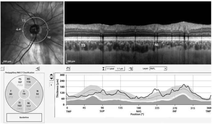
Choroidal thickness measurements were performed using an SD-OCT device (λ=870 nm, 40000A-scans/s and 3.9 μm axial resolu-tion) with software version 5.3 (Spectralis; Heidelberg Engineering, Heidelberg, Germany). RNFL imaging was performed using circular scans of a diameter of 3.4 mm around the optic disc. The scans were well centered at the optic nerve head. Spectralis OCT included RNFL thickness in six sectors: nasal (N), superonasal (NS), superotemporal (TS), temporal (T), inferotemporal (TI) and inferonasal (NI), as well as global RNFL thickness (G) (Figure 1).
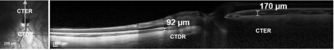
For measurements of peripapillary choroidal thickness, a 360-de-gree 3.4 mm diameter peripapillary circle scan was performed using the standard protocol for RNFL assessment. Choroidal thickness was measured manually from the outer portion of the hyperrelective line corresponding to the RPE to the inner surface of the sclera (Figure 2). Peripapillary choroidal thicknesses adjacent to the elevated disc rim and the depressed sector directly opposite to the elevated disc rim were evaluated. In the control group, measurements were taken of the superotemporal and inferonasal peripapillary choroidal thickness in which the superotemporal choroid region corresponded to the choroid adjacent to elevated disc rim in the tilted disc group and the inferonasal choroid corresponded to the choroid adjacent to the depressed disc rim, which is directly opposite the elevated disc rim location, in the tilted disc group (Figure 1). The choroid was measured by two independent graders (MO, SI) using the tools found on the Spectralis OCT analysis software. The intraclass correlation coei-cients for overall peripapillary choroidal thickness were used to assess agreement between both graders.
TS= superotemporal; NI= inferonasal.
The study was conducted under a protocol approved by the Konya Selcuk University Medical Faculty Ethics Committee and was in accor-dance with the ethical standards stated in the 1964 Declaration of Helsinki. Informed consent was obtained from all the patients after explanation of the procedures.
All statistical analysis of the data were performed using commer-cial software (Statistical Package for Socommer-cial Science, version 15.0; SPSS Inc., Chicago, IL, USA). Statistical analysis was performed by using the Independent t-test (parametric). Pearson correlation coeicient was used to evaluate the correlation between the RNFL thickness and the global indices of visual ield. Results were considered statistically signiicant when p<0.05. Results are represented as mean ± SD.
RESULTS
The recordings of 35 patients were assessed and six were exclu-ded from the study. Four cases were excluexclu-ded because the sclera was invisible in OCT, and two cases were excluded due to unreliability for visual ield analysis. A total of 58 subjects were included in the study. The tilted disc group included 29 eyes of 29 individuals, aged between 19 and 67 years(mean age, 45.3 ± 13.2). The control group included 29eyes of 29 individuals aged between 21 and 64 years (mean age, 48.3 ± 12.1).
Fundus examination in all eyes showed the existence of tilted optic disc with mainly inferior nasal crescent (Figure 1). The most common form of tilted disc we found was superotemporal rim eleva-tion (n=17, 58.6%), followed by temporal rim elevaeleva-tion (n=7, 24.1%). None of our cases had evident posterior staphylomas on fundoscopic examination.
Mean best-corrected visual acuity was 0.15 ± 0.13 logMAR for the tilted disc group and 0.08 ± 0.06 logMAR for the control group. The axial length value for the tilted disc group was 25.1 ± 2.3 mm and 24.8 ± 3.4 mm for the control group. There were no diferences between the tilted disc and control groups in regard to mean BCVA and axial lenght (p=0.09 and p=0.087, respectively). The mean sphe-rical equivalent for the tilted disc group was -1.94 ± 2.56 D, whereas the control group mean was -0.86 ± 1.96 D (p=0.076). All patients had normal color vision.
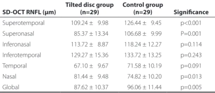
The RNFL thickness analysis showed that the superotemporal and superonasal sectors were signiicantly thinner in the tilted disc group than those of the control group (p<0.001 and p=0.001, respectively; Table 1). The RNFL thickness of the nasal sector was signiicantly higher in the tilted disc group (p=0.013); however, the global RNFL was signi-icantly lower in the tilted disc group (p=0.005).
Peripapillary choroidal thicknesses are presented in table 2. The analysis showed that the mean choroidal thickness next to the eleva-ted disc rim was 193.75 ± 37.87 µm and the mean choroidal thickness opposite this location was 125.48 ± 24.68 µm; the diference was statistically signiicant between these topographical regions in the same eye (p<0.001). The control group had a mean superotemporal peripapillary choroidal thickness of 209.37 ± 27.98 µm and an infe-ronasal choroidal thickness of 198.34 ± 17.08 µm; the diference was not signiicant between these topographical regions in the same eye
(p=0.075). There was no signiicant diference in superotemporal cho-roidal thickness between the tilted disc and control groups (p=0.079); however, inferonasal choroidal thickness was signiicantly lower in the tilted disc group (p<0.001).
Eighteen eyes (62%) presented visual ield defects, most frequen-tly found on the superotemporal quadrant (n=10), whereas 11 eyes had normal visual ields. There was generalized reduction in retinal sensitivity in all quadrants. Visual ield analysis revealed a pattern standard deviation (PSD) of 4.20 ± 1.47 for the tilted disc group, which was signiicantly higher (p<0.001) than the control group (1.65 ± 0.60). Mean deviation (MD) was signiicantly lower (p<0.001) in the tilted disc group than the control group, with a mean value of -7.09 ± 3.45, whereas the control group had a MD of -2.63 ± 1.29. There was no signiicant correlation (correlation coeicient, p value) for MD and the RNFL thicknesses at all quadrants except TI (0.373, p=0.046) and for PSD and the RNFL thicknesses at all quadrants except TI (0.458, p=0.013). There was a signiicant correlation for PSD the global RNFL thickness (0.428, p=0.020).
Regarding the manual measurement of peripapillary choroidal thickness, the intraclass correlation coeicients showed agreement between the two observers (p<0.001) and ranged from moderate (0.67, next to the elevated disc rim) to very high (0.96, superotemporal).
DİSCUSSİON
This study used SD-OCT to demonstrate that tilted optic disc is asso ciated with abnormal patterns of both peripapillary RNFL and choroidal thickness. The most common form of tilted disc in our stu-dy was inferonasal disc rotation with superotemporal rim elevation, which is similar to previous reports(1,5,14). We found that a signiicant diference exists in the choroidal thickness between the elevated and depressed sectors of the optic disc, in which there is a thinner cho -roid next to the depressed part of the disc. Furthermore, we found that eyes with inferior tilted disc show superior RNFL defects when analyzed by OCT imaging. We also conirmed that global RNFL was signiicantly lower in the tilted disc group. These indings show that OCT technology may be more sensitive for the detection of RNFL defects in abnormal optic disc morphologies.
The choroid inferior to the optic disc and the choroid nasal to the macula were reported to be thinner than all other sectors, and those variations were attributed to the natural anatomical architecture of normal eyes(15). This regional diference observed in choroidal thickness may be imputable to the development of the eye. Evidence from embryology indicates that the optic issure is located in the inferior aspect of the optic cup, which is the last part of the globe to close during eye formation. A thinner choroid may be important because it may make this area more susceptible to retinal and choroidal disea-ses(15). This idea is favored by eyes with glaucoma wherein the choroid was reported to be thinner and the observation of more severe ield defects in the thinner hemiield in glaucomatous eyes(16). Reported disorders such as subretinal neovascularization and chorioretinal de-generative changes in the tilted disc syndrome(3) may be associated with thinner choroid next to the depressed part of the optic disc. It CTER= peripapillary choroidal thickness adjacent to elevated disc rim; CTDR= peripapillary choroidal thickness adjacent to depressed disc rim.
is unclear why the choroid is thinner in the discoloration area than in the other quadrants. However, it may correspond to the absence of retinal pigment epithelium and photoreceptors and attenuation of the choroid(17).
Brito et al.(14) reported that the most common RNFL defect was lo cated on the superotemporal sector of the Spectralis OCT, and the most commonly involved perimetric quadrant was also supero-temporal. They speculated that this was the opposite of what would be expected. We likewise found superotemporal visual ield defects in cases with superotemporal RNFL defect; however, the results of analysis of correlations between MD and PSD were not completely consistent. We therefore concluded that visual ield defects in indivi-duals with tilted optic disc might be multifactorial in origin. Further-more, it would be beneicial to study visual ields and document the factors inluencing the test parameters in subjects with tilted optic disc. In this study, OCT examination found that RNFL thickness was signiicantly thinned in the superior quadrant in patients with tilted disc syndrome. This may be attributed to the fact that the density of nerve ibers entering the optic disc in tilted disc syndrome is lower in this sector(18).
The diferences may be due to hypoplasia associated with tilted disc. There is little knowledge regarding the underlying etiology of the embryonic aberrations(19). Simon et al.(20) reported that intraute-rine injury to the retina, optic nerve, chiasm or posterior visual pa-thway may cause hypoplasia of the optic nerve. Similarly Apple et al.(5) speculated that localized absence of ganglion cells and their failure to make synaptic connections in the lateral geniculate body might result in hypoplastic development of other supportive tissue. Consequently, the disc becomes tilted because of the imbalance in the number of ganglion cells and supportive tissue within the optic nerve. In addition, when the visual ields of eyes with tilted optic disc were compared to control groups, there was generalized reduction in retinal sensitivity in all quadrants(21). This generalized reduction in sensitivity has lead to the opinion that tilted disc may represent a difusely hypoplastic optic nerve(22).
It is an uneasy issue whether a patient with tilted disc and ocular hypertension has glaucomatous optic neuropathy and RNFL dama-ge. A commonly observed problem with tilted disc is the detection of early glaucomatous damage. Both are associated with visual
ield defects, and tilted disc morphology is a inding in optic disc rim eva luation; its presence makes identiication of the usual signs of glaucomatous damage, such as pathological cupping, diicult to detect(23,24). We may say that the presence of a small visual ield defect in a patient with an inferior tilted disc and superior RNFL loss is most probably related to tilted disc morphology instead of glaucomatous optic neuropathy; however, this argument needs to be proven by large-scale studies comparing visual ields in both glaucoma and til-ted disc groups. In addition to clinical examination, realization of OCT in conjunction with perimetry can help in the diagnosis and monito-ring of glaucoma in cases of concomitant optic disc tilt. OCT appears to have the ability to distinguish normal from glaucomatous optic nerve heads, but it is not clear that these indings can be generalized to tilted disc and glaucoma(25). Witmer et al.(2) partly supported this notion as they reported that new technologies for imaging the optic nerve head and RNFL currently had a limited role in diagnosing and monitoring tilted disc. There could be OCT error due to the abnormal anatomy of the nerve, as tilted discs can mimic other conditions; however, this diagnostic error is best prevented by clinicians who are familiar with the clinical manifestations of the anomaly. Nevertheless, in a study of patients with glaucoma and tilted discs, OCT was shown to be accurate in measuring the RNFL thickness and corresponded well to Humphrey visual ield defects(26).
Eyes with tilted discs were more miopic than controls. However, none of our cases had evident posterior staphylomas on fundoscopic examination. Furthermore, we recruited cases with a spherical equi-valent lower than 6.00 D of myopia to reduce the possible efects of myopic chorioretinopathy on peripapillary RNFL. To avoid RNFL changes due to mechanical tissue stretching, we included cases with an axial length measurement within a certain range (22-26 mm).
OCT imaging of the optic nerve head and RNFL rely on validated normative data; however, validated normative datasets for tilted disc do not currently exist. Therefore, considering these data, imaging the optic disc with OCT could be a limitation of our study.
This study demonstrated a clinical characterization of the main tilted disc morphologies that may be helpful in diferentiating a tilted disc from other altered disc morphologies. Additionally,the presence of optic disc tilt should be taken into account when interpreting peripapillary RNFL and choroidal thicknesses by SD-OCT. It would be beneicial to explore the comparison between glaucoma and tilted disc groups in additional studies.
ACKNOWLEDGMENTS
The authors thank Mehmet Yemenici and Zehra Yalcin for techni-cal assistance in obtaining OCT images
REFERENCES
1. Young SE, Walsh FB, Knox DL. The tilted disc syndrome. Am J Ophthalmol. 1976;82(1): 16-23.
2. Witmer MT, Margo CE, Drucker M. Tilted optic disks. Surv Ophthalmol. 2010;55(5):403-28. 3. Giufre G. Chorioretinal degenerative changes in the tilted disc syndrome. Int
Oph-thalmol. 1991;15(1):1-7.
4. Stur M. Congenital tilted disc syndrome with parafoveal subretinal neovascularization. Am J Ophthalmol. 1988;105(1):98-9.
5. Apple DJ, Rabb MF, Walsh PM. Congenital anomalies of the optic disc. Surv Ophthalmol. 1982;27(1):3-41.
6. Prost M, DeLaey JJ. Choroidal neovascularization in tilted disc syndrome. Int Ophthal-mol.1988;12(2):131-5.
7. Leys AM, Cohen SY. Subretinal leakage in myopiceyes with a posterior staphyloma or tilted disc syndrome. Retina. 2002;22(5):659-65.
8. Moschos MM, Margetis I, Papadimitriou S, Tzeni Z, Moschos MN. Clinical and multifo-cal-electroretinographic indings of congenital tilted disc syndrome associated with choroidal neovascularization: a case report. Doc Ophthalmol. 2007;115(2):121-4. 9. Law SK, Tamboli DA, Giaconi J,CaprioliJ. Characterization of retinal nerve iber layer
in nonglaucomatous eyes with tilted discs. Arch Ophthalmol. 2010;128(1):141-2. 10. Rauscher FM, Sekhon N, Feuer WJ,Budenz DL. Myopia afects retinal nerve iber layer
measurements as determined by optical coherence tomography. J Glaucoma. 2009; 18(7):501-5.
Table 1. RNFL thickness: tilted disc and control group
SD-OCT RNFL (µm)
Tilted disc group (n=29)
Control group
(n=29) Signiicance Superotemporal 109.24 ± 09.98 126.44 ± 09.45 p<0.001 Superonasal 085.37 ± 13.34 106.68 ± 09.99 P=0.001 Inferonasal 113.72 ± 08.87 118.24 ± 12.27 p=0.114 Inferotemporal 129.27 ± 15.36 133.72 ± 13.25 p=0.243
Temporal 067.10 ± 09.67 071.58 ± 10.19 p=0.091 Nasal 081.44 ± 09.48 074.82 ± 10.20 p=0.013 Global 087.62 ± 10.37 096.06 ± 11.44 p=0.005
Table 2. Average peripapillary choroidal thickness (µm)
Sectors Tilted disc group Control group p-value Superotemporal (CTER) 193.75 ± 37.87 209.37 ± 27.98 p=0.079
Inferonasal (CTDR) 125.48 ± 24.68 198.34 ± 17.08 p<0.001
p-value p<0.001 p=0.075
11. Hwang YH, Yoo C, Kim YY. Characteristics of peripapillaryretinal nerve iber layer thickness in eyes with myopic optic disctilt and rotation. J Glaucoma. 2012;21(6):394-400. 12. Pasol J. Neuro-ophthalmic disease and optical coherence tomography: glaucoma
look alikes. Curr Opin Ophthalmol. 2011;22(2):124-32.
13. Spaide RF, Koizumi H, Pozzoni MC. Enhanced depth imaging spectral-domain optical coherence tomography. Am J Ophthalmol. 2008;146(4):496-500.
14. Brito PN, Vieira MP, Falcao MS, Olinda FS, Fernando RF. Optical coherence tomography study of peripapillary retinal nerve iber layer and choroidal thickness in eyes with tilted optic disc. J Glaucoma. 2013 Feb 19 [Ahead of print].
15. Tanabe H, Ito Y, Terasaki H. Choroid is thinner in inferior region of optic disks of normal eyes. Retina. 2012;32(1):134-9.
16. Yin ZQ, Vaegan, Millar TJ, Beaumont P, Sarks S. J Glaucoma. Widespread choroidal insuiciency in primary open-angle glaucoma. 1997;6(1):23-32.
17. Dorrell D. The tilted disc. Br J Ophthalmol. 1978;62(1):16-20.
18. Moschos MM, Triglianos A, Rotsos T, Papadimitriou S, Margetis I, Minogiannis P, et al. Tilted disc syndrome: an OCT and mfERG study. Doc Ophthalmol. 2009;119(1):23-8. 19. Giufre G. Hypothesis on the pathogenesis of the papillary dysversion syndrome. J Fr
Ophtalmol. 1985;8(8-9):565-72.
20. Simon J, Aaby A, Drack A. Optic nerve hypoplasia. In: American Academy of Ophthal-mology, Basic and Clinical Science Course, Section 6, Pediatric Ophthalmology and Strabismus. San Francisco: AAO; 2008-2009. p.362-3.
21. Gurlu VP, Alimgil ML, Benian O. Topographical analysis of the visual ield in tilted disk syndrome. Retina. 2002;22(3):366-8.
22. Tay E, Seah SK, Chan SP, Lim AT, Chew SJ, Foster PJ, et al. Optic disk ovality as an index of tilt and its relationship to myopia and perimetry. Am J Ophthalmol. 2005;139(2):247-52. 23. Brazitikos PD, Safran AB, Simona F,Zulauf M. Threshold perimetry in tilted disc
syn-drome. Arch Ophthalmol. 1990;108(12):1698-700.
24. Vuori ML, Mantyjarvi M. Tilted disc syndrome may mimic false visual ield deteriora-tion. Acta Ophthalmol. 2008;86(6):622-5.
25. Medeiros F, Zangwill L, Bowd C, Weinreb RN. Comparison of the GDx VCC scanning laser polarimeter, HRT II confocal scanning laser ophthalmoscope, and stratus OCT optical coherence tomography for the detection of glaucoma. Arch Ophthalmol. 2004;122(6):827-37.
26. Yu S, Tanabe T, Hangai M, Morishita S, Kurimoto Y, Yoshimura N. Scanning laser pola-rimetry with variable corneal compensation and optical coherence tomography in tilted disk. Am J Ophthalmol. 2006;142(3):475-82.