Original Article
3 8 8 Arq Bras Oftalmol. 2014;77(6):388-91 http://dx.doi.org/10.5935/0004-2749.20140096
INTRODUCTION
Trypan blue (TB) is used to facilitate the creation of a continuous curvilinear capsulorhexis during cataract surgery(1,2). Several methods have been used to assess the toxic efects of TB on corneal cells: the concentration-related efect of TB on corneal cell viability; the efects of duration of exposure to TB on cell survival; the efect of the vehicle on the degree of TB toxicity; and the damage caused by (experimen-tal) manipulation of cells or endothelia(3).
Several experimental and clinical studies have tested the cyto-toxicity of TB on various ocular anterior segment structures during cataract surgery, and all have demonstrated good biocompatibility
for conventional intracameral application of TB 0.1%. However, in vitro studies have demonstrated toxicity of TB to corneal endothelium and corneal ibroblasts at higher concentrations and longer exposure times(4-7). Briely, at commonly used concentrations, both during ca-taract surgery and in corneal tissue banks, TB is harmless to corneal cells. However, at higher concentrations or longer exposures, extre-me caution is warranted.
Oxidative stress is believed to be a central mechanism of the cellular damage afecting all organs and tissues. Oxygen-derived free radicals appear to be responsible for injury to various organs, and apoptosis mediated by oxidative stress is well established(8-11). Considering the
Evaluation of the safety of intracameral trypan blue injection on corneal tissue using
oxidative stress parameters and apoptotic activity: an experimental study
Avaliação da segurança de injeção de azul de tripan intracameral no tecido da córnea utilizando
parâmetros de estresse oxidativo e atividade apoptótica: um estudo experimental
Ali AkAl1, TurgAy ulAs2, TugbA goncu1, MehMeT FATih Adibelli1, sezen kocArslAn3, MuhAMMeT eMin guldur3, MusluM guler4, uFuk ozkAn5,
MehMeT dusunur1, Tuncer deMir6
Submitted for publication: July 31, 2014 Accepted for publication: October 12, 2014
Study conducted at Harran University School of Medicine, Department of Ophthalmology.
1 Department of Ophthalmology, Faculty of Medicine, Harran University, Sanliurfa, Turkey. 2 Department of Internal Medicine, Faculty of Medicine, Harran University, Sanliurfa, Turkey. 3 Department of Pathology, Faculty of Medicine, Harran University, Sanliurfa, Turkey. 4 Department of Ophthalmology, Balikligol Training and Research Hospital, Sanliurfa, Turkey. 5 Department of Biochemistry, Harran University, Faculty of Medicine, Sanliurfa, Turkey. 6 Department of Physiology, Faculty of Medicine, Gaziantep University, Gaziantep, Turkey.
Funding: No specific financial support was used for this study.
Disclosure of potential conflicts of interest: None of the authors have any potential conflicts of interest to disclose.
Corresponding author: Ali Akal. Harran University School of Medicine. Department of Ophthalmo logy Yenisehir Campus Sanliurfa 63000 Turkey E mail: [email protected] ABSTRACT
Purpose: The present experimental study aimed to investigate the effects of intracameral trypan blue (TB) on oxidative stress parameters and apoptosis in cor neal tissue.
Methods: Thirty rats were randomly assigned to three groups of 10 rats each: the sham group (Group 1); control group (Group 2); and treatment group (Group 3). The control group was administered 0.01 cc of balanced salt solution. The treatment group was administered 0.006 mg/0.01 cc of TB. The total antioxidant status (TAS) and total oxidant status (TOS) in corneal tissue and blood were measured and the oxidative stress index (OSI) was calculated. Finally, corneal tissue histopathology was evaluated using staining for caspase-3 and -8, and apoptotic activity was examined. Results: The TAS, TOS and OSI levels in the blood samples were not significantly different (p>0.05 for all). Compared with the sham and control groups, the TOS and OSI levels in corneal tissue were significantly different in the treatment group (p<0.05 for all). No significant difference was observed between the sham group and the control group (p>0.05). Immunohistochemical staining for caspase-3 and caspase-8 demonstrated higher apoptotic activity in the TB group than in the sham and control groups.
Conclusion: The present study showed that intracameral TB injection is safe systematically but may be toxic to corneal tissue, as demonstrated using oxidative stress parameters and histopathological evaluation.
Keywords: Trypan blue/administration & dosage; Injections; Intraocular; Cornea; Oxidative stress; Apoptosis; Animals; Rats
RESUMO
Objetivo: Este estudo experimental tem como objetivo investigar os efeitos do azul de tripan intracameral (TB) sobre parâmetros de estresse oxidativo e apoptose no tecido da córnea.
Métodos: Trinta ratos foram divididos aleatoriamente em três grupos de 10 ani mais cada: grupo simulação (Grupo 1); grupo controle (Grupo 2); e grupo tratamento (Grupo 3). No grupo controle foi administrado 0,01 cc de solução salina balanceada (BSS). No grupo tratamento foi administrado 0,006 mg/0,01 cm de TB. O estado antioxidante total (TAS) e estado oxidante total ( TOS) no tecido da córnea e sangue foram medidos e o índice de estresse oxidativo (OSI) foi calculado. Finalmente, histopatologia do tecido da córnea foi avaliada por meio da coloração para caspase-3 e -8; atividade apoptótica também foi examinada.
Resultados: Os níveis de TAS, TOS e OSI das amostras de sangue não foram significa-tivamente diferentes (p>0,05 para todos). Em comparação com os grupos simulação e controle, os níveis de TOS e OSI no tecido da córnea foram significativamente diferentes no grupo tratamento (p<0,05 para todos). Não houve diferença significativa entre o grupo simulção e o grupo controle (p>0,05). A coloração imuno-histoquímica com a caspase-3 e caspase-8 demonstrou maior atividade apoptótica no grupo tratamento do que nos grupos controle e simulação.
Conclusão: Este estudo mostrou que a injeção intracameral TB é segura sistemati-camente, mas pode ser tóxica ao tecido da córnea, como demonstrado através de parâmetros de estresse oxidativo e avaliação histopatológica.
Akal A, et al.
389 Arq Bras Oftalmol. 2014;77(6):388-91 controversial safety results of TB on corneal tissue mentioned above,
the current study aimed to further investigate the possible toxic efects of TB using oxidative stress parameters and the degree of apoptosis in corneal tissue.
METHODS
A
NIMALSANDEXPERIMENTALGROUPSThirty adult male Wistar Albino rats, aged 4-8 weeks and weighing 180-200 g each, were used. Animals were housed under continuous observation in appropriate cages under ambient temperature (21 ± 2°C) and humidity (60% ± 5%) with a 12-h light:dark cycle. The animals were housed ive per cage and fed a commercial standard diet with water ad libitum. All experiments in the present study were perfor-med in accordance with the “Principles of Laboratory Animal Care” and were approved by the Ethical Committee on Human and Animal Research at Harran University, Sanliurfa, Turkey.
Rats were randomly assigned to three groups of 10 rats each as follows: the sham group (Group 1) (n=10); the control group (Group 2) (n=10), which was administered 0.01 cc balanced sterile salt solution (BSS); and the treatment group (Group 3) (n=10), which was adminis-tered 0.006 mg/0.01 cc TB.
The total antioxidant status (TAS) and total oxidant status (TOS) were measured in corneal tissue and blood, and the oxidative stress index (OSI) was calculated. Corneal tissue histopathology was evalua-ted using caspase-3 and caspase-8 staining, and apoptotic activity was assessed.
S
URGICALPROCEDURESANDTISSUEPREPARATIONIntracameral injection technique
Before the intracameral injection, the rats were aseptically anes-thetized with an intramuscular injection of 50 mg/kg ketamine (Ke-talar; Parke Davis, Eczacibasi, Istanbul, Turkey) and 10 mg/kg xylazine (Rompun; Bayer AG, Leverkusen, Germany). Topical anesthetic drops (0.5% proparacaine) were applied to the rats’ eyes 10 min before injection. The globe was then detached from the edge of the limb using horizontal angled conjunctival forceps with teeth; anterior chamber injections, tangential to the limbus, were then administered at 3 o’clock using an insulin syringe [0.30 x 8 mm (30-G x 5/16’’) (Ayset Medical Products Industry Co., Adana, Turkey)] under guidance using a YZ20T9 microscope (Nanjing, Redsun Optical Co., Ltd. Jiangsu, Chi-na). Group 1 received no injection; Group 2 received intracameral BSS (0.01 cc) [Industrıa Farmaceutica Galenica Senese Materino d’Arbia (SI), Italy]; and Group 3 received intracameral TB (0.006 mg/0.01 cc) (Bio-Blue, Biotech Ophthalmics Pvt. Ltd, Gujarat, India). After 7 days, two rats in Group 2 and one rat in Group 3 had died.
After 7 days, rats were again anesthetized before enucleation. To reach the back of the globe, pressure was applied to the edge of the limb using conjunctival forceps and enucleation was performed using corneoscleral scissors. Following enucleation, the bulbus oculi was removed, after which all animals were euthanized by exsangui-nation. Round corneal specimens were removed via a limbal incision using a 15° corneal blade; the histopathological specimens were placed in 10% formaldehyde and the biochemical specimens were placed into dry boxes. Last, a 3-cm midline abdominal incision was made and 1.5 cc of blood was drawn from the inferior vena cava. The tissue and blood samples were stored at -70°C until measurement of TAS and TOS activity.
Repeatability of the experimental results: With regard to the re-peatability of this experimental result (i.e., whether it is suiciently re peatable in diferent trials), the experimental protocol does not in-volve any diferent techniques compared with previously published studies, which will help to improve the stability and repeatability of the experimental results.
C
ASPASE-3
AND-8
STAININGThe samples were ixed with 10% formaldehyde and 4 µm thick spe-cimens obtained from parain blocks were stained using a standard streptavidin-biotin immunoperoxidase method with anti-caspase-3 (cleaved) (Clone: N/A, Catalog No: PP 229 AA, Biocare Medical) and -caspase-8 antibodies (Clone: C502S, Catalog No: GTX59555, Gene Tex). Tonsillar tissue was used as a positive control, and all specimens were evaluated using light microscopy (Olympus BX51TF, Olympus Corporation, Tokyo, Japan). Also, immunohistochemical staining was evaluated semi-quantitatively using a scale from 0 to 3, on which ne gative staining was rated 0, weak staining was rated 1, moderate staining was rated 2 and intense staining was rated 3.
TAS
The TAS of supernatant fractions was determined using a novel automated measurement method developed by Erel(12). This method produces the hydroxyl radical, the most potent biological radical. In the assay, ferrous ion solution, present in Reagent 1, is mixed with hydrogen peroxide, present in Reagent 2. The resulting radicals, such as a brown-colored dianisidinyl radical cation produced by the hydro-xyl radical, are very potent. Using this method, the anti-oxidative efect of the sample against the potent free radical reactions, which are initiated by the produced hydroxyl radical, is measured. The assay yields precision values <3%. The results are expressed as nmol Trolox equivalents/mg protein.
TOS
The TOS of supernatant fractions was determined using a novel automated measurement method developed by Erel(13). The oxi-dants in the sample oxidize the ferrous ion-o-dianisidine complex to a ferric ion. The oxidation reaction is enhanced by glycerol in the reaction medium. The ferric ion creates a colored complex with xylenol orange in an acidic medium. The color intensity can be mea-sured spectrophotometrically and is related to the total amount of oxidant molecules present in the sample. The assay is calibrated using hydrogen peroxide, and the results are expressed in terms of nmol H2O2 equivalents/mg protein.
OSI
The OSI was deined as the ratio of TOS to TAS levels. For calcula-tions, TAS units were converted to mmol/L, and the OSI was calcu-lated according to the following formula: OSI (arbitrary units) = TOS (μmol H2O2 equivalents/L)/TAS (mmol Trolox equivalents/L)(12,13).
S
TATISTICALANALYSISAll statistical analyses were performed using SPSS for Windows software, version 17.0 (SPSS, Chicago, IL, USA), and nonparametric in -dependent group comparisons were performed. Data were expressed as medians, minimums and maximums. The Kruskal-Wallis was used for multiple comparisons between groups and the Mann-Whitney test was used if statistical signiicance was found. A two-sided p value <0.05 was considered to indicate statistical signiicance.
RESULTS
Evaluation of the safety of intracameral trypan blue injection on corneal tissue using oxidative stress parameters and apoptotic activity: an experimental study
390 Arq Bras Oftalmol. 2014;77(6):388-91
viscosurgical device, before washout(14). Most studies investigating potential TB toxicity have examined the efects of TB on corneal cells in vitro(1,3,4,15,16).Panda et al. included 205 eyes of 205 patients that had undergone phacoemulisiication of cataracts or corneal opacities(17). The authors revealed that TB (0.06%) dye-assisted capsulorhexis was successfully completed in all eyes and the patients were followed up at day 1, day 7, and one and three months postoperatively. Also, some studies found good biocompatibility at incubation times of up to 15 min and concentrations up to 0.40%, indicating that TB is safe for corneal endothelial cells(3,4,15,18-21). Chung et al. also evaluated the sa-fety of TB 1% for assisting visualization of the anterior capsule during phacoemulsiication of a mature cataract, and found it to be safe(22). Moreover, Norn reported the perioperative use of this dye (at 0.1%) for the staining of corneal endothelium in anterior segment surgery. No ocular complications were found during eight years of follow-up. Additionally, Melles et al. advocated the use of TB (0.1 mL of a 0.06% solution) to visualize the anterior capsule in cataract surgery(1,15).
Although the safety of TB is well established, it is not without re ports of toxicity. Several animal studies have indicated that TB has teratogenic and carcinogenic potential(16,23,24). In animal studies, in-crea sing the concentration from that used clinically (0.006 mg) to 0.017% resulted in a 38% reduction in endothelial cell viability using generic TB and 15% using the proprietary TB. When the concentration was increased to 0.05%, viability decreased by 55% with generic TB, and by 30% with the proprietary TB. A study performed using corneal ibroblasts and endothelial cells in human donor eyes demonstrated a TB-induced dose-dependent cytotoxic efect after incubation times of up to 24 h. High TB concentrations and long incubation times have also been shown to exhibit signiicant cytotoxicity in human cultured corneal endothelial cells(14,25).
Given these indings, controversy exists over whether TB is safe for corneal tissue. In our study, we detected no systemic efects using oxidative stress parameters from blood samples; however, caspase-3 and -8 staining demonstrated increased apoptotic activity in the corneal tissue samples, and levels of oxidative stress markers were also increased.
CONCLUSIONS
Our evaluation showed that intracameral TB is safe systemically but may be toxic to corneal tissue, as conirmed by histopathological evaluation. However, the present study was performed in animals and future clinical studies are needed to support these indings in hu -mans.
Table 1. Biochemical oxidative stress parameters
Sham group (n=10) Control group (n=8) Trypan blue group (n=9) p-value TAS (mmol Trolox equivalents/L) 01.00 (00.90, 01.13) 01.00 (00.90, 01.48) 01.00 (00.91, 01.18) 0.890
TOS (μmol H2O2 equivalents/L) 28.65 (16.20,53.12) 39.15 (15.21, 69.42) 22.13 (15.21, 70.34) 0.112
OSI (arbitrary units) 02.85 (01.79, 06.10) 04.29 (03.00, 06.10) 03.24 (02.11, 05.96) 0.109 The variables expressed as median (minimum, maximum). The Kruskal-Wallis test was used; TAS= total antioxidant status; TOS= total oxidant status;OSI= oxidative stress index.
Table 2. Corneal tissue oxidative stress parameters
Sham group (n=10) Control group (n=8) Trypan blue group (n=9) p-value
TAS (mmol Trolox equivalents/L) 0.28 (0.14, 0.44) 0.52 (0.17, 0.97) 0.25 (0.16, 0.29)¥, £ 0.068
TOS (μmol H2O2 equivalents/L) 5.13 (2.67, 9.51) 4.18 (1.38, 8.25) 4.01 (1.98, 4.73)
¥, £ 0.125
OSI (arbitrary units) 1.27 (1.01, 1.71) 1.35 (1.02, 1.96) 1.82 (1.48, 2.04)¥, £ 0.002
The variables expressed as median (minimum, maximum). Kruskal-Wallis and Mann-Whitney U tests were used; TAS= total antioxidant status; TOS= total oxidant status; OSI= oxidative stress index; Trypan blue vs sham¥= p=0.001; Trypan blue vs control£= p=0.012.
Table 3. Immunohistochemical staining for caspase-3
0 + ++ +++ Total
Sham group 9 1 - - 10
Control group 7 1 - - 08
Trypan blue group 4 4 1 - 09
Table 4. Immunohistochemical staining for caspase-8
0 + ++ +++ Total
Sham group 9 1 - - 10
Control group 6 2 - - 08
Trypan blue group 3 4 2 - 09
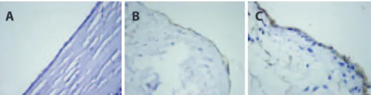
Figure 1. Immunohistochemical staining of the endothelial cells for caspase-3. Negative
(A), weak (B), and moderate (C) (caspase-3 × 400).
A B C
A B C
Figure 2. Immunohistochemical staining of the endothelial cells for caspase 8. Negative
(A), weak (B), and moderate (C) (caspase-8 × 400).
DISCUSSION
The present study yielded intriguing results and, to our knowled-ge, is the irst report of the safety of intracameral TB in corneal tissue using oxidative stress parameters and apoptotic activity. The present study showed that although intracameral TB does not exert a syste-mic efect, it might be locally toxic to corneal tissue.
Akal A, et al.
391 Arq Bras Oftalmol. 2014;77(6):388-91 REFERENCES
1. Melles GR, de Waard PW, Pameyer JH, Houdijn Beekhuis W. Trypan blue capsule staining to visualize the capsulorhexis in cataract surgery. J Cataract Refract Surg. 1999; 25(1):7-9.
2. Jacobs DS, Cox TA, Wagoner MD, Ariyasu RG, Karp CL; American Academy of Ophthal-mology; Ophthalmic Technology Assessment Committee Anterior Segment Panel. Capsule staining as an adjunct to cataract surgery: a report from the American Aca -demy of Ophthalmology. Ophthalmology. 2006;113(4):707-13.
3. van Dooren BT, Beekhuis WH, Pels E. Biocompatibility of trypan blue with human corneal cells. Arch Ophthalmol. 2004;122(5):736-42.
4. van Dooren BTH, de Waard PWT, Poort-van Nouhuys H, Beekhuis WH, Melles GR. Cor-neal endothelial cell density after trypan blue capsule staining in cataract surgery. J Cataract Refract Surg. 2002;28(4):574-5.
5. Dada VK, Sharma N, Sudan R, Sethi H, Dada T, Pangtey MS. Anterior capsule staining for capsulorhexis in cases of white cataract: comparative clinical study. J Cataract Refract Surg. 2004;30(2):326-33.
6. Jacob S, Agarwal A, Agarwal A, Agarwal S, Chowdhary S, Chowdhary R, Bagmar AA. Trypan blue as an adjunct for safe phacoemulsiication in eyes with white cataract. J Cataract Refract Surg. 2002;28(10):1819-25.
7. Pohanish RP, editor. Sittig’s handbook of toxic and hazardous chemical carcinogens 5th ed. Norwich, NY: William Andrew; 2008. p.2528.
8. Ulas T, Buyukhatipoglu H, Kirhan I, Dal MS, Ulas S, Demir ME, et al. Evaluation of oxidative stress parameters and metabolic activities of nurses working day and night shifts. Rev Esc Enferm USP. 2013;47(2):471-6.
9. Yalcin S, Ulas T, Eren MA, Aydogan H, Camuzcuoglu A, Kucuk A, et al. Relationship between oxidative stress parameters and cystatin C levels in patients with severe pree clampsia. Medicina (Kaunas). 2013;49(3):118-23.
10. Ma JQ, Ding J, Zhang L, Liu CM. Hepatoprotective properties of sesamin against CCl4 induced oxidative stress-mediated apoptosis in mice via JNK pathway. Food Chem Toxicol. 2014;64:41-8.
11. Tanaka Y, Komatsu T, Shigemi H, Yamauchi T, Fujii Y. BIM(EL) is a key efector molecule in oxidative stress-mediated apoptosis in acute myeloid leukemia cells when combi-ned with arsenic trioxide and buthionine sulfoximine. BMC Cancer. 2014;14:27. 12. Erel O. A new automated colorimetric method for measuring total oxidant status. Clin
Biochem. 2005;38(12):1103-11.
13. Erel O. A novel automated method to measure total antioxidant response against po tent free radical reactions. Clin Biochem. 2004;37(2):112-9.
14. Thaler S, Hofmann J, Bartz-Schmidt KU, Schuettauf F, Haritoglou C, Yoeruek E. Methyl blue and aniline blue versus patent blue and trypan blue as vital dyes in cataract surgery: capsule staining properties and cytotoxicity to human cultured corneal endothelial cells. J Cataract Refract Surg. 2011;37(6):1147-53.
15. Norn MS. Per operative trypan blue vital staining of corneal endothelium. Eight years’ follow up. Acta Ophthalmol (Copenh). 1980;58(4):550-5.
16. Veckeneer M, van Overdam K, Monzer J, Kobuch K, van Marle W, Spekreijse H, van Meurs J. Ocular toxicity study of trypan blue injected into the vitreous cavity of rabbit eyes. Graefes Arch Clin Exp Ophthalmol. 2001;239(9):698-704.
17. Panda A, Krishna SN, Dada T. Outcome of phacoemulsiication in eyes with cataract and cornea opacity partially obscuring the pupillary area. Nepal J Ophthalmol. 2012; 4(2):217-23.
18. Sperling S. Evaluation of the endothelium of human donor corneas by induced di-lation of intercellular spaces and trypan blue. Graefes Arch Clin Exp Ophthalmol. 1986; 224(5):428-34.
19. Chang YS, Tseng SY, Tseng SH, Chen YT, Hsiao JH. Comparison of dyes for cataract surgery. Part 1: cytotoxicity to corneal endothelial cells in a rabbit model. J Cataract Refract Surg. 2005;31(4):792-8.
20. Kothari K, Jain SS, Shah NJ. Anterior capsular staining with trypan blue for capsulorhe-xis in mature and hypermature cataracts; a preliminary study. Indian J Ophthalmol. 2001;49(3):177-80.
21. Yoeruek E, Spitzer MS, Tatar O, Aisenbrey S, Bartz-Schmidt KU, Szurman P. Safety proile of bevacizumab on cultured human corneal cells. Cornea. 2007;26(8):977-82. 22. Chung CF, Liang CC, Lai JS, Lo ES, Lam DS. Safety of trypan blue 1% and indocyanine
green 0.5% in assisting visualization of anterior capsule during phacoemulsiication in mature cataract. J Cataract Refract Surg. 2005;31(5):938-42.
23. Chung KT. The signiicance of azo-reduction in the mutagenesis and carcinogenesis of azo dyes. Mutat Res. 1983;114(3):269-81.
24. Ema M, Kanoh S. [Studies on the pharmacological bases of fetal toxicity of drugs. (II). Efect of trypan blue on the pregnant rats and their ofspring]. Nihon Yakurigaku Zasshi. 1982;79(5):369-81.