428
DOI:
10.1590/0004-282X20160031
IMAGES IN NEUROLOGY
Primary central nervous system lymphoma
(PCNSL)
Linfoma primário do sistema nervoso central
Elizabete Maria Pereira de Andrade Caires
1, Fernando Freua
2, Felipe D’Almeida Costa
3, Fernanda Lemos
Moura
1, Marcos Aurélio Peterlevitz
2, Antônio Alberto Zambon
2Primary Central Nervous System Lymphoma (PCNLS), an
uncommon variant of extranodal non-Hodgkin lymphoma,
involves brain, leptomeninges, eyes or spinal cord
1.
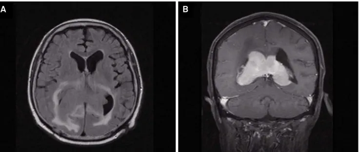
A 63-year-old man with two months of cognitive impairment
was submitted to brain MRI that revealed a expansive lesion
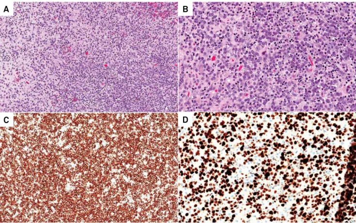
in corpus callosum (Figure 1A and 1B). Biopsy was performed
and histopathology showed a difuse large-B-cell lymphoma
(Figure 2). Cytarabine, metrotrexate and rituximab
2was
per-formed and after two months, patient returns to the emergency
with cerebelar ataxia and somnolence. New brain MRI detected
disease progression. Brain radiotherapy was indicated with
ad-ditional boost of radiotherapy to the tumor bed. After 1 month,
patient evolved with seizure and a lumbar puncture revealed
meningeal dissemination. It was decided palliative care.
1A. C. Camargo Cancer Center, Departamento de Oncologia Clínica, São Paulo SP, Brasil; 2A. C. Camargo Cancer Center, Departamento de Neurologia Clínica, São Paulo SP, Brasil; 3A. C. Camargo Cancer Center, Departamento de Patologia Clínica, São Paulo SP, Brasil.
Correspondence: Fernando Freua; Rua Conselheiro Brotero, 1505 / cj 41; 01232-011 São Paulo SP, Brasil; E-mail: [email protected]
Conflict of interest: There is no conflict of interest to declare. Received 26 August 2015; Accepted 04 January 2016.
Figure 1.
FLAIR sequence (A) showing expansive mass located in the topography of the fornix and splenium of corpus callosum
and extending to the lateral ventricles. Presents intense and homogeneous staining in T1 sequence coronal (B).
429
Elizabete Maria Pereira de Andrade Caires et al. Primary central nervous system lymphoma (PCNSL)
A
B
C
D
Figure 2.
Histologically, the central nervous system parenchyma was diffusely infiltrated by discohesive cells (A, H&E, 100x).
At higher power, the neoplastic cells were round and hyperchromatic, with scant cytoplasm and one or two prominent nucleoli
(B, H&E, 200x). Immunohistochemically, these cells were strongly positive for CD20 (C, anti-CD20 antibody, 100x) with a high
proliferative index (D, Ki-67 antibody, 200x). These findings, together with the clinical and radiological data, supported the
diagnosis of primary CNS diffuse large B-cell lymphoma.
References
1. Rubenstein J, Ferreri AJ, Pittaluga S. Primary lymphoma of the central nervous system: epidemiology, pathology and current approaches to diagnosis, prognosis and treatment. Leuk Lymphoma. 2008;49(1):43-51. doi:10.1080/10428190802311441