Brain regions supporting verbal memory
improvement in healthy older subjects
Regiões do cérebro relacionadas à melhora da memória verbal em idosos saudáveis
Eliane C. Miotto1, Joana B. Balardin1, Cary R. Savage2, Maria da Graça M. Martin3, Marcelo C. Batistuzzo1, Edson Amaro Junior3, Ricardo Nitrini1
ABSTRACT
Despite growing interest in developing cognitive training interventions to minimize the aging cognitive decline process, no studies have attempted to explore which brain regions support the application of semantic strategies during verbal memory encoding. Our aim was to investigate the behavioral performance and brain correlates of these strategies in elderly individuals using fMRI in healthy older subjects.
Method:Subjects were scanned twice on the same day, before and after, directed instructions to apply semantic strategies during the encoding of word lists.Results:Improved memory performance associated to increased semantic strategy application and brain activity in the left inferior and middle and right medial superior prefrontal cortex were found after the directed instructions. There was also reduced activation in areas related to strategy mobilization.Conclusion:Improved memory performance in older subjects after the application of semantic strategies was associated with functional brain reorganization involving regions inside and outside the typical memory network.
Keywords:older subjects, semantic strategies, fMRI, episodic memory.
RESUMO
Apesar do crescente interesse em intervenções de treinamento cognitivo para minimizar o declínio cognitivo do envelhecimento, nenhum estudo explorou quais regiões do cérebro estão relacionadas à aplicação de estratégias semânticas durante a codificação da memória verbal. Nosso objetivo foi investigar o comportamento e correlatos cerebrais associados a essas estratégias usando fMRI em idosos saudáveis. Método: Os sujeitos foram examinados no mesmo dia, antes e depois, de instruções dirigidas para aplicar estratégias semânticas durante a codificação de palavras.Resultados:Melhora da memória relacionada ao uso de estratégias semânticas e aumento da atividade no córtex prefrontal inferior e medial esquerdo e medial superior direito foram encontrados após as instruções. Também houve redução de ativação em áreas de mobilização de estratégias. Conclusão: A melhora da memória em idosos após o uso de estratégias semânticas estava associada à reorganização cerebral funcional envolvendo regiões dentro e fora da rede de áreas cerebrais típicas da memória.
Palavras-chave:idosos, estratégias semânticas, fMRI, memória episódica.
The aging process is associated with progressive decline in cognitive abilities such as memory, attention and
exec-utive function1,2,3. Therefore, older subjects commonly
experience functional and cognitive losses which can affect quality of life, social interaction, and occupational perform-ance. More recently, there has been a great concern with benign cognitive decline and the risk for developing mild cognitive impairment (MCI) or dementia progression. As a consequence, there has been growing interest to explore non-pharmacological clinical interventions, particularly cog-nitive training, to minimize this process or improve some cognitive skills in older adults4.
Episodic memory is the most commonly affected type of memory in aging5,6. The term
“episodic memory”refers to a system responsible for the encoding, storing, and retrieving of personally experienced and highly temporally specific information7. Combined evidence from cognitive psychology,
neuropsychological and functional neuroimaging studies suggested that efficient episodic memory encoding might engage information processing within specific cortical regions that provides a critical input to medial temporal cor-tex. Task-related fMRI studies supplied further information regarding the functional neuroanatomy of these cognitive processes. For example, engagement of portions of the
dor-1Departamento de Neurologia, Faculdade de Medicina, Universidade de São Paulo, Sao Paulo SP, Brazil; 2Center for Health Behavior Neuroscience, Kansas University, Kansas, EUA;
3Departamento de Radiologia, Faculdade de Medicina, Universidade de São Paulo, Sao Paulo SP, Brazil.
Correspondence:Departamento de Neurologia, Faculdade de Medicina, Universidade de São Paulo; Av. Dr. Enéas de Carvalho Aguiar, 255; 05403-900 São Paulo SP, Brasil; E-mail: [email protected]
Conflict of interest:There is no conflict of interest to declare.
Received 17 January 2014; Received in final form 16 June 2014; Accepted 07 July 2014.
DOI:10.1590/0004-282X20140120
solateral prefrontal and the posterior parietal cortices
have been reported during working memory tasks8, which
generally modulates episodic memory performance by promoting efficient encoding through manipulation and monitoring of information to subsequent retrieval9. The left
ventrolateral prefrontal (VLPFC) cortex has been implicated in the access to the semantic memory system in the
tem-poral pole10, which is involved in modulating the depth
and organization of material at encoding.
Encoding strategies, used to enhance episodic memory performance are also reduced in aging possibly due to changes in prefrontal structures and their connections with posterior cortical areas such as temporal and parietal cortex which are involved in episodic memory encoding and retrieval11. The participation of the prefrontal cortex (PFC)
in executive strategic processes, learning and episodic mem-ory is well documented by brain lesion and functional neu-roimaging studies12,13. One of the encoding strategies known
to improve episodic verbal memory is semantic categoriza-tion. In typical verbal learning tasks, participants study lists of words and subsequently recall the studied items in the order they come to mind without external cues. But when the studied material is composed by lists of related words, participants have a tendency to recall semantically related items consecutively. Although differing accounts have attrib-uted such effects to associative automatic versus strategic controlled mechanisms operating at encoding, retrieval, or both14,15, this effect, named semantic clustering16, has been
used as an index of strategic episodic learning. The left inferior prefrontal cortex (IPFC) mediates semantic proces-sing of information17,18. The dorsolateral prefrontal cortex
(DLPFC) supports updating, sequencing, and monitoring operations, all features of working memory, along with atten-tion19. The orbitofrontal cortex (OFC) plays an important
role in initiating relevant behavior, suppressing irrelevant responses and in the early mobilization of efficient strategies in novel situations20. Therefore, the PFC plays an important
role at the encoding, organization and strategy creation during memory functioning and this role seems to increase with age5.
It has been suggested that older adults are less likely than young adults to use efficient strategies when performing memory tasks although this difference can be reduced by cognitive training21. However, direct evidence of the neural
mechanisms and brain regions related to memory improve-ment in healthy older adults are scarce. Yet, brain imaging studies can contribute to our understanding of the aging brain. To date, only few studies have investigated the brain correlates after cognitive training in healthy older subjects. In one such study using positron emission tomography
(PET), older adults’ brain activity was assessed during
encoding before and after the loci training − a mnemonic
method to encode words which requires visualization of a
series of items in familiar locations22. Older adults showed
greater activity after training in occipitoparietal cortex related to improved performance. Using the same loci method in an 8-week cognitive training, older participants showed increased cortical thickness, as measured by mag-netic resonance imaging (MRI), in the right orbitofrontal gyrus, insula, and fusiform gyrus23
. In a functional MRI ( fMRI) study, 16 older adults received two training sessions, in different days, associating pleasantness, personal
rel-evance, and sentence creation to encode words21. There
was increased activity during encoding in the medial super-ior frontal gyrus, right precentral gyrus, and left caudate after training. Other recent studies have examined the neural correlates of cognitive training in both healthy aging and MCI subjects. In one of them, increased activation was found after a 6-week cognitive training in the MCI group encom-passing a large network including the frontal, temporal and parietal regions whereas healthy controls showed mostly areas of decreased activation following training24. In
another recent brain imaging study25, MCI and healthy
con-trols were scanned as they encoded and retrieved object–
location associations – a visuospatial memory task.
Although the fMRI analyses were restricted to the hip-pocampus, following training, the MCI group showed increased left and right hippocampi activity at encoding and retrieval, respectively and the healthy older controls showed mainly decreased activation after training in the right hippocampus.
Despite these previous results, to our knowledge, there are no studies exploring which brain regions, particularly involving the frontal networks, are engaged during the applica-tion of semantic organizaapplica-tional strategies in healthy older sub-jects and what are their impact on memory performance. Therefore, the aims of the current study were to investigate1
the brain correlates of semantic strategy application during
verbal memory encoding in older adults using fMRI, and2
the effects of this strategy on memory performance.
METHOD
Participants
or psychiatric disease, cognitive complaints or deficits iden-tified by the neuropsychological tests based on their norms. Exclusion criteria included the use of any psychotropic med-ication (except antidepressants in stable doses) within the previous 6 months, and previous or current use of illegal psy-choactive drugs. In addition, the exclusion criteria included vascular lesions, hippocampus or cerebral atrophy identified by the structural MRI, uncontrolled hypertension, unstable medical conditions (i.e., diabetes, hypothyroidism, vitamin B12 or folate deficiency), and presence of any metal in their body other than dental fillings. The study was approved by the local ethics committee (CAPPesq 06/03336).
Study design and fMRI experimental paradigm partici-pants were scanned using a block design during the encod-ing of word lists: 16 unrelated words (UR) and 16 semantically related words (SR) in terms of semantic cat-egories, alternated with a fixation baseline (+) (Figure 1). The current paradigm was based on the California Verbal Learning Test (CVLT)26and validated in healthy adults and
patients with brain lesions using fMRI19,27. For the UR
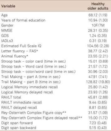
con-dition, the words presented were not semantically related making it difficult to apply the semantic organizational strat-egies. For the SR condition, the words were semantically related into four categories and were presented in a rando-mized mixed order (Figure 1). The word lists were generated from 32 categories of words with four words in each cat-egory. The paradigm was tested extensively in pilot studies in order to develop word lists that prompted significant Table 1.Participants’clinical and neuropsychological
characteristics. Results are expressed as mean±standard deviation.
Variable Healthy
older adults
Age 68.12 (1.19)
Years of formal education 10.94 (1.30)
Gender 10F/7M
MMSE 28.31 (0.35)
GDS 1.24 (0.35)
IADLs3 0.31 (0.19)
Estimated Full Scale IQ 104.56 (2.28)
Letter fluency–FAS* 38.77 (2.42)
Animal fluency* 23.55 (2.21)
Stroop task–color card (time in sec.) 15.01 (0.69) Stroop task–Word card (time in sec.) 21.57 (1.72) Stroop task–color/word card (time in sec.) 30.96 (2.03) Trail Making–part A (time in sec.) 47.91 (7.41) Trail Making–part B (time in sec.) 128.82 (19.80) Logical Memory immediate recall 25.80 (1.42) Logical Memory delayed recall 23.93 (1.26)
RAVLT total 45.81 (2.88)
RAVLT immediate recall 9.44 (0.65)
RAVLT delayed recall 8.81 (0.65)
Rey-Osterrieth Complex Figure copy** 31.57 (1.09) Rey-Osterrieth Complex Figure delayed recall** 15.00 (1.72)
Digit span forward 7.23 (0.48)
Digit span backward 5.15 (0.43)
MMSE: Mini-Mental State Examination; GDS: Geriatric Depression Scale; Instrumental Activities of Daily Living Scale (Pfeffer); Estimated Full Scale IQ based on the Weschler Adult Intelligence Scale III; RAVLT: Rey Auditory Verbal Learning Test, sum of words recalled across the 5 consecutive trials; Letters selected to prompt word generation; *Total number of words recalled in one minute; **Total score during unconstrained recall.
Fix1 Fix2 Fix3 Fix4 Fix5 Fix6 Fix7
+ SR + UR + SR + UR + UR + SR + 12s 33s 12s 33s 12s 33s 12s 33s 12s 33s 12s 22s 12s
Categories SR UR
Body parts Eye Lemon
(Land animals) Penguin Comb Cleaning supplies Dustpan Ship (Water animals) Oyster Cake
Detergent Fork
Squid Piano
Beaver Shirt
Finger Salt
Cod Doll
Sponge Blocks
Deer Table
Ankle Paper
Bucket Water
Salmon Window
Nose Paint
Wolf Cable
SR: semantically related words; UR: unrelated words.
differences in semantic clustering. In addition, the inclusion of categories that shared“fuzzy”semantic relationships (e.g., land animals and water animals) made the task more chal-lenging and minimized ceiling effects for clustering. Scanning was carried out twice: before and after a brief per-iod of directed instructions and practice (30-min) to apply the semantic organizational strategies. In the first fMRI ses-sion (before the directed instructions), subjects were instructed to look at the words presented onto the screen and try to remember as many words as they could, without a specific instruction to apply any strategy. After the first fMRI session, they were taken to a different room and sub-mitted to a training consisted of teaching them to apply semantic organizational strategies to a set of different word lists. Subjects were taught to organize a set of different word lists into categories and then to memorize and retrieve the words according to each category. The instructions for the orientation training were as follows:“Notice that the words appearing on the screen can be grouped into four different categories. By grouping the words together into these related categories, you will remember more words. For example, in this list you can group the words into body parts, land ani-mals, cleaning supplies and water animals. The words can be grouped into four categories. Be sure and group the words
together into categories this time as you learn them”.
Immediately after and on the same day, they were taken to the scanner and instructed to look at novel words pre-sented onto the screen and apply the semantic organiza-tional strategy. As in the first fMRI session, following the scan, participants were prompted to recall as many words as they could remember in any order and this measure was used as free recall. It should be noted that for all condi-tions including those in the first and second fMRI sessions we used novel words in order to avoid practice effects. Semantic clustering index scores were defined as consec-utive recall of two words from the same category. They reflected the proportion of clustered responses out of the total possible clusters for that condition defined as [clusters/ (words recalled-categories recalled)]. Differences between scanning sessions in free recall scores and semantic clustering index were examined using a paired-sample t-test. All stimuli were visually presented on a screen and synchronized with the scanner via an optic relay triggered by the radiofrequency pulse. The presentation order of the words in each list was ran-domized and conditions (SR, UR) were counterbalanced within scans and across participants.
Image acquisition and fMRI analysis scans were per-formed on a Philips 3 tesla Achieva MRI scanner (8ch head coil). The fMRI acquisition was based on T2*-weighted echo planar (EPI) images for the whole brain with the following parameters: TR=3,000 ms, TE=30 ms, 41 slices, 3 mm
iso-tropic voxel size, FOV=240 mm and matrix 80¾78, with 94
volumes, discarding the initial four scans in each run to
allow steady-state magnetization to be approached. There were two runs, each run with 32 words and a total number of words equal to 64 (all novel words). Each run lasted 282 s (4min42s), in which were collected 94 volumes covering all the brain.
FSL package (version 4.1, FMRIB Analysis Group, www. fmrib.ox.ac.uk/fsl) was used to analyze the fMRI data. Preprocessing included movement correction (MCFLIRT), spatial smoothing using a 5 mm Gaussian kernel, and spatial normalization to MNI-152 template with a 12 DoF affine registration (FLIRT). The general linear model (GLM) with residual autocorrelation correction (FILM) was applied to obtain statistical activation maps. The design matrix was composed of regressors according to the experimental con-ditions (SR and UR). For each condition, the haemodynamic response function in GLM was determined considering the convolution of experimental designs with a gamma function (SD=3 s, mean lag=6 s). The group activation maps and ses-sion comparisons were obtained using the mixed-effects
model. Contrast images for each encoding condition (SR.
fixation, UR.fixation) in the first and second fMRI sessions were created for each subject. In a preliminary analysis, the contrasts SR.fixation and UR.fixation were entered into a
second-level analysis [FMRIB’s Local Analysis of Mixed
Effects-FLAME stage 128to test if there were a main effect
of list (SR, UR) or a list session (spontaneous, directed) inter-action, in order to identify regions whose activation increased or decreased between sessions, more for the SR contrast than the NR contrast. There were no brain regions that showed a significant interaction or a main effect of list. Therefore, because the focus of the present study was on identifying session-related activation that are associated to strategic behaviour, the remaining analyses focused only in the contrast SR.fixation, giving the sensitivity of the SR word list in prompting measurable semantic clustering scores. These contrast images were then used for between-sessions comparisons (paired-sample t-test) in order to obtain the brain activation pattern change over time. All the statistical images were thresholded by using Gaussian random field-based cluster inference with a threshold of Z.2.3 at the voxel level and a corrected cluster significance threshold of P,0.05.
RESULTS
Behavioural results
directed instructions and the total number of words recalled only after training for the SR condition (r=0.652, p=0.002). In terms of word free recall, there was a significant change only for the SR condition (t=-3.665, p=0.002) with more words recalled after the directed instructions.
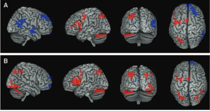
Neuroimaging results are presented in Figure 2. Participants exhibited increased activation in the 2nd fMRI
session compared to the 1stfMRI for the contrast SR
. base-line in clusters encompassing left inferior frontal gyrus, mid-dle frontal gyrus and precentral gyrus, left intraparietal sulcus, angular gyrus, left cerebellum and right medial super-ior frontal gyrus and antersuper-ior cingulate. When comparing the 1st
.2ndfMRI, decreased activation was observed in the
bilateral posterior cingulate and left precuneus, right medial superior frontal gyrus and anterior cingulate, right inferior
parietal lobule and middle and superior temporal cortex, right dorsal superior frontal gyrus, and bilateral orbitofrontal cortex (Figures 2 and 3; Table 3).
Results for the UR.baseline showed increased activa-tion in the 2nd fMRI session in left superior frontal gyrus
and precentral gyrus, bilateral precuneus and inferior pari-etal lobule, bilateral medial superior frontal gyrus and right anterior cingulate. Decreased activation was observed in the right superior frontal gyrus, medial orbital gyrus and anterior cingulate, bilateral lingual gyrus, left cuneus and posterior cingulate, and bilateral precuneus (Figures 2 and 3; Table 3).
DISCUSSION
The present study investigated the neural correlates of semantic strategy application during memory encoding in older adults using fMRI. The main behavioral results showed a significant increase in semantic strategy application after the directed instructions and practice to apply these strategies, suggesting that older adults are able to learn and implement efficient strategies. In addition, participants recalled more words after the directed instructions for the SR condition and there was a significant correlation between semantic strat-egy scores and words recalled for the SR condition, a condition where this strategy can be used. These findings are similar to the ones found in healthy adult subjects19and other studies
using different cognitive training in elderly subjects21,22 and
show that after a brief period of practice to apply efficient Table 2.Performance on free recall, semantic clustering
index and recognition for each condition during the first and second fMRI sessions. Results are expressed as mean ±standard deviation.
1stsession 2ndsession
Free recall
SR* 7.53 (0.575) 9.41 (0.810)
UR* 3.59 (0.613) 3.29 (0.391)
Semantic clustering index 0.242 (0.044) 0.452 (0.038) Number of clusters (max 12) 3.23 (0.465) 4.58 (0.675) Recognition
Hit rate SR 0.92 (0.022) 0.82 (0.036) Hit rate UR 0.83 (0.036) 0.87 (0.026) Corrected rejection rate 0.92 (0.029) 0.89 (0.023) *Total number of words recalled; SR: semantic related list; UR: semantic unrelated list.
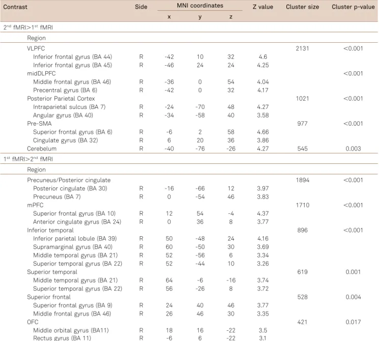
Table 3.Significant clusters highlighted when comparing the BOLD response between the spontaneous and directed use of learning strategies during word list encoding compared to fixation baseline.
Contrast Side MNI coordinates Z value Cluster size Cluster p-value
x y z
2ndfMRI.1stfMRI
Region
VLPFC 2131 ,0.001
Inferior frontal gyrus (BA 44) R -42 10 32 4.6
Inferior frontal gyrus (BA 45) R -46 24 24 4.25
midDLPFC ,0.001
Middle frontal gyrus (BA 46) R -36 0 54 4.04
Precentral gyrus (BA 6) R -42 0 32 4.17
Posterior Parietal Cortex 1021 ,0.001
Intraparietal sulcus (BA 7) R -24 -70 48 4.27
Angular gyrus (BA 40) R -34 -58 40 3.58
Pre-SMA 977 ,0.001
Superior frontal gyrus (BA 6) R -6 2 58 4.66
Cingulate gyrus (BA 32) R 6 20 36 3.86
Cerebelum R -40 -76 -26 4.27 545 0.003
1stfMRI.2ndfMRI
Region
Precuneus/Posterior cingulate 1894 ,0.001
Posterior cingulate (BA 30) R -16 -66 12 3.97
Precuneus (BA 7) R 0 -54 46 3.83
mPFC 1710 ,0.001
Superior frontal gyrus (BA 10) R 12 54 -4 4.37
Anterior cingulate gyrus (BA 24) R 0 36 8 3.77
Inferior temporal 896 ,0.001
Inferior parietal lobule (BA 39) R 50 -48 24 4.16
Supramarginal gyrus (BA 40) R 60 -50 30 3.69
Middle temporal gyrus (BA 21) R 52 -56 6 3.34
Superior temporal gyrus (BA 22) R 52 -44 10 3.26
Superior temporal 619 0.001
Middle temporal gyrus (BA 21) R 64 -6 -16 3.74
Superior temporal gyrus (BA 22) R 56 -26 8 3.72
Superior frontal 528 0.004
Superior frontal gyrus (BA 9) R 24 40 46 3.77
Middle frontal gyrus (BA 46) R 26 46 30 3.35
OFC 421 0.017
Middle orbital gyrus (BA11) R 18 16 -22 3.5
Rectus gyrus (BA 11) R -6 6 -22 3.1
FMRI: functional magnetic resonance imaging; VLPFC: left ventrolateral prefrontal; DLPPC: dorsolateral prefrontal cortex; BA: Brodmann area; SMA: supplementary motor area; PFC: prefrontal cortez; OFC: orbitofrontal cortex.
Figure 3.Brain regions with significant differences for the contrasts A 1st
.2ndfMRI and B 1st
strategies older subjects can substantially improve their mem-ory performance.
Brain imaging results for the SR condition, in which semantic processing and active organization in working memory were most demanded in relation to the UR con-dition, indicated increased activation in the second fMRI session compared to the first in the left inferior and middle frontal gyrus, precentral gyrus, intraparietal sulcus, angular gyrus, cerebellum and in the right medial superior frontal gyrus and anterior cingulate. These findings are in line with studies of healthy older subjects employing longer periods of cognitive training showing increased activity after training in bilateral prefrontal regions including the inferior and medial frontal gyrus21,23,25. Previous results suggested that the left
middle and inferior frontal gyrus and cerebellum support young adults’ verbal encoding strategies19,27. In addition,
there was greater activity in the right medial superior frontal gyrus and anterior cingulate. Semantic categorization strat-egy training has been associated to increased brain activity in the right middle and inferior frontal gyri in healthy adults19. Taken together these findings suggest that semantic
organizational strategy application is associated with bilat-eral PFC increased activity regardless of age.
Decreased activation was noted in bilateral OFC, right medial and dorsal superior frontal gyrus, inferior parietal lobule, anterior and posterior cingulate, left precuneus, mid-dle and superior temporal cortex. Here again, these results are similar to studies with healthy young adults showing decreased activation in the OFC after training or instruction to apply semantic organizational strategies19,27,28. Previous
investigations indicated that the OFC mediates memory pro-cesses by mobilizing effective strategies in novel situa-tions18,29. It is possible that, after the instructions to apply
the semantic strategies, this ability became consolidated resulting in OFC decreased activation. Areas of reduced activity were also found in the right DLPFC, medial frontal cortex and anterior cingulate after the instructions to apply the semantic strategies. These findings were possibly related to the reduced attentional demands after these instructions. These cerebral regions have previously been associated with attention and monitoring processes and patients with lesions in these areas are usually impaired in tasks requiring sustained attention and working memory12.
Participants supressed the responses of mPFC, precu-neous and bilateral posterior cinulate encoding after the explicit orientation to apply the verbal learning strategy compared to the first fMRI session. In the context of task induced deactivation studies, these regions, along inferior parietal lobule, have been consistently reported as nodes
of the default mode network (DMN) of the brain30.
Although the identification of a possible DMN was originally identified as a phenomenon whereby a set of regions exhib-ited greater blood flow or BOLD signal during baseline
resting or fixation conditions than during task performance, subsequent studies demonstrated similar patterns of results when tasks with different cognitive demands were
con-trasted31. Recent studies have shown, for example, that
mPFC activity is greater during performance of a relatively
easy 0-back task than a 2-back working memory task32,
and that suppressing activity in DMN regions during working memory encoding, was predictive of subsequent
better performance33. In our study, when an increased
working memory demand was imposed by the direct application of the encoding strategy, healthy older adults exhibited a suppression of these regions (i.e. less activation). The exact nature of the DMN suppression, however, is not fully understood. The most common theoretical interpreta-tion has been that “task-induced deactivations” reflect the occurrence of spontaneous internally directed thought pro-cesses such as autobiographical memory retrieval and internally oriented mindwandering during unconstrained baseline conditions34.
For the UR condition, where application of the semantic strategy proved to be difficult because the words were not related, increased activation after the directed instructions was found in the left superior frontal and gyrus, bilateral pre-cuneus and inferior parietal lobule, bilateral medial portion of superior frontal gyrus and right anterior cingulate. It is possible that these activations were associated with the attempt of the subjects to apply the semantic strategies, even though this would prove to be difficult given that the words were unrelated. It should be noted that participants were not instructed as to which order each condition was presented. The results of our study should be interpreted in the con-text of some potential limitations. First, we did not control for repetition effects in the MRI. However, in a previous work
of our group35 we adopted an approach used by Belleville
and cols.24 to evaluate the fMRI task repetition effect on
brain activation using the same word-list encoding para-digm. In this study35the activations related to the first and
second runs acquired in the first fMRI session were com-pared, so each subject was used as its own control. The results of this analysis demonstrated no significant brain activation changes between the runs, suggesting that repeat-ing the task without the instructions and practice to apply the strategies did not produce measurable repetition or prac-tice effects on brain activation. Another limitation is the lack of a placebo-control condition, which is a challenge in beha-vioral intervention studies.
In conclusion, the present results demonstrated the engagement of bilateral PFC and posterior areas during semantic strategy application in a verbal encoding paradigm indicating the recruitment of new, in addition to the typical memory encoding network, consistent with functional reor-ganization also found in healthy adult subjects19,21,22. These
prac-tice to apply semantic strategies are sufficient to alter older adults’ brain activity and behavioural performance. Future longitudinal studies are needed to determine whether this
effect persists or changes across time and if it can also be observed in patients with mild cognitive impairment and other cerebral degenerative diseases.
References
1. Fillit HM, Butler RN, O’Connell AW, et al. Achieving and maintaining cognitive vitality with aging. Mayo Clin Proc 2002;77:681-696.
2. Verhaeghen P, Geraerts N, Marcoen A. Memory complaints, coping, and well-being in old age: a systemic approach. Gerontologist 2000;40:540-548.
3. Bischkopf J, Busse A, Angermeyer MC. Mild cognitive impairment - a review of prevalence, incidence and outcome according to current approaches. Acta Psychiatr Scand 2002;106:403-414.
4. Gross AL, Parisi JM, Spira AP, et al. Memory training interventions for older adults: a meta-analysis. Aging Ment Health 2012;16:722-734.
5. Dennis NA, Hayes SM, Prince SE, Madden DJ, Huettel SA, Cabeza R. Effects of aging on the neural correlates of successful item and source memory encoding. J Exp Psychol Learn Mem Cogn 2008;34:791-808.
6. Fairchild JK, Scogin FR. Training to Enhance Adult Memory (TEAM): an investigation of the effectiveness of a memory training program with older adults. Aging Ment Health 2010;14:364-373.
7. Tulving E. Elements of episodic memory. Oxford [Oxfordshire]: New York: Clarendon Press; Oxford University Press; 1983:351.
8. Champod AS, Petrides M. Dissociation within the frontoparietal network in verbal working memory: a parametric functional magnetic resonance imaging study. J Neurosci 2010;30:3849-3856.
9. Baddeley A. Working memory: theories, models, and controversies. Annu Rev Psychol 2012;63:1-29.
10. Paller KA, Wagner AD. Observing the transformation of experience into memory. Trends Cogn Sci (Regul Ed) 2002;6:93-102.
11. Craik FIM. Levels of processing: past, present. and future? Memory 2002;10:305-318.
12. Stuss DT. Functions of the frontal lobes: relation to executive functions. J Int Neuropsychol Soc 2011;17:759-765.
13. Shallice T, Fletcher P, Frith CD, Grasby P, Frackowiak RS, Dolan RJ. Brain regions associated with acquisition and retrieval of verbal episodic memory. Nature 1994;368:633-635.
14. Gershberg FB, Shimamura AP. Impaired use of organizational strategies in free recall following frontal lobe damage. Neuropsychologia 1995;33:1305-1333.
15. Alexander MP, Stuss D, Gillingham S. Impaired list learning is not a general property of frontal lesions. J Cogn Neurosci 2009;21:1422-1434.
16. Bousfield WA. The occurrence of clustering in the recall of randomly arranged associates. J Gen Psychol 1953;49:229-240.
17. Demb JB, Desmond JE, Wagner AD, Vaidya CJ, Glover GH, Gabrieli JD. Semantic encoding and retrieval in the left inferior prefrontal cortex: a functional MRI study of task difficulty and process specificity. J Neurosci 1995;15:5870-5878.
18. Savage CR, Deckersbach T, Heckers S, et al. Prefrontal regions supporting spontaneous and directed application of verbal learning strategies: evidence from PET. Brain 2001;124:219-231.
19. Miotto EC, Savage CR, Evans JJ, et al. Bilateral activation of the prefrontal cortex after strategic semantic cognitive training. Hum Brain Mapp 2006;27:288-295.
20. Bechara A, Tranel D, Damasio H, Damasio AR. Failure to respond autonomically to anticipated future outcomes following damage to prefrontal cortex. Cereb Cortex 1996;6:215-225.
21. Kirchhoff BA, Anderson BA, Barch DM, Jacoby LL. Cognitive and neural effects of semantic encoding strategy training in older adults. Cereb Cortex 2012;22:788-799.
22. Nyberg L, Sandblom J, Jones S, et al. Neural correlates of training-related memory improvement in adulthood and aging. Proc Natl Acad Sci USA 2003;100:13728-13733.
23. Engvig A, Fjell AM, Westlye LT, et al. Effects of memory training on cortical thickness in the elderly. Neuroimage 2010;52:1667-176.
24. Belleville S, Clément F, Mellah S, Gilbert B, Fontaine F, Gauthier S. Training-related brain plasticity in subjects at risk of developing Alzheimer’s disease. Brain 2011;134:1623-1634.
25. Hampstead BM, Stringer AY, Stilla RF, Giddens M, Sathian K. Mnemonic strategy training partially restores hippocampal activity in patients with mild cognitive impairment. Hippocampus 2012;22:1652-1658.
26. Donders J. Performance discrepancies on the California Verbal Learning Test - Second Edition (CVLT-II) in the standardization sample. Psychol Assess 2006;18:458-463.
27. Miotto EC, Savage CR, Evans JJ, et al. Semantic strategy training increases memory performance and brain activity in patients with prefrontal cortex lesions. Clin Neurol Neurosurg 2013;115:309-316.
28. Woolrich MW, Behrens TEJ, Beckmann CF, Jenkinson M, Smith SM. Multilevel linear modelling for FMRI group analysis using Bayesian inference. Neuroimage 2004;21:1732-1747.
29. Nobre AC, Coull JT, Frith CD, Mesulam MM. Orbitofrontal cortex is activated during breaches of expectation in tasks of visual attention. Nat Neurosci 1999;2:11-12.
30. Toro R, Fox PT, Paus T. Functional coactivation map of the human brain. Cereb Cortex 2008;18:2553-2559.
31. Raichle ME, MacLeod AM, Snyder AZ, Powers WJ, Gusnard DA, Shulman GL. A default mode of brain function. Proc Natl Acad Sci USA 2001;98:676-682.
32. Leech R, Kamourieh S, Beckmann CF, Sharp DJ. Fractionating the default mode network: distinct contributions of the ventral and dorsal posterior cingulate cortex to cognitive control. J Neurosci 2011;31:3217-3224.
33. Anticevic A, Repovs G, Barch DM. Resisting emotional interference: brain regions facilitating working memory performance during negative distraction. Cogn Affect Behav Neurosci 2010;10:159-173.
34. Buckner RL, Andrews-Hanna JR, Schacter DL. The brain’s default network: anatomy, function, and relevance to disease. Ann N Y Acad Sci 2008;1124:1-38.