Atherosclerotic Plaque in Patients with Zero Calcium Score at
Coronary Computed Tomography Angiography
Fabíola Santos Gabriel,
1,3Luiz Flávio Galvão Gonçalves,
2,3Enaldo Vieira de Melo,
1Antônio Carlos Sobral Sousa,
1,4,5Ibraim Masciarelli Francisco Pinto,
6Sara Melo Macedo Santana,
3Carlos José Oliveira de Matos,
1,4Maria Júlia
Silveira Souto,
1,4Flávio Mateus do Sacramento Conceição,
1,4Joselina Luzia Menezes Oliveira
1,4,5,6,7Núcleo de Pós-Graduação em Medicina da Universidade Federal de Sergipe (UFS),1 São Cristóvão, SE - Brazil Centro de Pesquisas da Fundação São Lucas,2 Aracaju, SE - Brazil
Clínica de Medicina Nuclear de Diabetes – CLIMEDI,3 Aracaju, SE - Brazil
Departamento de Medicina - Universidade Federal de Sergipe (UFS),4 São Cristóvão, SE - Brazil
Centro de Ensino e Pesquisa e Laboratório de Ecocardiografia (ECOLAB) do Hospital e Fundação São Lucas,5 Aracaju, SE - Brazil Instituto Dante Pazzanese de Cardiologia,6 São Paulo, SP - Brazil
Centro de Ensino e Pesquisa da Fundação São Lucas,7 Aracaju, SE - Brazil
Mailing Address: Fabíola Santos Gabriel •
Av. Dr. Francisco Moreira, 220, Bl. F, Apt. 04. Postal Code 49047-335, Ponto novo, Aracaju, SE – Brazil
E-mail: [email protected]
Manuscript received May 01, 2017, revised manuscript November 12, 2017, accepted November 22, 2017
DOI: 10.5935/abc.20180063
Abstract
Background: In view of the high mortality for cardiovascular diseases, it has become necessary to stratify the main risk factors and to choose the correct diagnostic modality. Studies have demonstrated that a zero calcium score (CS) is characteristic of a low risk for cardiovascular events. However, the prevalence of individuals with coronary atherosclerotic plaques and zero CS is conflicting in the specialized literature.
Objective: To evaluate the frequency of patients with coronary atherosclerotic plaques, their degree of obstruction and associated factors in patients with zero CS and indication for coronary computed tomography angiography (CCTA).
Methods: This is a cross-sectional, prospective study with 367 volunteers with zero CS at CCTA in four diagnostic imaging centers in the period from 2011 to 2016. A significance level of 5% and 95% confidence interval were adopted.
Results: The frequency of atherosclerotic plaque in the coronary arteries in 367 patients with zero CS was 9.3% (34 individuals). In this subgroup, mean age was 52 ± 10 years, 18 (52.9%) were women and 16 (47%) had significant coronary obstructions (> 50%), with involvement of two or more segments in 4 (25%) patients. The frequency of non-obese individuals (90.6% vs 73.9%, p = 0.037) and alcohol drinkers (55.9% vs 34.8%, p = 0.015) was significantly higher in patients with atherosclerotic plaques, with an odds ratio of 3.4 for each of this variable.
Conclusions: The frequency of atherosclerotic plaque with zero CS was relatively high, indicating that the absence of calcification does not exclude the presence of plaques, many of which obstructive, especially in non-obese subjects and alcohol drinkers. (Arq Bras Cardiol. 2018; 110(5):420-427)
Keywords: Cardiovascular Diseases/mortality; Plaque, Atherosclerotic; Coronary Artery Disease/diagnosis; Calcium Signaling; Coronary, Angiotomography; Risk Factors.
Introduction
Coronary artery disease (CAD) are the leading cause of death in the world, including in Brazil. Many methods for CAD diagnosis, risk stratification of patients and indication of revascularization are currently available.1
One of the greatest challenges of routine cardiology practice is to determine the best method to detect subclinical CAD. Coronary computed tomography angiography (CCTA) is a predominantly anatomical test with excellent diagnostic
accuracy in detecting obstructive and nonobstructive lesions as compared with coronary angiography, which is considered the gold standard method for this purpose. Also, CCTA may provide relevant information regarding atheroma composition according to radiological density.2,3
The role of coronary calcification, identified by calcium score (CS), used for classification of patients into a higher risk for cardiovascular events, is well known. Although individuals with a zero CS may also have atherosclerotic plaques,4,5 its
presence has not been associated with increased risk for future cardiovascular events.4
Therefore, the main aim of this study is to evaluate the frequency of coronary atherosclerotic plaque, its degree of obstruction and associated factors in patients with zero CS and clinical indication for CCTA.
Methods
Subjects
This was a cross-sectional, analytical, prospective study carried out from April 2011 to November 2016. Subjects were consecutively and not randomly selected, and subjected to CCTA by referral from their assistant physicians in four diagnostic imaging centers, two public centers: Instituto Dante Pazzanese de Cardiologia de São Paulo-SP e Hospital Universitário do Campus da Saúde Dr. João Cardoso Nascimento da Universidade Federal de Sergipe and two private centers: Hospital Primavera e Clínica de Medicina Nuclear e Diabetes-CLIMEDI.
Data on cardiovascular risk factors were collected from each participant. Chest pain was classified according to the Diamond and Forrester method, and most patients were classified as at intermediate risk for CAD.
Patients with no calcium in the coronary arteries (zero CS) were included. Patients who had undergone percutaneous or surgical myocardial revascularization, patients with history of acute coronary syndrome or cardiomyopathy of ischemic cause, and those who declined to participate were excluded. The tests performed at private institutions were free of charge for both patients and investigators.
The study was approved by the research ethics committee (CAAE identification number 0289.0.107.000-11).
CS and CCTA of coronary arteries
CCTA of coronary arteries were performed using a 64-slice (or greater) scanner of the following models and manufacturers: Aquilion64™ - Toshiba™ Medical Systems Corporation, Otawara, Japan and Discovery STE VCT - General Electric Company, Connecticut, USA.
Non-contrast computed tomography for CS analysis was carried out using a longitudinal scan coverage from the level of the tracheal bifurcation to the superior border of cardiac silhouette, including the whole diaphragm for evaluation of the whole cardiac area. For CS examination, a field of view (FOV) of 200 mm was used, slice thickness 2.5-3 mm and interval 1.25-1.5 mm, 2 x 32 x 0,6 mm collimaton, rotation time 350 msec, tube current up to 600 mAs.
The study was conducted in two phases: in the first phase, CS was determined by the Agatston score;6 calcification was
defined as the presence of a lesion with an area greater than 1 mm2, and peak intensity equal to or greater than
130 Hounsfield Units (HU), which was automatically identified and marked with color by the software. The presence of coronary plaques and extension of stenosis was evaluated in patients with zero CS.
In the second phase, CCTA was performed using the CS parameters for FOV construction, voltage 120 kv, and
400 miliamperes. Up to 1.5mL/kg iopamidol was administered intravenously to patients still positioned on the table. Iopamidol is a nonionic, iodinated contrast, administered at concentrations of 350-370 mg/mL and rate of 4.5-5.5 mL/s (Ultravist® 370, Bayer HealthCare and Pharmaceuticals, Berlin, Germany; HenetiX® 350, Guerbet, Paris, France).
An oral betablocker was administered within 24 hours before the test, or intravenously on the day of the test in patients with sinus rhythm and heart rate (HR) > 70 bpm. The system uses HR values monitored during the exam to establish the parameters for imaging acquisition, such as the helical pitch (relationship between table distance traveled in one 360° X-ray tube rotation, slice thickness and the number of detector rows), speed of gantry rotation, and exposure time, to achieve the best possible temporal resolution.
Images were sent to the workstation for analysis of coronary arteries by three experienced observers. The presence of atherosclerotic plaque was examined in vessels with a luminal diameter larger than 2 mm, divided into 15 segments.7
Extension of stenosis was estimated by calculating the area of the narrowest part of the lumen in relation to the area of the lumen immediately distal to the same segment. Plaques detected by the CCTA were classified into nonobstructive and obstructive
lesions, with a reduction ≥ 50% of the lumen in the latter.
Data analysis
Quantitative variables were described as mean and standard deviation. Kolmogorov-Smirnov test was used to test normality of the sample. The Student’s t-test was used for independent groups, according to data normality. Absolute and relative frequencies were used for categorical variables. For between-group comparisons of these variables, the chi-square test of the Fisher’s exact test was used as appropriate.
Differences were considered statistically significant when
probabilities were lower than 5% (p ≤ 0.05) and power of 0.80.
For analysis of independent predictors for the presence of plaque, a manual backwards selection (Backward:Wald
method) for logistic regression was used. A p ≤ 0.25 was
considered for an initial selection and the variable was maintained in the model when p < 0.05. The outcome variable presence of plaque was adjusted for age, sex, smoking, diabetes mellitus, systemic arterial hypertension, dyslipidemia, family history, obesity and alcohol consumption.
Statistical analyses of results were performed using the SPSS software for Windows version 20.0 (IBM® Corporation,
Somers, USA).
Results
Clinical characteristics of the sample
In the study period, 1,639 patients were subjected to CCTA at the four participating centers; 619 of them had zero CS. However, 252 were excluded due to lack of clinical data or refusal to participate in the study. Patients were referred to CCTA
Table 1 – Clinical characteristics of patients with zero calcium score in diagnostic imaging centers in Sao Paulo and Aracaju, Brazil, from 2001 to 2016
Variable n† %
Mean age (years) * 367 53.7 ± 10.5
Female sex 233/367 63.5
Systemic arterial hypertension 211/367 57.5
Dyslipidemia 180/367 49.3
Diabetes mellitus 55/367 15.0
Body mass index (kg/m²) 316 27.3 ± 4.4
Obesity 77/316 24.4
Family history of CAD 187/364 51.4
Alcohol consumption 135/367 36.8
Smoking 51/366 13.9
Atypical chest pain † 138/342 40.4
Typical chest pain † 85/341 24.9
CAD: coronary artery disease; (*): Values in mean ± standard deviation; other values expressed as simple frequency (%); (†): “n” different from total population due
to missing data in the records
Of 367 patients, 211 (57.7%) patients were hypertensive, 180 (49.3%) dyslipidemic and 55 (15.0%) diabetic. Mean age was 53.7 (±10.5) years and 63.5% were women. Clinical data
of patients with zero CS according to the presence or absence of atherosclerotic plaque at CCTA are described in Table 1.
Frequency of atherosclerotic plaque in coronary arteries
was 9.3% (34/367); 95%CI 6.3 – 12.3. In this group, mean age was 52 ± 10 years and 18 (52.9%) were women (Table 2).
A detailed analysis revealed the presence of obstructive
lesions (larger than 50% of vessel lumen) in 47% (16/34) of
cases, distributed as follows: a) in one segment – 12 patients; b) in two segments – 3 patients; and c) in more than two segments – 1 patient (Figure 1). In the subgroup of patients with nonobstructive lesions (18/34), 15 and 3 patients, respectively, had one and three coronary segments affected (Table 3).
The most affected artery was the anterior descending,
16 (35.56%) in its proximal segment, 10 (22.22%) in the middle segment, and 2 (4.44%) in the distal one.
It is worth mentioning that analysis of atheroma in the
CCTA with contrast phase revealed that 3/34 (8.8%) patients
had plaques with some degree of calcification that were not detected by the CS (Figure 2).
Clinical features of patients with zero CS, classified by
the presence or absence of atherosclerotic plaques in coronary arteries
In patients with coronary artery plaque, most patients were
obese (90.6% vs. 73.9%; BMI: 25.9 ± 3.3 k/m2vs. 27.5 ± 4.4 k/m2;
p = 0.046) and alcohol drinkers (55.9% vs. 34.8%) (Table 2).
The other variables were not different between the groups. Non-adjusted odds ratio of the factors associated with the presence of atherosclerotic plaque in patients with zero
CS were 2.3 (95%CI = 1.1 – 4.8; p = 0.018) for alcohol consumption and 3.4 (95%CI = 1.0 – 11.5; p = 0.049) for
absence of obesity (Table 4). Finally, analysis of contingency table for adjusted odds ratio confirmed higher OR values for
alcohol drinkers (OR = 3.4; 95%CI = 1.1 – 5.19; p = 0.018) and non-obese patients (OR = 3.4; 95%CI = 1.0 – 11.7;
p = 0.047) (Table 5).
Discussion
The main finding of the present study was the considerable
presence (9.3%) of obstructive (≥ 50%) coronary atherosclerotic
plaques in patients with zero CS.
Clinical features found to be associated with the presence of plaques were alcohol consumption and absence of obesity, in contrast to other risk factors usually associated with CAD, such as: diabetes mellitus, systemic arterial hypertension and dyslipidemia.8
Data on the literature have shown variable prevalence of atherosclerotic plaque in individuals with zero CS. In a study conducted in Isfahan (Iran), 385 patients with zero CS
were studied, and 16 of them (4.1%) had atherosclerotic
plaque at CCTA.5 In another study involving symptomatic
and asymptomatic patients showed that only symptomatic
subjects with zero CS had atherosclerotic plaque (8.4%).9
According to the CONFIRM study, in patients with zero CS,
13% had nonobstructive atherosclerotic lesions, and 3.5% had obstructive lesion greater than 50%.4 A multicentric
cohort study in which Brazil participates (a CORE64 sub-study) confirmed that a zero CS does not exclude the need for revascularization. With a sample of 291 patients
(72 with zero CS), 19% had stenosis ≥50%, and 13% of them
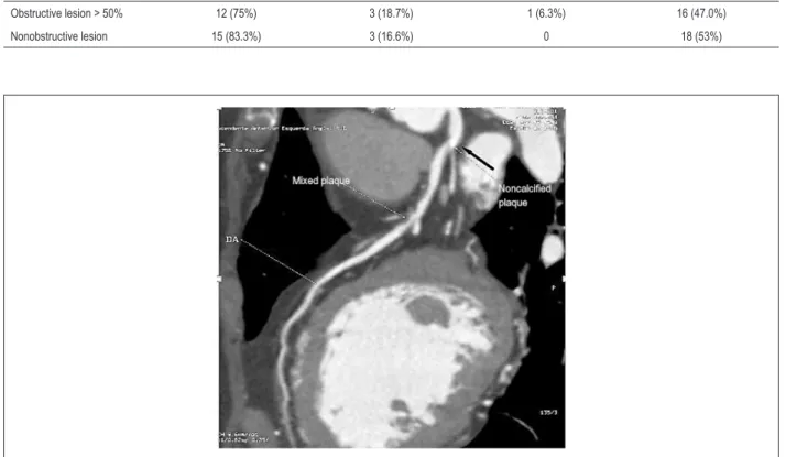
Figure 1 – Noncalcified plaque with zero calcium score. Thirty-eight-year old woman; A and B) multiplanar reconstructions showing considerable lumen reduction in anterior descending artery (DA); C) Tridimensional reconstruction showing impairment in DA (yellow arrow).
Table 2 – Distribution of clinical characteristics of patients with zero calcium score with and without atherosclerotic plaque in four diagnostic imaging centers in Sao Paulo and Aracaju, Brazil, from 2001 to 2016
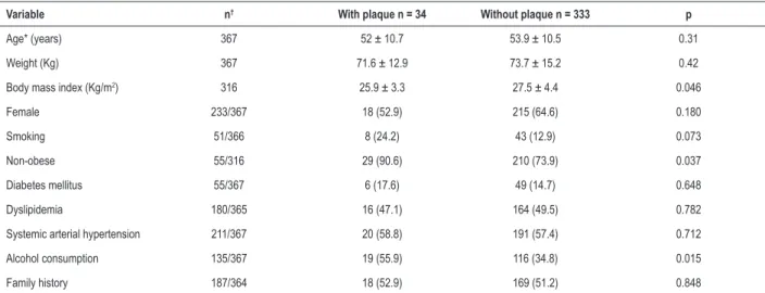
Variable n† With plaque n = 34 Without plaque n = 333 p
Age* (years) 367 52 ± 10.7 53.9 ± 10.5 0.31 Weight (Kg) 367 71.6 ± 12.9 73.7 ± 15.2 0.42 Body mass index (Kg/m2) 316 25.9 ± 3.3 27.5 ± 4.4 0.046
Female 233/367 18 (52.9) 215 (64.6) 0.180
Smoking 51/366 8 (24.2) 43 (12.9) 0.073
Non-obese 55/316 29 (90.6) 210 (73.9) 0.037 Diabetes mellitus 55/367 6 (17.6) 49 (14.7) 0.648 Dyslipidemia 180/365 16 (47.1) 164 (49.5) 0.782 Systemic arterial hypertension 211/367 20 (58.8) 191 (57.4) 0.712 Alcohol consumption 135/367 19 (55.9) 116 (34.8) 0.015 Family history 187/364 18 (52.9) 169 (51.2) 0.848
(*): Values as mean ± standard deviation; other values expressed as simple frequency (%); p-value obtained by the chi-square test for associations; (†): “n” different from total population due to missing data in the records.
Also, studies involving patients with chest pain in the emergency department have shown frequencies of
atherosclerotic plaques with zero CE of up to 39%,11-13
although this is a different population from those attending outpatient services. It is of note, however, that our sample population was composed of patients referred to CCTA from their assistant physicians. As reported in international studies, we also found that the presence of atherosclerotic plaque cannot be ruled out in patients with zero CS.
In our study, only the variables alcohol consumption and absence of obesity were associated with higher risk of atherosclerotic plaque, in contrast to classical risk factors for CAD (diabetes mellitus, systemic arterial hypertension and dyslipidemia). Interestingly, higher BMI was associated with absence of atherosclerotic lesion. Previous studies have suggested obesity as a protective factor for CAD, the so-called obesity paradox.14 Nevertheless, such paradox
is not concerned to abdominal obesity, which has been
associated with CAD and considered more pathological than subcutaneous fat accumulation.14-16 In our study, we
did not measure abdominal circumference, which may have influenced the consistency of results. Besides, obese patients included in many studies that indicated obesity as a protective factor were younger, which may be a source of bias.17
Alcohol consumption has also yielded diverging results. While some studies have indicated alcohol consumption as a risk factor for CAD, others have pointed out its beneficial effects, such as studies performed with wine and its component resveratrol.18-20 Resveratrol is known for its
antioxidant and anti-inflammatory effects, in addition to promote the synthesis of HDL in the liver and inhibit LDL production, thereby preventing LDL oxidation and reducing the risk of cardiovascular diseases.21 In this regard, further
Figure 2 – Presence of calcification in zero calcium score. Female patient, 67 years old; Black arrow - Partially calcified plate in anterior descending ostium (AD), not detected by calcium score, followed by noncalcified plaques in proximal and middle thirds (white arrows)
Table 3 – Distribution of atherosclerotic lesions at coronary computed tomography angiography in patients with zero calcium score
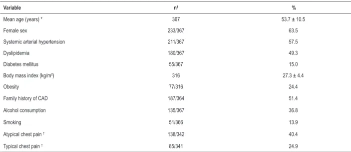
Variable One vessel affected Two vessels affected Two or more vessels affected Total n = 34
Obstructive lesion > 50% 12 (75%) 3 (18.7%) 1 (6.3%) 16 (47.0%) Nonobstructive lesion 15 (83.3%) 3 (16.6%) 0 18 (53%)
Limitations
Some inherent limitations deserve to be mentioned – first, as previously described, patients were referred to CCTA with CS from their assistant physicians, and the possibility of a selection bias cannot be excluded; second, coronary risk stratification of patients was not performed before their inclusion and data on risk factors were obtained by questionnaires; third, sample was collected in four different centers and, although the tests were performed following similar protocols, some characteristics are particular of each service which may have cause a bias in the analysis; fourth, since we studied patients with clinical indication for CCTA, our sample differed from asymptomatic patients without positive ischemic test, who would be referred to CS alone, and in whom coronary calcification would predict cardiovascular events.
Conclusions
The frequency of atherosclerotic plaque in patients with zero CS was relatively high, indicating that in patients with clinical indication for CCTA, the absence of coronary
calcification does not exclude atherosclerotic plaque or obstructive lesion, especially in obese and alcohol drinkers.
Author contributions
Conception and design of the research: Gabriel FS, Gonçalves LFG, Pinto IMF, Oliveira JLM; Acquisition of data: Gabriel FS, Gonçalves LFG, Santana SMM, Matos CJO, Conceição FMS, Souto MJS; Analysis and interpretation of the data: Gabriel FS, Gonçalves LFG, Melo EV, Sousa ACS, Oliveira JLM; Statistical analysis: Gabriel FS, Melo EV; Writing of the manuscript: Gabriel FS, Gonçalves LFG, Sousa ACS, Pinto IMF, Oliveira JLM; Critical revision of the manuscript for intellectual content: Gabriel FS, Gonçalves LFG, Melo EV, Sousa ACS, Pinto IMF, Santana SMM, Matos CJO, Conceição FMS, Oliveira JLM, Souto MJS.
Potential Conflict of Interest
Table 4 – Factors associated with the presence of plaque† (diagnostic imaging centers in Sao Paulo and Aracaju, Brazil, from 2001 to 2016)
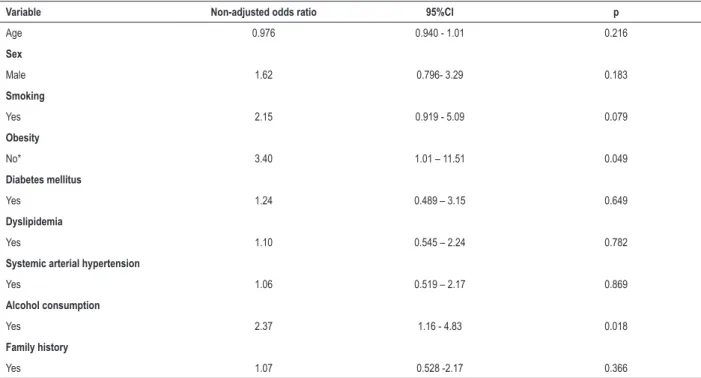
Variable Non-adjusted odds ratio 95%CI p
Age 0.976 0.940 - 1.01 0.216
Sex
Male 1.62 0.796- 3.29 0.183
Smoking
Yes 2.15 0.919 - 5.09 0.079
Obesity
No* 3.40 1.01 – 11.51 0.049
Diabetes mellitus
Yes 1.24 0.489 – 3.15 0.649
Dyslipidemia
Yes 1.10 0.545 – 2.24 0.782
Systemic arterial hypertension
Yes 1.06 0.519 – 2.17 0.869
Alcohol consumption
Yes 2.37 1.16 - 4.83 0.018
Family history
Yes 1.07 0.528 -2.17 0.366
Outcome variable: presence of plaque; other variables described in the table are associated factors; (*): presence of obesity was used as reference for the variable obesity, CI: confidence interval; (†): adjusted for age, sex, smoking, diabetes mellitus, systemic arterial hypertension, dyslipidemia, family history, obesity and alcohol consumption.
Table 5 – Factors associated with the presence of plaque† after model adjustment in diagnostic imaging centers in Sao Paulo and Aracaju, Brazil, from 2001 to 2016
Variable Adjusted odds ratio 95%CI p
Alcohol consumption 3.46 1.16 - 5.19 0.018
Non-obese* 3.45 1.01 – 11.7 0.047
Outcome variable: presence of plaque; other variables described in the table are associated factors; (*): presence of obesity was used as reference for the variable non-obese, CI: confidence interval; (†): adjusted for age, sex, smoking, diabetes mellitus, systemic arterial hypertension, dyslipidemia, family history, obesity and alcohol consumption.
Sources of Funding
There were no external funding sources for this study.
Study Association
This article is part of the thesis of master submitted by Fabíola Santos Gabriel o, from Universidade Federal de Sergipe.
Ethics approval and consent to participate
1. Cesar LA, Ferreira JF, Armaganijan D, Gowdak LH, Mansur AP, Bodanese LC et al; Sociedade Brasileira de Cardiologia. Guideline for stable coronary artery disease. Arq Bras Cardiol. 2014;103(2 Suppl 2):1-56. doi: http:// dx.doi.org/10.5935/abc.2014S004
2. Abdulla J, Abildstrom SZ, Gotzsche O, Christensen E, Kober L, Torp-Pedersen C. 64-multislice detector computed tomography coronary angiography as potential alternative to conventional coronary angiography: a systematic review and meta-analysis. Eur Heart J. 2007;28(24):3042-50. doi: 10.1093/ eurheartj/ehm466.
3. Schroeder S, Kuettner A, Wojak T, Janzen J, Heuschmid M, Athanasiou T, et al. Non-invasive evaluation of atherosclerosis with contrast enhanced 16 slice spiral computed tomography: results of ex vivo investigations. Heart. 2004;90(12):1471-5. doi: 10.1136/hrt.2004.037861.
4. Villines TC, Hulten EA, Shaw LJ, Goyal M, Dunning A, Achenbach S, et al; CONFIRM Registry Investigators. Prevalence and severity of coronary artery disease and adverse events among symptomatic patients with coronary artery calcification scores of zero undergoing coronary computed tomography angiography. J Am Coll Cardiol. 2011;58(24):2533-40. doi: 10.1016/j.jacc.2011.10.851.
5. Moradi M, Varasteh E. Coronary atherosclerosis evaluation among Iranian patients with zero coronary calcium score in computed tomography coronary angiography. Adv Biomed Res. 2016 Feb 8;5:24. doi: 10.4103/2277-9175.175920.
6. Agatston AS, Janowitz WR, Hildner FJ, Zusmer NR, Viamonte M Jr, Detrano R. Quantification of coronary artery calcium using ultrafast computed tomography. J Am Coll Cardiol. 1990;15(4):827-32. doi: https://doi. org/10.1016/0735-1097(90)90282-T.
7. Sousa AG. [Percutaneous cardiovascular intervention procedures in Brazil (1992-1993). Report of the National Registry-National Center for Cardiovascular Interventions]. Arq Bras Cardiol. 1994;62(4):217-23. PMID: 7998847.
8. Oliveira JL, Hirata MH, Sousa AG, Gabriel FS, Hirata TD, Tavares IS, et al. Male gender and arterial hypertension are plaque predictors at coronary computed tomography angiography. Arq Bras Cardiol. 2015;104(5):409-16. doi: http://dx.doi.org/10.5935/abc.20150028.
9. Akram K, O’Donnell RE, King S, Superko HR, Agatston A, Voros S. Influence of symptomatic status on the prevalence of obstructive coronary artery disease in patients with zero calcium score. Atherosclerosis. 2009;203(2):533-7. doi: 10.1016/j.atherosclerosis.2008.07.008.
10. Gottlieb I, Miller JM, Arbab-Zadeh A, Dewey M, Clouse ME, Sara L, et al. The absence of coronary calcification does not exclude obstructive coronary artery disease or the need for revascularization in patients referred for conventional coronary angiography. J Am Coll Cardiol. 2010;55(7):627-34. doi: 10.1016/j.jacc.2009.07.072.
11. Henneman M, Schuijf JD, Pundziute G, van Werkhoven JM, van der Wall EE, Jukema JW, et al. Noninvasive evaluation with multislice computed tomography in suspected acute coronary syndrome. J Am Coll Cardiol. 2009;52(3):216-22. doi: 10.1016/j.jacc.2008.04.012.
12. Pursnani A, Chou ET, Zakroysky P, Deaño RC, Mamuya WS, Woodard PK, et al. Use of coronary artery calcium scanning beyond coronary computed tomographic angiography in the emergency department evaluation for acute chest pain. The ROMICAT II trial. Circ Cardiovasc Imaging. 2015;8(3):pii: e002225. doi: 10.1161/CIRCIMAGING.114.002225.
13. Rubinshtein R, Gaspar T, Halon DA, Goldstein J, Peled N, Lewis BS. Prevalence and extent of obstructive coronary artery disease in patients with zero or low calcium score undergoing 64-slice cardiac multidetector computed tomography for evaluation of a chest pain syndrome. Am J Cardiol. 2007;(99):472-5. doi: 10.1016/j.amjcard.2006.08.060.
14. Parsa AF, Jahanshahi B. I Is the relationship of body mass index to severity of coronary artery disease different from that of waist to-hip ratio and severity of coronary artery disease? Paradoxical findings. Cardiovasc J Afr. 2015;26(1):13-6. doi: 10.5830/CVJA-2014-054.
15. Morricone L, Ferrari M, Enrini R, Inglese L, Giardini D, Garancini P, et al. The role of central fat distribution in coronary artery disease in obesity: comparison of nondiabetic obese, diabetic obese, and normal weight subjects. Int J Obes Relat Metab Disord. 1999;23(11):1129-35. PMID: 10578202. Erratum in: Int J Obes Relat Metab Disord. 2000;24(4):525.
16. Empana JP, Ducimetiere P, Charles MA, Jouven X. Sagittal abdominal diameter an risk of sudden death in asymptomatic middle-aged men in Paris Prospective Study I. Circulation. 2004;110(18):2781-5. doi: 10.1161/01. CIR.0000146395.64065.BA.
17. Rubinshtein R, Halon DA, Jaffe R, Shahla J, Lewis BS. Relation between obesity and severity of coronary artery disease. Am J Cardiol. 2006;97(9):1277-80. doi: 10.1016/j.amjcard.2005.11.061.
18. Renaud S, Lorgeril M. Wine, alcohol, platelets, and the French paradox for coronary heart disease. Lancet. 1992;339(8808):1523-6. doi: https://doi. org/10.1016/0140-6736(92)91277-F.
19. Maisch B. Alcoholic cardiomyopathy: the result of dosage and individual predisposition. Herz. 2016;41(6):484-93. doi: 10.1007/s00059-016-4469-6.
20. Gaziano JM, Buring JE, Breslow JL, Goldhaber SZ, Rosner B, VanDenburgh M, et al. Moderate alcohol intake, increased levels of highdensity lipoprotein and its subfractions, and decreased risk of myocardial infarction. N Engl J Med. 1993; 329(25):1829-34. doi: 10.1056/ NEJM199312163292501.
21. Sautter CK, Denardin S, Alves AO, Mallmann CA, Penna NG, Hecktheuer LH. Determinação de resveratrol em sucos de uva no Brasil. Ciênc Tecnol Aliment. 2005;25(3):437-42. doi: http://dx.doi.org/10.1590/S0101-20612005000300008.