Case Report
Becker muscular dystrophy (BMD) integrates dystrophy occurring due to genetic mutations that express the dystrophin protein in chromosome X. The onset of neuromuscular symptoms usually precedes the impairment of cardiac function, and may conversely happen by heart failure (HF). Physical training is well established in HF, however, when combined with BMD, it is controversial and without any scientific basis.
This study presents the case of a patient with BMD associated with HF in cardiac transplant waiting list undergoing a physical training program.
Physical Training in Becker Muscular Dystrophy Associated with
Heart Failure
Jean Marcelo Roque
1,2, Vitor Oliveira Carvalho
1,2, Lucas Nóbilo Pascoalino
1,2, Silvia Ayub Ferreira
1, Edimar Alcides
Bocchi
1, Guilherme Veiga Guimarães
1,2Laboratório de Insuficiência Cardíaca e Transplante - Instituto do Coração HCFMUSP1, Laboratório de Atividade Física e Saúde - Centro de
Práticas Esportivas - USP2, São Paulo, SP - Brazil
Keywords
Heart failure; muscular dystrophy, Duchenne; exercise; electromyography.
Mailing address: Guilherme Veiga Guimarães •
Rua Dr. Baeta Neves, 98 - Pinheiros - 05444-050 - São Paulo, SP - Brazil E-mail: [email protected]
Manuscript received June 22, 2010; revised manuscript received October 21, 2010, accepted on November 05, 2010.
Fibrillations and complex repetitive discharges are commonly seen in muscles at rest, especially in the proximal and paraspinal muscles5.
Physical activity in BMD is controversial because high intensity training and eccentric muscular activity may be harmful in patients with dystrophy; however, low-intensity resistance exercise is able to improve strength, endurance and fatigue, with no deleterious effects on muscles6. However, when it comes to patients with BMD associated with HF, these data are not entirely clear.
Thus, the purpose of this report was to assess the strength and muscle electromyographic activity after a physical activity program in a patient with DMB awaiting heart transplant.
Case Report
Apparently healthy male individual, age 25, felt short of breath and fatigue in 2005, a fact which led him to seek medical care, where pulmonary congestion was detected during clinical evaluation. On that occasion, the patient was treated with diuretics and instructed to decrease physical activity until stabilization of the clinical picture for a better investigation. In return, the patient reported progressive difficulty in climbing stairs and getting up. During the interview, the patient reported a family history of three cousins with DMB, and was referred to a neurologist specializing in muscular dystrophies. With the clinical assessment associated to the high level of creatine kinase, the patient was referred to genetic testing, which confirmed the diagnosis of DMB.
In late 2007, the patient complained of fatigue on minimal exertion and was referred to cardiovascular evaluation. The echocardiogram showed a significant increase in heart size, mitral regurgitation and a left ventricular ejection fraction of 20.0%. The study of radionuclide ventriculography revealed a reduced function of left and right ventricles in moderate and severe degrees, respectively. Peak oxygen consumption, measured during the cardiopulmonary stress test on a treadmill was 10.3 ml/kg/min-1. The slope of the ventilation/carbon dioxide production (slope VE/VCO2) was 31.8. With the patient’s clinical picture and laboratory tests, HF functional class IV was diagnosed.
After six months from drug introduction and optimization (75 mg carvedilol, 100 mg losartan, 0.25 mg digoxin, 25 mg aldactone, 80 mg monocardil and 80 mg furosemide), the patient was reassessed, but his clinical picture did not progressed well. Thus, the clinical team decided to include
Introduction
Becker muscular dystrophy (BMD) is a dystrophy occurring due to mutations in the gene that expresses the dystrophin protein, located in chromosome X1. Dystrophin is present in the muscle cytoskeleton and its failure causes sarcolemma instability and disruption. This seems to be the main factor of myopathy occurring in DMB, resulting in progressive muscle weakness2.
The impairment of cardiac function occurs in most cases of BMD, with the myocardium affected by the myopathic process. Its progression is unpredictable and it plays a determining role in the survival of such patients1. Typically, the onset of neuromuscular symptoms precedes cardiac involvement, but in some cases, this may happen in reverse. The severity of cardiac dysfunction in these patients may lead to heart transplant3.
In the electromyographic (EMG) assessment of patients with DMB, myopathic changes are found as potentially small and polyphase motor units with an early recruitment4.
Case Report
Roque et al Becker and heart failure
Arq Bras Cardiol 2011;97(6):e128-e131 him in the single queue of candidates for heart transplantation.
Subsequently, the patient asked the medical staff to provide orientation for physical activity, after which he was released and referred to the Laboratory of Physical Activity and Health for physical rehabilitation.
In early 2008, muscle strength tests were performed in the right quadriceps and the electromyographic activity of the vastus medialis and lateralis muscles of the same member were assessed before and after training. To measure the strength, the patient remained sitting at a height where his feet would not touch the ground, and bending his knees at 90° (dominant), the patient would then extend them. Three motions were made after verbal command to the patient, and the largest movement was accepted. Measurement was performed with a dynamometer model Crow AT (Filizola). In the same motion of knee extension, the surface electromyographic amplitude of the vastus medialis and lateralis muscles was measured. For this measurement, a two-channel electromyographic conditioning module (Phoenix USB 2 V4.01 R8) was used. Surface electrodes were used to capture the electrical activity of the muscles, as recommended by the Surface EMG for the Non-Invasive Assessment of Muscles7.
From April to August 2008, an exercise training program was started on a bi-weekly basis for a period of three months in confinement, temperature controlled at 22°C. Aerobic exercise was performed on a treadmill and the patient was instructed to control the walking speed between 11 and 13 of the Borg Scale8, with initial duration of two minutes, progressing gradually to 12 minutes. Resistance training was performed on a weight-lifting equipment branded NAKAGYM with a weight degree ranging from one to 21 kg. At first, we performed a series of three repetitions of assisted active concentric movement and resistance active eccentric movement with minimal load, progressing to unassisted active movement, keeping the same number of sets, repetitions and load. The following exercises were performed: Leg Extension, Leg Curl, Lat Pulldown, Peck Deck, and Chest Press. To monitor heart rate, a Polar pulse frequency meter was used.
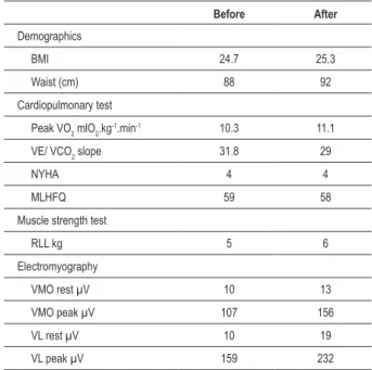
This protocol was approved by the Ethics Committee and the patient signed a consent form. The results of this case study revealed that, despite the clinically stable and unchanged condition, there was a slight increase in peak VO2, muscle strength, and electromyographic activity (Table 1).
Discussion
In this study, we have described, in an unprecedented manner, the effect of exercise training on skeletal muscle strength and electromyographic activity in a patient with BMD associated with HF.
While assessing right quadriceps strength after training, there was an increase of 20.0% compared to the pre-training condition. A previous study has shown in patients with DMB, trained on a cycle ergometer, an improvement of 40.0% of the quadriceps extension strength8. However, in this study, patients had only DMB without impaired cardiac function associated, showing that physical training program for patients with BMD associated with HF may have a positive impact on muscle
performance. In addition, it has shown that after training there was an increase of 47.0% in peak VO2
7. In the HF-ACTION study, we observed a 4.0% gain in peak VO2 among patients who underwent physical activity, however, the expectation of improvement was 10.0% after training9.
Our study of BMD associated with HF showed a 10.0% improvement in peak VO2 after the training period, a result that is usually considered relevant. The differences found in these studies are probably due to musculoskeletal weakness, cardiac and initial physical condition impairment, i.e., improvement of physical capacity is inversely proportional to the patient’s limitations. In addition, patients underwent cardiopulmonary exercise testing before and after training with different times, indicating that such differences should be interpreted with caution.
In a previous study, the electromyographic amplitude analysis before and after training of the vastus medialis oblique muscle revealed an increase in microvolt, both at rest and at peak muscle contraction, demonstrating that muscle strength in BMD suffers mild and compensated changes, but it is preserved in most muscles, at least at the onset of the disease10. Muscle strength gains and increased electromyographic activity, measured after training in this study, may be suggestive of a potential slowdown in the process expected in the DMB.
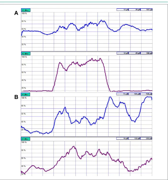
Our case study revealed similar results obtained from the vastus lateralis (Figures 1A and 1B) and that the physical activity in DMB associated with HF promoted increased oxygen consumption, muscular strength and a slight elevation of the electromyographic activity. Further studies may elucidate in more details the electromyographic and cardiac alterations in DMB associated with HF.
Table 1 - Patient data
Before After
Demographics
BMI 24.7 25.3
Waist (cm) 88 92
Cardiopulmonary test
Peak VO2 mlO2.kg
-1.min-1 10.3 11.1
VE/ VCO2 slope 31.8 29
NYHA 4 4
MLHFQ 59 58
Muscle strength test
RLL kg 5 6
Electromyography
VMO rest µV 10 13
VMO peak µV 107 156
VL rest µV 10 19
VL peak µV 159 232
VO2 - oxygen consumption; NYHA - New York Heart Association; MLHFQ - Minnesota Living with Heart Failure Questionnaire; RLL - right lower limb; VMO - vastus medialis oblique; VL - vastus lateralis.
Case Report
Roque et al Becker and heart failure
Arq Bras Cardiol 2011;97(6):e128-e131
Figure 1 -A - pre-training; Channel A1 - vastus medialis oblique (VMO); Channel A2 - vastus lateralis (VL). B - post-training; Channel A1 - vastus medialis oblique (VMO); Channel A2 - vastus lateralis (VL).
Case Report
Roque et al Becker and heart failure
Arq Bras Cardiol 2011;97(6):e128-e131
References
1. Finsterer J, Stöllberger C. The heart in human dystrophinopathies. Cardiology. 2003;99(1):1-19.
2. Campbell KP. Three muscular dystrophies: loss of cytoskeleton-extracellular matrix linkage. Cell. 1995;80(5):675-9.
3. Hoogerwaard EM, Voogt WG, Wilde AM, van der Wouw PA, Bakker E, van Ommen GI, et al. Evolution of cardiac abnormalities in Becker muscular dystrophy over a 13-year period. J Neurol. 1997;244(10):657-63.
4. Kopeć J, Emeryk-Szajewska B. “Functional-QEMG” a new reliable method
in daily routine investigation: electromyography. Clin Neurophysiol. 2002;42(8):495-506.
5. Liguori R, Fuglsang-Frederiksen A, Nix W, Fawcet PR, Andersen K. Electromyography in myopathy. Neurophysiol Clin. 1997;27(3):200-3. 6. Sveen ML, Jeppesen TD, Hauerslev S, Kober L, Krag TO, Vissing J. Endurance
training improves fitness and strength in patients with Becker muscular dystrophy. Brain. 2008;131(Pt 11):2824-31.
7. Hermens HJ, Freriks B, Disselhorst-Klug C, Rau G. Development of recommendations for SEMG sensors and sensor placement procedures. J Electromyogr Kinesiol. 2000;10(5):361-74.
8. Carvalho VO, Bocchi EA, Guimarães GV. The Borg scale as an important tool of self-monitoring and self-regulation of exercise prescription in heart failure patients during hydrotherapy: a randomized blinded controlled trial. Circ J. 2009;73(10):1871-6.
9. O’Connor CM, Whellan DJ, Lee KL, Keteyian SJ, Cooper LS, Ellis SJ, et al. Efficacy and safety of exercise training in patients with chronic heart failure HF-ACTION randomized controlled trial. JAMA. 2009;301(14):1439-50. 10. Monti RJ, Roy RR, Edgerton VR. Role of motor unit structure in defining
function. Muscle Nerve. 2001;24(7):848-66.