Role of Monocyte Count and
Neutrophil-to-Lymphocyte Ratio in Survival of Oral Cancer Patients
Saurabh Bobdey
1Balasubramaniam Ganesh
1Prabhashankar Mishra
1Aanchal Jain
11Department of Medical Records, Biostatistics and Epidemiology, Tata Memorial Hospital Parel, Mumbai, India
Int Arch Otorhinolaryngol 2017;21:21–27.
Address for correspondence Saurabh Bobdey, MD, DNB, Department of Medical Records, Biostatistics and Epidemiology, Tata Memorial Hospital, Parel, Mumbai 400012, India (e-mail: [email protected]).
Introduction
Oral cavity is one of the most frequent locations of squamous cell carcinoma in the head and neck region. Globally, an estimated 300,400 new cases of oral cavity cancer are diag-nosed each year and it is the eleventh leading cause of cancer worldwide. In the Indian subcontinent oral cancer is a major public health problem and ranks among the top three types of cancer in the country.1Almost 80,000 cases are diagnosed and 50,000 deaths occur annually due to oral cavity cancer in India. The 5-year survival rate for oral cancer in India is37%
(26–45).2This indicates that, despite improvement in health infrastructure and treatment modalities, prognosis from oral cancer remains poor as compared with other Asian countries.2Therefore, a low-cost, standardized, reliable, and reproducible prognostic marker for oral cancer patients is desirable to facilitate individualized treatments and, thus, better outcomes for the patients.
Inflammation seems to play a critical role in the develop-ment and progression of numerous cancers.3Tumor develop-ment and progression induced by an inflammatory response is thought to be mediated by an interaction between Keywords
►
oral cancer
►
prognosis
►
survival
►
monocyte
►
neutrophil-to-lymphocyte ratio
Abstract
Introduction
In
fl
ammation seems to play a critical role in the development and
progression of numerous cancers. Peripheral blood leukocyte count is an easily
assessable parameter of systemic in
fl
ammatory response.
Objective
The aim of this study was to investigate whether the pretreatment
leukocyte counts can predict the prognosis of patients with oral cavity cancer.
Methods
Medical records of 471 oral cavity cancer patients diagnosed between
January 2007 and December 2008 were retrospectively analyzed. Receiver operating
characteristic curve analysis and Cox proportional hazards analyses were applied to
evaluate the associations of leukocyte counts with overall survival.
Results
The overall
fi
ve year
’
s survival of the cohort was found to be 49.4%. On
univariate analysis, elevated monocyte count (
500/mm3) and
neutrophil-to-lympho-cyte ratio (NLR) (
>
2.38) were associated with poor overall survival (OS) (
p
¼
0.001 and
0.000, respectively). Multivariate Cox proportional hazard analysis showed that higher
monocyte and NLR levels were signi
fi
cant independent predictors of worse OS (HR
¼
1.385, 95% CI
¼
1.049 - 1.829;
p
<
0.05 and HR
¼
1.392, 95% CI
¼
1.045 - 1.855;
p
<
0.05, respectively). The advanced overall stage and lymph nodal involvement were
also independent indicators for poor OS.
Conclusions
Higher pretreatment monocyte and NLR levels are independent
predic-tors of poor prognosis for patients with oral cavity cancer. Thus, these easily accessed
variables can serve as a potent marker to predict the outcomes of oral cancer patients.
received February 17, 2016 accepted June 18, 2016 published online August 16, 2016
DOI http://dx.doi.org/ 10.1055/s-0036-1587318. ISSN 1809-9777.
Copyright © 2017 by Thieme-Revinter Publicações Ltda, Rio de Janeiro, Brazil
proinflammatory cytokines and pathways including nuclear factor-kappa B (NF-κB) and signal transducer & activator of transcription 3 (STAT3).4 It has been postulated that the increased release of proinflammatory cytokines produces a systemic inflammatory response reflected in changes in circulating markers of inflammation, such as C-reactive protein and white blood cells.5 There are several lines of evidence to date suggesting that the total white blood cell count as well as its components, such as neutrophils, lym-phocytes, monocytes, and the neutrophil-to-lymphocyte ratio (NLR) can predict survival in a variety of malignancies, including oral cavity,6,7breast cancer,8gastric cancer,9 hepa-tocellular carcinoma,10 Hodgkin’s lymphoma11 and lung cancer.12 However, evidence for the use of hematologic markers of inflammation as predictors of clinical outcome in oral cavity cancer patients is limited.6,7Given this back-ground, the main objective of our study was to assess the role of total and differential leukocyte counts in overall survival of oral cavity cancer patients. The study had the approval of the Research Ethics Committee of the hospital.
Methods
We retrospectively analyzed the medical records of 471 pathologically proven oral cavity cancer patients, residents of Mumbai, diagnosed between January 01, 2007 to Decem-ber 31, 2008, and who had not received any prior treatment. We retrospectively collected data from medical records on routine laboratory measurements of white blood cells (WBC) performed prior to onset of treatment, including the counts of neutrophils, lymphocytes, and monocytes. All patients were staged according to the sixth edition of the UICC/AJCC TNM classification system.13,14In addition, we also collected data on age at diagnosis, sex, primary tumor site, and pretreat-ment tumor staging. We retrieved type of treatpretreat-ment received and status of the patient (Alive/ Dead) at the end offive years from the date of diagnosis from medical records.
Receiver operating characteristic (ROC) curve analysis to select the most appropriate cut-off points for the counts of total WBC, neutrophil, lymphocyte, and monocyte, and NLR was performed to stratify patients at a high risk of malignancy-related death. We selected the score at the point with both maximum sensitivity and specificity as the best cut-off value. In survival analysis, overall survival time was defined as time from diagnosis until death; the follow-up of patients still alive has been censored at their latest date of follow-up. We generated survival curves by the Kaplan–Meier method and compared them by the log-rank test. We applied the Cox proportional hazards model for univariate and multivariate (backward meth-od) analysis to identify prognostic factors. We performed statis-tical analyses using SPSS software v17.0 (SPSS, Chicago, IL).
Results
The patients’characteristics are summarized in►Table 1. As shown, the median age was 50 years (range: 25–85 years), and the percentage of males and females were 69.9% and 30.1%, respectively. Out of the 471 patients, 347 (73.67%) were
diagnosed at late stages (III and IV) and the other 124 patients (26.33%) were at early stages (I and II). The median follow-up period was 22 months (range, 0 to 98 months). Thefive-year overall survival of the cohort was 49.4% and stage-wise survival rate for TNM stage I, II, III, and IV patients was found to be 86.4%, 68%, 57.9%, and 33.6% (p<0.00), respectively.
We determined the cut-off value of total WBC, neutrophil, lymphocyte, monocyte counts, and neutrophil lymphocyte ratio (NLR) for survival outcomes by ROC curve analyses. We selected the NLR cut-off point of 2.38 for the survival analyses and divided all patients into either high (NLR2.38) or low (NLR<2.38) NLR groups. Additionally, to ensure comparability with other studies, we also conducted an analysis using NLR cut-off of 5; however, only 72 (15.3%) patients had NLR of5. Similarly, we selected as the optimal cut-off points for survival analyses, a total WBC count of 7.900109/L, neutrophil count of 4.900
109/L,
lymphocyte count of 1.980109/L, and monocyte count of
0.500109/L.
We performed a univariate analysis of age, gender, overall stage, lymph node involvement, treatment modali-ty, total WBC count, neutophil count, lymphocyte count, monocyte count, and NLR to assess their role as prognostic factors for overall survival. Univariate analysis revealed that lower WBC count (<7.900109/L;
p¼0.026), mono-cyte count (< 0.500109/L;
p¼0.001), and NLR level
Table 1 Patients’characteristics
Parameter No. of patients Percentage (%)
Age
mean 50 Years –
Range 25–85 Years –
Sex
Male 329 69.9
Female 142 30.1
T Classification
T1 41 8.7
T2 142 30.1
T3 56 11.9
T4 232 49.3
N Classification
N0 208 44.2
Nþ 263 55.8
M Classification
M0 469 99.6
M1 2 0.4
Tumor Stage, AJCC
I 32 6.8
II 92 19.5
III 76 16.1
IV 271 57.5
(<2.38;p<0.001) were associated with superior survival, but when we used traditional NLR cut-off of 5, we did not find it to be significantly associated with survival (p¼0.091). In addition, advanced disease (TNM stage, IIIþIV; p< 0.001) and lymph nodal involvement (p<0.001) were found to be associated with poorer prog-nosis. Other factors such as age, gender, neutrophil count, lymphocyte count, and treatment modality were not sig-nificantly associated with survival of oral cavity cancer patients.
We included all the characteristics, such as age, gender, overall stage, lymph node involvement, treatment modality, total WBC count, neutophil count, lymphocyte count, monocyte count, and NLR in the multivariate analysis. The results showed that higher NLR level (HR¼1.392, 95% CI¼1.045 - 1.855; p< 0.001), monocyte count (HR¼1.385, 95% CI¼1.049 -1.829; p< 0.001), advanced stage disease (HR¼1.921, 95% CI¼1.193 - 3.092; p<0.001), and lymph node involvement (HR¼1.715, 95% CI¼1.200 - 2.451;p<0.001) were indepen-dent predictors for poor overall survival of oral cavity cancer patients (►Table 2).
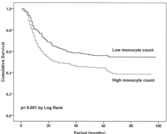
Thefive-year survival rate of patients with high monocyte count (0.500109/L) was found to be 43.9%, which was
significantly (p<0.001) less than the 55.4% survival rate for patients with lower monocyte count (►Fig. 1). Similarly, we found five yearś survival rate for patients with higher (2.38) and lower NLR was 42.2% and 58.6%, respectively (p<0.001) (►Fig. 2). We categorized patient characteristics
including hematological parameters as high / low NLR and monocyte counts (►Table 3). Patients with high NLR level (2.38) had a higher incidence of advanced T, N, and overall stages (p¼0.001,p¼0.015, andp¼0.003, respec-tively). Lifestyle factors such as tobacco chewing, smoking and alcohol consumption did not appear to be associated with NLR or monocyte count (p>0.05).
Fig. 1 Kaplan–Meier survival curves of cumulative survival rates in patients with oral cavity cancer classified into two groups according to monocyte count.
Table 2 Univariate and multivariate analysis of prognostic factors for overall survival in patients with oral cavity
Univariate Multivariate
Parameter HR
(95% CI)
p HR
(95% CI)
p
Age,65 years 1.088
(0.745 - 1.589)
0.664 – –
Sex, (male) 1.244
(0.928 - 1.669)
0.145 – –
TNM stage (IIIþIV) 3.155
(2.168 - 4.591)
0.000 1.921
(1.19 3 - 3.092)
0.007
Lymph node mets 2.492
(1.876 - 3.310)
0.000 1.715
(1.200 - 2.451)
0.003
Neutrophil4.900 109/L 1.290
(0.989 - 1.683)
0.060 – –
Lymphocyte1.980 109/L 1.224
(0.941 - 1.592)
0.131 – –
Monocyte0.500 109/L 1.558
(1.190 - 2.040)
0.001 1.385
(1.049 - 1.829)
0.021
WBC7.900 109/L 1.352
(1.037 - 1.764)
0.026 0.164
Neutrophil/ lymphocyte ratio (NLR),2.38 1.676
(1.271 - 2.209)
0.000 1.392
(1.045 - 1.855)
0.024
Neutrophil/ lymphocyte ratio (NLR),5 1.348
(0.954 - 1.904)
0.091 – –
Treatment (single/ multi-modality) 0.924 (0.712–1.200)
0.555 – –
Abbreviations: CI, confidence interval; HR, hazard ratio.Signi
Discussion
The total and differential white blood cell (WBC) count has been historically used as a marker of infection and infl am-mation. Nonetheless, its role has gone beyond the assessment of infectious processes and it has become an important prognostic measurement of outcomes in cancer treatment. Thus, while a link between inflammation and cancer has been known for more than a century, compelling recent evidence have suggested a strong association between pretreatment peripheral inflammatory cells and prognosis in different kinds of cancers.6–12 As part of the functional relevance, inflammatory responses lead to chronic oxidative stress and generate oxygen free radicals, which have been shown to stimulate cancer initiation, promotion, and progres-sion.15,16Here, we have performed a retrospective medical record based study on oral cavity cancer to evaluate the prognostic values of total WBC, neutrophil, lymphocyte, monocyte counts, and NLR together with the other clinical factors. Our results confirmed the previous findings that factors such as absence of nodal involvement and early stage, were associated with favorable prognosis for oral cavity cancer patients.17,18 More importantly, we found that an elevated neutrophil lymphocyte ratio (NLR) and monocyte count were significantly associated with poorer overall sur-vival and were independent of other variables to predict the prognosis for oral cavity cancer patients. These results were in consonance with other published studies that implicate role of monocyte count (Tsai et al,n¼213)6and NLR (Perisanidis et al,n¼97)7in prognostication of oral cavity cancer pa-tients. However, these studies relied on a relatively smaller sample and the cut-off value for monocyte count was based on a median value of circulating monocyte count. In the present study, to exclude empirical bias, we used ROC curve to determine the optimal cut-off value to predict risk of malignancy related death.
In the present study, we found that monocyte count was an independent prognostic factor for patients with oral cavity
cancer. There is substantial evidence that, in advanced cancer, the host systemic immune response is an important inde-pendent predictor of outcome, and that pretreatment meas-urements of the systemic inflammatory immune response can be used to independently predict cancer survival.6,19 Sasaki and colleagues20,21studied the pre-operative absolute monocyte count in patients who had liver resection due to hepatocellular carcinoma, as well as in patients who under-went hepatic surgery due to colorectal metastasis and found that pretreatment absolute monocyte count was an indepen-dent prognostic indicator of tumor recurrence and survival in patients with hepatocellular carcinoma. Similarly, absolute monocyte count has been reported to be independent prog-nostic indicator for breast cancer,8gastric cancer,9Hodgkin’s lymphoma,11Colorectal cancer,21and Ovarian cancer.22
The exact underlying mechanism explaining the association between the elevated number of monocytes and unfavorable cancer prognosis is unclear. However, a possible explanation can be that monocytes secrete various proinflammatory cytokines, such as interleukin (IL)-1, IL-6, IL-10, and TNF-α, which have been associated with shorter survival and worse prognosis in malignances.23,24 Moreover, monocytes upon stimulation are known to release monocyte chemo-attractant protein (MCP-1)-1 and mediate tumor-associated macrophage infi ltra-tion in solid tumors, which could produce a variety of chemo-kines such as TGF-α, TNF-α, IL-1, and IL-6 to promote tumorigenesis, angiogenesis, and distant metastasis of malig-nant tumors.24,25Further, studies have linked monocyte with an increased number of bone marrow-derived myelomonocytic cells. These cells infiltrate the tumor and differentiate into tumor-associated macrophages, which in turn release many angiogenic factors and have been shown to be associated with poor prognosis in cancers.24,26,27
The other main finding of our analysis was that high pretreatment NLR was significantly associated with poor survival in oral cavity cancer patients. This result is in accordance with previous observations on the association between NLR and a variety of cancers such as lung cancer, colorectal cancer, cholangiocarcinoma, and Pancreatic cancer.28–32 More specifically, in Head and Neck cancers, elevated pretreatment NLR has been shown to be significantly associated with worse survival in two studies of nasopharyn-geal cancer patients33,34 and only one study of oral cavity cancer patients.7In all three studies, NLR cut-off was taken on the basis of either median value or ROC analysis and none of them have considered traditional NLR cut-off of 5.7,33,34In our study, oral cancer survival was also found to be associated with an ROC-based NLR cut-off of 2.38, but failed to achieve statistical significance with the traditional NLR cut-off of 5. This can be due to the small sample size, from which few patients had an NLR above 5.
Thus, to establish a ROC-based lower NLR cut-off for predict-ing survival in oral cancer patients larger, multicenter prospec-tive studies are needed. In our study, we also assessed the prognostic value of the individual components of NLR, that is, neutrophil and lymphocyte count. Individually, however, neither was significantly associated with survival of oral cavity cancer patients. Similarly, Perisanidis et al also reported a significant Fig. 2 Kaplan–Meier survival curves of cumulative survival rates in patients
relationship between NLR and oral cancer survival but not with its individual components.7It has been suggested that, in cancer patients, NLR is superior to other individual leukocyte param-eters.35This superiority of NLR can be attributed to the stability of NLR compared with the absolute counts that could be altered by various physiological, pathological, and physical factors. Moreover, NLR may represent the two opposing inflammatory and immune pathways that exist together in cancer patients.7 Therefore, NLR can be considered as the balance between pro-tumor inflammatory status and anti-tumor immune status. Patients with elevated NLR have a relative neutrophilic leukocy-tosis and lymphocytopenia, which denotes that the balance is inclined in favor of pro-tumor inflammatory and is associated with poor outcome.34,35
The mechanisms underlying the association of high NLR and poor outcome of cancer patients are poorly understood. One potential mechanism underlying the prognostic impact of NLR may be an association of high NLR with inflammation. An elevated NLR has been associated with an increase in the peritumoral infiltration of macrophages and an increase in interleukin (IL-17).36 Neutrophils and other cells such as macrophages have been reported to secrete tumor growth promoting factors, including vascular endothelial growth factor,37 hepatocyte growth factor,38 IL-6,39 IL-8,40 matrix metalloproteinases,41and elastases,42 and, thus, likely con-tribute to a stimulating tumor microenvironment. It is a consensus that the adaptive immune system carries out immune surveillance and can eliminate newborn tumors,
Table 3 Baseline clinical characteristics of the 471 Oral cavity carcinoma patients according to high/ low Neutrophil lymphocyte
ratio (NLR) and monocyte count
Characteristic NLR<2.38 (n¼199)
NLR2.38 (n¼272)
p LM Count
<500 (109/L) (n¼206)
HM Count 500 (109/L) (n¼265)
p
Mean age (years) 50.2012.01 51.8412.48 0.152 50.3711.53 51.7512.85 0.225
Tobacco, no. (%)
Non-chewers 82 (41.2) 120 (44.1) 0.528 92 (44.7) 110 (41.5) 0.493
Chewers 117 (58.8) 152 (55.9) – 114 (55.3) 155 (58.5) –
Non-smokers 144 (72.4) 212 (77.9) 0.164 153 (74.3) 203 (76.6) 0.559
Smokers 55 (27.6) 60 (22.1) – 53 (25.7) 62 (23.4) –
Alcohol, no. (%)
Non-drinker 156 (78.4) 229 (84.2) 0.108 171 (83.0) 214 (80.8) 0.530
Drinker 43 (21.6) 43 (15.8) – 35 (17.0) 51 (19.2) –
T-classification
T1–T2 105 (52.8) 78 (28.7) 0.000
86 (47.0) 97 (53.0) 0.256
T3–T4 94 (47.2) 194 (71.3) – 120 (58.3) 168 (63.4) –
N-classification
N0 101 (50.8) 107 (39.5) 0.015 99 (48.1) 109 (41.3) 0.143
Nþ 98 (49.2) 164 (60.5) – 107 (51.9) 155 (58.7) –
Overall stage
I–II 75 (37.7) 49 (18.0) 0.000 63 (30.6) 61 (23.0) 0.064
III–IV 124 (62.3) 223 (82.0) – 143 (69.4) 204 (77.0) –
Treatment
Uni-modality 98 (49.2) 127 (46.7) 0.583 100 (48.5) 125 (47.2) 0.767
Dual/ Multi-modality 101 (50.8) 145 (53.3) – 106 (51.5) 140 (52.8) –
Hematological Parameters
Red blood cell, X106/lL 4.60
0.64 4.570.70 0.145 4.520.69 4.530.67 0.888
Hemoglobin, g/dL 12.82.1 12.72.2 0.528 12.72.2 12.72.2 0.987
Hematocrit, % 38.65.9 38.26.4 0.464 38.36.2 38.46.2 0.884
MCV, fL 84.410.0 85.89.6 0.144 85.19.3 85.210.1 0.925
MCH, pg/cell 28.03.8 28.53.7 0.165 28.33.7 28.33.9 0.994
MCHC, g/dL 33.11.3 33.21.3 0.688 33.21.3 33.11.3 0.640
Abbreviations: HM, high monocyte; LM, low monocyte; MCH, mean corpuscular hemoglobin; MCHC, mean corpuscular-hemoglobin concentration; MCV, mean corpuscular volume.ŦData are meansSD;
but effective adaptive immune responses are always sup-pressed in established tumors through several pathways, including inhibition of dendritic cell differentiation and acti-vation, infiltration of regulatory T cells.43 Lymphocytes are crucial components of adaptive immune system, and studies have reported infiltrating lymphocytes to indicate the gener-ation of an effective antitumor cellular immune response.44A low peripheral lymphocyte level may indicate a poorer lymphocyte-mediated immune response to tumor and sug-gests poor prognosis.45,46 NLR may be explained by the diverse effects of neutrophils and lymphocytes on tumor progression. In vitro studies have shown that the cytolytic activity of lymphocytes and natural killer cells was sup-pressed when cocultured with neutrophils, and the extent of suppression was proportionally enhanced to the addition of neutrophils,46–48implying that high NLR was associated with poor prognosis.33
Conclusion
We found that both NLR and monocyte counts are indepen-dent predictors of overall survival for patients with oral cavity cancer. Given the low cost, easy accessibility, and reproduc-ibility of a full blood count, both NLR and monocyte counts seem promising candidates for use in clinical practice. How-ever, thefindings of the study are based on a retrospective design in a single center, thus, further studies in either multicenter or prospective manner should be undertaken to validate and determine the clinical usages of NLR and monocyte count as prognostic markers for oral cavity cancer patients.
Conflict of Interest
The authors declare no conflict of interest in conducting this study.
References
1 Ferlay J, Shin HR, Bray F, Forman D, Mathers C, Parkin DM. GLOBOCAN 2012, Cancer incidence and mortality worldwide: IARC CancerBase No. 10. Lyon, France: International Agency for Research on Cancer; 2012. Available at http://globocan.iarc.fr. Accessed on Dec 12, 2015
2 Sankaranarayanan R, Swaminathan R, Lucas E. Cancer survival in Africa, Asia, Caribbean and Central America: SURVCAN. Lyon, France: IARC Scientific publication international agency for research on cancer; 2010
3 Weitzman SA, Gordon LI. Inflammation and cancer: role of phago-cyte-generated oxidants in carcinogenesis. Blood 1990;76(4): 655–663
4 Jarnicki A, Putoczki T, Ernst M. Stat3: linking inflammation to epithelial cancer: More than a“gut”feeling? Cell Div 2010;5:14 5 Kao SC, Pavlakis N, Harvie R, et al. High blood
neutrophil-to-lymphocyte ratio is an indicator of poor prognosis in malignant mesothelioma patients undergoing systemic therapy. Clin Cancer Res 2010;16(23):5805–5813
6 Tsai YD, Wang CP, Chen CY, et al. Pretreatment circulating mono-cyte count associated with poor prognosis in patients with oral cavity cancer. Head Neck 2014;36(7):947–953
7 Perisanidis C, Kornek G, Pöschl PW, et al. High neutrophil-to-lymphocyte ratio is an independent marker of poor disease-specific survival in patients with oral cancer. Med Oncol 2013; 30(1):334–335
8 Hornychova H, Melichar B, Tomsova M, Mergancova J, Urminska H, Ryska A. Tumor-infiltrating lymphocytes predict response to neo-adjuvant chemotherapy in patients with breast carcinoma. Cancer Invest 2008;26(10):1024–1031
9 Bruckner HW, Lavin PT, Plaxe SC, Storch JA, Livstone EM. Absolute granulocyte, lymphocyte, and moncyte counts. Useful determi-nants of prognosis for patients with metastatic cancer of the stomach. JAMA 1982;247(7):1004–1006
10 Chew V, Chen J, Lee D, et al. Chemokine-driven lymphocyte infiltration: an early intratumoural event determining long-term survival in resectable hepatocellular carcinoma. Gut 2012; 61(3):427–438
11 Koh YW, Kang HJ, Park C, et al. The ratio of the absolute lympho-cyte count to the absolute monolympho-cyte count is associated with prognosis in Hodgkin’s lymphoma: correlation with tumor-asso-ciated macrophages. Oncologist 2012;17(6):871–880
12 Tibaldi C, Vasile E, Bernardini I, Orlandini C, Andreuccetti M, Falcone A. Baseline elevated leukocyte count in peripheral blood is associated with poor survival in patients with advanced non-small cell lung cancer: a prognostic model. J Cancer Res Clin Oncol 2008;134(10):1143–1149
13 Greene FL. The American Joint Committee on Cancer: updating the strategies in cancer staging. Bull Am Coll Surg 2002;87(7): 13–15
14 Greene FL, Sobin LH. The staging of cancer: a retrospective and prospective appraisal. CA Cancer J Clin 2008;58(3):180–190 15 Nakamura Y, Gindhart TD, Winterstein D, Tomita I, Seed JL,
Colburn NH. Early superoxide dismutase-sensitive event pro-motes neoplastic transformation in mouse epidermal JB6 cells. Carcinogenesis 1988;9(2):203–207
16 Hussain SP, Aguilar F, Amstad P, Cerutti P. Oxy-radical induced mutagenesis of hotspot codons 248 and 249 of the human p53 gene. Oncogene 1994;9(8):2277–2281
17 Baatenburg de Jong RJ, Hermans J, Molenaar J, Briaire JJ, le Cessie S. Prediction of survival in patients with head and neck cancer. Head Neck 2001;23(9):718–724
18 Sayed SI, Sharma S, Rane P, et al. Can metastatic lymph node ratio (LNR) predict survival in oral cavity cancer patients? J Surg Oncol 2013;108(4):256–263
19 Roxburgh CS, McMillan DC. Role of systemic inflammatory response in predicting survival in patients with primary operable cancer. Future Oncol 2010;6(1):149–163
20 Sasaki A, Iwashita Y, Shibata K, Matsumoto T, Ohta M, Kitano S. Prognostic value of preoperative peripheral blood monocyte count in patients with hepatocellular carcinoma. Surgery 2006;139(6): 755–764
21 Sasaki A, Kai S, Endo Y, et al. Prognostic value of preoperative peripheral blood monocyte count in patients with colorectal liver metastasis after liver resection. J Gastrointest Surg 2007;11(5): 596–602
22 Bishara S, Griffin M, Cargill A, et al. Pre-treatment white blood cell subtypes as prognostic indicators in ovarian cancer. Eur J Obstet Gynecol Reprod Biol 2008;138(1):71–75
23 Anand M, Chodda SK, Parikh PM, Nadkarni JS. Abnormal levels of proinflammatory cytokines in serum and monocyte cultures from patients with chronic myeloid leukemia in different stages, and their role in prognosis. Hematol Oncol 1998; 16(4):143–154
24 Pollard JW. Tumour-educated macrophages promote tumour pro-gression and metastasis. Nat Rev Cancer 2004;4(1):71–78 25 Hefler L, Tempfer C, Heinze G, et al. Monocyte chemoattractant
26 Dirkx AE, Oude Egbrink MG, Wagstaff J, Griffioen AW. Monocyte/ macrophage infiltration in tumors: modulators of angiogenesis. J Leukoc Biol 2006;80(6):1183–1196
27 Lewis CE, Pollard JW. Distinct role of macrophages in different tumor microenvironments. Cancer Res 2006;66(2):605–612 28 Halazun KJ, Aldoori A, Malik HZ, et al. Elevated preoperative
neutro-phil to lymphocyte ratio predicts survival following hepatic resection for colorectal liver metastases. Eur J Surg Oncol 2008;34(1):55–60 29 Walsh SR, Cook EJ, Goulder F, Justin TA, Keeling NJ.
Neutrophil-lymphocyte ratio as a prognostic factor in colorectal cancer. J Surg Oncol 2005;91(3):181–184
30 Gomez D, Morris-Stiff G, Toogood GJ, Lodge JP, Prasad KR. Impact of systemic inflammation on outcome following resection for intra-hepatic cholangiocarcinoma. J Surg Oncol 2008;97(6):513–518 31 Sarraf KM, Belcher E, Raevsky E, Nicholson AG, Goldstraw P, Lim E.
Neutrophil/lymphocyte ratio and its association with survival after complete resection in non-small cell lung cancer. J Thorac Cardiovasc Surg 2009;137(2):425–428
32 Wang DS, Luo HY, Qiu MZ, et al. Comparison of the prognostic values of various inflammation based factors in patients with pancreatic cancer. Med Oncol 2012;29(5):3092–3100
33 He JR, Shen GP, Ren ZF, et al. Pretreatment levels of peripheral neutrophils and lymphocytes as independent prognostic factors in patients with nasopharyngeal carcinoma. Head Neck 2012; 34(12):1769–1776
34 An X, Ding PR, Wang FH, Jiang WQ, Li YH. Elevated neutrophil to lymphocyte ratio predicts poor prognosis in nasopharyngeal carcinoma. Tumour Biol 2011;32(2):317–324
35 Azab B, Bhatt VR, Phookan J, et al. Usefulness of the neutrophil-to-lymphocyte ratio in predicting short- and long-term mortality in breast cancer patients. Ann Surg Oncol 2012;19(1):217–224 36 Motomura T, Shirabe K, Mano Y, et al. Neutrophil-lymphocyte
ratio reflects hepatocellular carcinoma recurrence after liver transplantation via inflammatory microenvironment. J Hepatol 2013;58(1):58–64
37 McCourt M, Wang JH, Sookhai S, Redmond HP. Proinflammatory mediators stimulate neutrophil-directed angiogenesis. Arch Surg 1999;134(12):1325–1331, discussion 1331–1332
38 McCourt M, Wang JH, Sookhai S, Redmond HP. Activated human neutrophils release hepatocyte growth factor/scatter factor. Eur J Surg Oncol 2001;27(4):396–403
39 Jabłońska E, Kiluk M, Markiewicz W, Piotrowski L, Grabowska Z, Jabłoński J. TNF-alpha, IL-6 and their soluble receptor serum levels and secretion by neutrophils in cancer patients. Arch Immunol Ther Exp (Warsz) 2001;49(1):63–69
40 Schaider H, Oka M, Bogenrieder T, et al. Differential response of primary and metastatic melanomas to neutrophils attracted by IL-8. Int J Cancer 2003;103(3):335–343
41 Shamamian P, Schwartz JD, Pocock BJ, et al. Activation
of progelatinase A (MMP-2) by neutrophil elastase, cathepsin G, and proteinase-3: a role for inflammatory cells in tumor
invasion and angiogenesis. J Cell Physiol 2001;189(2):
197–206
42 Scapini P, Nesi L, Morini M, et al. Generation of biologically active angiostatin kringle 1-3 by activated human neutrophils. J Immu-nol 2002;168(11):5798–5804
43 Mantovani A, Allavena P, Sica A, Balkwill F. Cancer-related infl am-mation. Nature 2008;454(7203):436–444
44 Rabinowich H, Cohen R, Bruderman I, Steiner Z, Klajman A. Functional analysis of mononuclear cells infiltrating into tumors: lysis of autologous human tumor cells by cultured infiltrating lymphocytes. Cancer Res 1987;47(1):173–177
45 Cho H, Hur HW, Kim SW, et al. Pre-treatment neutrophil to lymphocyte ratio is elevated in epithelial ovarian cancer and predicts survival after treatment. Cancer Immunol Immunother 2009;58(1):15–23
46 Yamanaka T, Matsumoto S, Teramukai S, Ishiwata R, Nagai Y, Fukushima M. The baseline ratio of neutrophils to lymphocytes is associated with patient prognosis in advanced gastric cancer. Oncology 2007;73(3Y4):215–220
47 Petrie HT, Klassen LW, Kay HD. Inhibition of human cytotoxic T lymphocyte activity in vitro by autologous peripheral blood granulocytes. J Immunol 1985;134(1):230–234