OR
IGI
N
A
L
R
E
S
E
A
R
C
H
Is optoelectronic plethysmography a valid instrument
to measure inspiratory capacity?
A pletismograia optoeletrônica é um instrumento válido para medir a capacidade
inspiratória?
¿Tiene validez para medir la capacidad inspiratoria la pletismografía optoelectrónica?
Danielle Soares Rocha Vieira1, Daniele Aparecida Gomes Pereira2, Mariana Hofman Barbosa3, Verônica
Franco Parreira2
Mailing address: Verônica Franco Parreira – Av. Antônio Carlos, 6627 – Pampulha – CEP: 31270-901 – Belo Horizonte (MG), Brazil – E-mail: [email protected] Presentation: May 2014 – Accepted for publication: Apr. 2015 – Financing source: PROCAD NF – CAPES, CNPq and FAPEMIG – Conlict of interests: nothing to declare – Approved by the Ethics Committee of UFMG (ETIC 0258.0.203.000-10).
Study developed at the Physical Therapy Department of Universidade Federal de Minas Gerais – Belo Horizonte (MG), Brazil.
1Full Professor, Department of Physical Therapy, Universidade Federal de Santa Catarina (UFSC) – Araranguá (SC) Brazil. 2Full Professor, Department of Physical Therapy, Universidade Federal de Minas Gerais (UFMG) – Belo Horizonte (MG), Brazil. 3PhD student, Rehabilitation Science Program, Universidade Federal de Minas Gerais (UFMG) – Belo Horizonte (MG), Brazil.
ABSTRACT | The aim of this study was to evaluate the concurrent validity of the optoelectronic plethysmography (OEP) to measure inspiratory capacity (IC) at rest and during submaximal exercise in healthy subjects. Twelve subjects (6 Male/6 Female; 23.8±1.34 yrs) with normal body mass index and lung function completed the study protocol. Participants were assessed at rest and during a 12-minute submaximal exercise protocol on a cycle ergometer. IC maneuvers were simultaneously recorded by OEP system and by a spirometer at rest and during exercise. The percentage of discrepancy between measurements, linear regression analysis and Bland-Altman method were used for data analysis. The study was approved by the institution ethics committee. It was considered 150 valid IC maneuvers for analysis (44 for resting and 106 for exercise). The percentage of discrepancy between the measurements were -9.6 (8.6%) at rest and -4.6 (5.5%) during exercise. Regression analysis showed good linear associations between methods at rest (r2=0.90; p=0.0002) and during exercise (r2=0.96; p=0.0008). Bland-Altman analysis using data obtained during exercise showed a bias between the two methods of 0.13L. The limits of agreement indicate that the difference between methods can vary from -0.04L to 0.57L. Additionally, data was equally distributed between the upper and lower limits, demonstrating no systematic overestimation or underestimation of the IC by any of the instruments. In conclusion, this study demonstrated that the OEP is a valid evaluation system 155
to measure IC of healthy individuals at rest and during submaximal exercise.
Keywords | Plethysmography, Inspiratory Capacity, Validity of Tests, Exercise Test.
sistemática da CI. Em conclusão, o estudo demonstrou que a POE é um sistema válido para mensuração da CI de indivíduos saudáveis no repouso e durante o exercício submáximo.
Descritores | Pletismograia; Capacidade Inspiratória; Validade dos Testes; Teste de Esforço.
RESUMEN | Este estudio tiene como propósito evaluar la validez de la pletismografía optoelectrónica (OEP) para medir la capacidad inspiratoria (CI) en sujetos saludables tanto en reposo como durante ejercicio submáximo. Participaron de este estudio 12 sujetos (6 hombres y 6 mujeres; 23,8 ± 1,34 años de edad) con índice de masa corporal y función pulmonar normales. Los participantes fueron evaluados en reposo por 7 minutos y durante 12 minutos de ejercicio submáximo en bicicleta estática. Se grabaron simultáneamente por la OEP y por un espirómetro las maniobras de la CI tanto en reposo como durante el ejercicio. Se utilizaron el porcentaje de discrepancia en las mediciones, el análisis de regresión lineal y el método Bland-Altman para análisis
de datos del estudio, el que fue aprobado por el comité de ética de la institución. Se consideró 150 maniobras CI para el análisis, las cuales 44 fueron para reposo y 106 para ejercicio. El porcentaje de discrepancia entre las medidas fue del -9,6 (un 8,6%) en reposo y del -4,6 (un 5,5%) durante el ejercicio. El análisis de regresión lineal mostró asociaciones para los métodos en reposo (r2=0,90; p=0,0002) y durante el ejercicio (r2 = 0,96; p = 0,0008). Los datos obtenidos durante el ejercicio utilizándose el análisis Bland-Altman mostraron un sesgo de 0.13L entre los métodos. Los límites de concordancia indicaron que la diferencia entre los métodos puede tener variación desde -0.04L hasta 0.57L. Además, se distribuyeron por igual los datos entre los límites de concordancia, los cuales no mostraron sobreestimación o subestimación sistemática de la CI. En conclusión, este estudio mostró que el OEP es un sistema de evaluación valido para medir la CI de sujetos saludables tanto en reposo como durante ejercicio submáximo.
Palabras clave | Pletismografía; Capacidad Inspiratoria; Validez de las Pruebas; Prueba estática.
INTRODUCTION
Measurements are frequently reported without the description of their characteristics such as reliability and validity. If those characteristics are not appropriate, their ability to detect the efect of an independent variable may be low or nonexistent. Moreover, when the error measured is large, diferences between groups and changes over time cannot be detected. herefore, reporting the characteristics of a measure can improve the quality of rehabilitation research and clinic evaluation process1.
Optoelectronic Plethysmography (OEP) is an instrument able to measure changes in the total volume of the chest wall and its diferent compartments (pulmonary rib cage, abdominal rib cage and abdomen). OEP uses an optical measurement with a inite number of displacement points positioned on the chest wall surface, which allows the detection of small movements without the need of subject participation2-4. It is a noninvasive method with no
assumption of the number of degrees of freedom of the chest wall, it does not require using mouthpiece, nose clip or any device attached to the subject under analysis and it presents a relatively simple calibration procedure without using respiratory maneuvers requiring cooperation2,3,5. his instrument has been
used to assess changes in volume of the chest wall in diferent positions6-9 and experimental conditions,
including physical exercise9,10.
Regarding the investigation of OEP characteristics, a study that has recently evaluated the interater and intrarater reliability of the OEP showed that this system is a reliable tool to assess volumes of the chest wall in healthy individuals at rest and during submaximal exercise with intraclass correlation coeicient values above 0.75 and coeicient of variation of method error below 10%11.
he validity of the OEP to measure volume changes was evaluated in some studies using the comparison of tidal volume obtained by this system and by a spirometer or pneumotachometer. hose studies were conducted either with patients in intensive care unit settings, healthy subjects, patients with chronic obstructive pulmonary disease (COPD) or newborns6,8,9,12. Overall,
studies showed good linear relationship between the two methods evaluated, with r2 values greater than 0.896,8,9,12,13.
Additionally, the diference between the volumes obtained by distinct methods was usually less than 10%6,8,9,12,13 and
Bland-Altman analysis showed good agreement between the methods6,12. However, some of those studies evaluated
From the studies evaluating the validity of OEP, only one used the inspiratory capacity (IC) maneuver, published by Vogiatzis et al.9 IC consists in the
diference between the total lung capacity and the functional residual capacity (FRC) and has been mostly used to assess patterns of hyperinlation than direct measurements of FRC that needs more sophisticated techniques14.
Vogiatzis et al.9 compared IC maneuvers performed
using a spirometer with others using the OEP system during quiet breathing, maximal incremental exercise and recovery without analysing the maneuvers separately. here are no studies in the literature directing to investigate the validity of OEP during submaximal execise using IC maneuvers. herefore, the aim of this study was to evaluate the concurrent validity of the OEP system to measure IC at rest and during submaximal exercise subjects.
METHODOLOGY
Sample
Seventeen healthy subjects of both sexes were initially recruited. his methodological study was conducted at the research laboratory of the university. he following inclusion criteria were considered: age between 20 and 30 years, body mass index (BMI) with no indication of obesity (between 18.5 and 29.99kg/m2)15, no smoking
history, no symptoms of cold in the last four weeks, no respiratory disorders in lung function according to the predicted values16, no obvious thoracic deformity,
no reported heart disease or neuromuscular disorders and no orthopedic diseases that could compromise the physical exercise performance. he exclusion criteria were inability to understand and/or perform procedures. he study was approved by the Ethics Committee (ETIC 0258.0.203.000-10) and all subjects gave written consent form.
Measurements and procedures
Initially, weight and height of the subjects were assessed using a calibrated scale and a lung function test was performed (Vitalograph, Buckingham, England). Before the test, the spirometer was calibrated using a graduated one liter pump (Vitalograph, Buckinghan, England). he OEP system (BTS, Bioengineering,
Milan, Italy) was calibrated and the 89 markers (ive vertical lines, seven horizontal lines, two medium axillary lines and seven extra markers) were placed on the chest wall surface of the subject. Markers were positioned in anatomical structures between the sternal notch and the clavicles until the anterior superior iliac crest level. Forty-two markers were placed anteriorly, 37 posteriorly and 10 laterally. his displacement limited the boundaries between the pulmonary rib cage and the abdominal rib cage at the level of the xiphoid appendix and between the abdominal rib cage and the abdomen along the costal margin anteriorly and at the lowest point of the costal inferior margin posteriorly5.
Data collection with the OEP was conducted in two diferent conditions: seven minutes of quiet breathing and twelve minutes of exercise on a cycle ergometer where the intensity was 50% of peak workload predicted for the age17. Subjects used a mask in which
a pneumotach type Pitot tube was set. he mask was positioned adequately to avoid leaks. Participants were asked to perform 4 to 5 IC maneuvers after 7 minutes of quiet breathing, recorded simultaneously in the OEP and in the spirometer (slow vital capacity) of a gas analysis system (CPX Ultima, Miami FL, USA). During the exercise, IC maneuvers were required after 2 minutes of stabilization at the target load. At least 10 maneuvers per individual were assessed on the test. he gas analysis system was calibrated before the test by a three-liter pump (Hans Rudolph, St. Paul, MN, USA).
Before placing the markers, IC maneuvers were explained to participants and practiced until reproducible eforts were obtained (<10% for the largest acceptable value)17. After 4-6 stable breath cycles,
subjects were instructed to take a deep breath in and verbal encouragement was given to promote a maximal efort17,18. he maneuver was completed with a normal
non forced expiration19. Data collection followed the
recommendations of the American horacic Society (ATS) for exercise test using gas analysis metabolic system20. Subjects were instructed not to perform
physical activity 12 hours before tests21.
Variables Analyzed
Values of IC measured by the OEP (ICOEP) were compared with those measured by the spirometer
(ICSPIROMETER).
STATISTICAL ANALYSIS
Descriptive analyses were used to characterize the sample. he percentage of discrepancy between measurements, linear regression analysis and Bland-Altman method were used to compare the IC values obtained by OEP and by the spirometer.
he percentage of discrepancy between measurements was calculated by the following formula7,9,10,12:
% of discrepancay = ICSPIROMETER - IC OEP X 100
ICSPIROMETER
A linear regression equation was determined considering the IC from OEP as a dependent variable (Y) and the IC from the spirometer as an independent variable (X)22.
To use the Bland-Altman method it is necessary to have a sample size calculated considering the accuracy of the bias and limits of agreement, because, as the clinical decision should be based on limits of agreement, it is important that they are accurate. herefore, a sample size of approximately 100 subjects is recommended, which provides a conidence interval around ±0.34 standard deviation23,24.
he SPSS version 15.0 was used to calculate the linear regression analysis. he Bland-Altman analysis was performed using the GraphPad Prism 5.
RESULTS
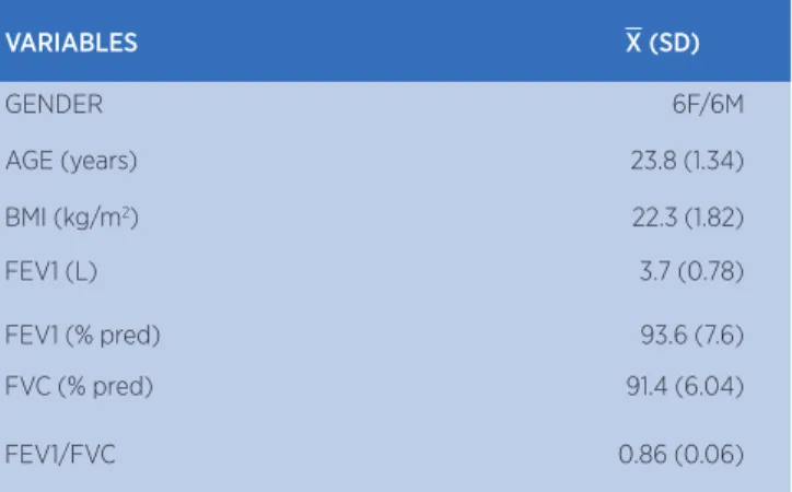
Initially, 17 subjects were evaluated, however, 5 presented abnormal lung function test and were excluded. herefore, the data of the remaining 12 subjects were analyzed. Table 1 presents the demographic, anthropometric and spirometric data of the individuals.
Table 1. Demographic, anthropometric and spirometric data of the individuals studied
VARIABLES X (SD)
GENDER 6F/6M
AGE (years) 23.8 (1.34)
BMI (kg/m2) 22.3 (1.82)
FEV1 (L) 3.7 (0.78)
FEV1 (% pred) 93.6 (7.6)
FVC (% pred) 91.4 (6.04)
FEV1/FVC 0.86 (0.06)
Data presented as mean (X) and standard deviation (SD)
M: male; F: female; BMI:body mass index; FEV1: forced expiratory volume in one second; FVC:
forced vital capacity
At rest, a total of 99IC maneuvers were collected and 56% were excluded due to irregularities. herefore, 44 maneuvers were included in the statistical analysis. During exercise, a total of 200IC maneuvers were evaluated but 47% were excluded, therefore, 106 maneuvers were analyzed.
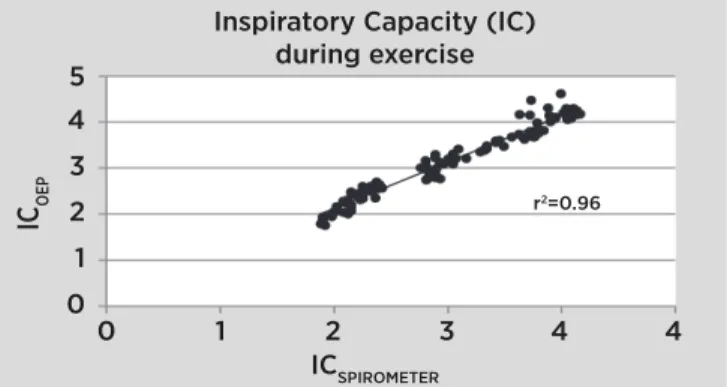
Figure 1 shows the results for the regression analysis of the IC values obtained with the OEP and the spirometer at rest. he r2 observed was of
0.90 (p=0.0002) for the related regression equation: CIOEP=1.16×(ICSPIROMETER)-0.15. he percentage of discrepancy between the measurements at rest was -9.6 (8.6%).
0 1 2 3 4 5
0 1 2
ICSPIROMETER ICOEP
Inspiratory Capacity (IC) at rest
3 4
r2=0.07
Figure 1. Results of regression analysis for inspiratory capacity obtained by OEP (ICOEP) and by spirometer (ICSPIROMETER) at rest
0 1 2 3 4 5
0 1 2
ICSPIROMETER ICOEP
Inspiratory Capacity (IC)
x cise
3 4 4
r2=0.96
Figure 2. Results of regression analysis for inspiratory capacity obtained by OEP (ICOEP) and by spirometer (ICSPIROMETER) during exercise
Figure 3 presents the results for the Bland-Altman analysis of IC values obtained during exercise and showed bias between methods of 0.13L. he limits of agreement indicate that the diference between the measures carried out by OEP and measurements made by the spirometer can vary from -0.04 L to 0.57 L. Additionally, the data was equally distributed between the upper and lower limits, thereby demonstrating that there was no systematic overestimation or underestimation of the IC by any of the instruments used.
0 1 2 3 4 5
0 1 2
(ICOEP–ICSP) /2
UL = upper limit LL = lower limit
IC
OEP
–IC
SP
(L)
3 4 5
Figure 3. Bland-Altman plot for inspiratory maneuvers obtained by OEP (ICOEP) and by the spirometer (ICSP)
DISCUSSION
his study aimed to verify the concurrent validity of the OEP system comparing IC maneuvers analyzed by this instrument and by the spirometer at rest and submaximal exercise. he coeicients of determination found were of high magnitude (>0.90) and the percentage of discrepancy between the methods was usually less than 10%. he Bland-Altman analysis of
values obtained during exercise showed a bias between measurements of 0.13 liters and none of the instruments tended to systematically overestimate or underestimate the values of IC.
he concurrent validity of the OEP was assessed by IC measurements. Tidal volume was not considered, as a pneumotach system synchronized with the OEP was not used. Only in one study the concurrent validity of the OEP was evaluated by IC maneuvers. Vogiatzis et al.9 compared IC measured by the OEP with the ones
obtained by a pneumotachometer in 20 patients with COPD. Regression analysis was performed considering the IC maneuvers obtained at rest and during incremental exercise on a cycle ergometer and resulted in the equation: ICOEP=0.65(ICSPIROMETER)+0.52, with r2 of 0.89. he percentage of discrepancy between the
methods was 3.8 (1.8%). Diferences in the experimental protocol in this population as well as the instrument used to measure the IC (a spirometer and mouthpiece in the study by Vogiatzis et al.9 and a metabolic system
gas analysis using a pneumotachometer type Pitot tube and mask in the present study) make the comparison between the studies diicult although both studies showed r2 higher than 0.80. Additionally, the maneuvers
of IC obtained at rest and during diferent phases of the exercise were evaluated together and the authors did not provide the number of IC maneuvers included in the analysis.
Bland-Altman analysis was done only with the 106 IC maneuvers obtained during exercise given that at least 100 measurements are recommended to achieve reliable results23,24. Regarding this analysis, the results
of this study are similar to the ones observed by Aliverti et al6. hese authors observed that the measurement
of lung volumes by OEP did not introduce systematic error to the measured values compared to the volumes obtained by a spirometer in healthy subjects evaluated at rest in the supine position. However, unlike the study by Aliverti et al.6, in which this analysis was conducted
for the tidal volume, in this study, it was calculated considering the IC values.
he discrepancy between the IC values obtained by the OEP and the gas analysis system observed in this study may be due to diferent potential sources of variation that should be considered as variations in humidity, pressure and temperature of the gas between the lung and the spirometer6,8. Furthermore,
changes may not necessarily be the same in both methods, because there can be displacement of blood from the chest and/or abdomen to the periphery and vice versa, which are measured by OEP8,25 but not by
the spirometer.
CONCLUSION
he results of this study demonstrate that the OEP is a valid evaluation system of healthy individuals at rest and during exercise regarding IC maneuvers. he OEP system has been extensively used in many studies, and reporting the characteristics of this instrument can contribute to improve the quality of rehabilitation research. Further studies are important to assess if similar results will be obtained in populations with cardiopulmonary dysfunction.
REFERENCES
1. Gadotti IC, Vieira ER, Magee DJ. Importance and clariication of measurement properties in rehabilitation. Rev Bras Fisioter. 2006;10(2):137-46.
2. Aliverti A. Opto-electronic plethysmography: new indings in chronic obstructive pulmonary disease. Int J Respir Care. 2008;(2):45-50.
3. Aliverti A, Pedotti A. Opto-electronic plethysmography. Monaldi Arch Chest Dis. 2003;59(1):12-6.
4. Iandelli I, Aliverti A, Kayser B, Dellaca R, Cala SJ, Duranti R, et al. Determinants of exercise performance in normal men with externally imposed expiratory low limitation. J Appl Physiol. 2002;92(5):1943-52.
5. Parreira VF, Vieira DS, Myrrha MA, Pessoa IM, Lage SM, Britto RR. Optoelectronic plethysmography: a review of the literature. Rev Bras Fisioter. 2012;16(6):439-53.
6. Aliverti A, Dellaca R, Pelosi P, Chiumello D, Pedotti A, Gattinoni L. Optoelectronic plethysmography in intensive care patients. Am J Respir Crit Care Med. 2000;161(5):1546-52.
7. Aliverti A, Dellaca R, Pelosi P, Chiumello D, Gatihnoni L, Pedoti A. Compartmental analysis of breathing in the supine and prone positions by optoelectronic plethysmography. Ann Biomed Eng. 2001;29(1):60-70.
8. Cala SJ, Kenyon CM, Ferrigno G, Carnevali P, Aliverti A, Pedotti A, et al. Chest wall and lung volume estimation by optical relectance motion analysis. J Appl Physiol. 1996;81(6):2680-9.
9. Vogiatzis I, Georgiadou O, Golemati S, Aliverti A, Kosmas E, Kastanakis E, et al. Patterns of dynamic hyperinlation
during exercise and recovery in patients with severe chronic obstructive pulmonary disease. Thorax. 2005;60(9):723-9. 10. Aliverti A, Stevenson N, Dellaca RL, Lo Mauro A, Pedotti
A, Calverley PM. Regional chest wall volumes during exercise in chronic obstructive pulmonary disease. Thorax. 2004;59(3):210-6.
11. Vieira DS, Hofman M, Pereira DA, Britto RR, Parreira VF. Optoelectronic plethysmography: intra-rater and inter-rater reliability in healthy subjects. Respir Physiol Neurobiol. 2013;189(3):473-6.
12. Dellaca RL, Ventura ML, Zannin E, Natile M, Pedotti A, Tagliabue P. Measurement of total and compartmental lung volume changes in newborns by optoelectronic plethysmography. Pediatr Res. 2010;67(1):11-6.
13. Kenyon CM, Cala SJ, Yan S, Aliverti A, Scano G, Duranti R, et al. Rib cage mechanics during quiet breathing and exercise in humans. J Appl Physiol. 1997;83(4):1242-55.
14. Duranti R, Filippelli M, Bianchi R, Romagnoli I, Pellegrino R, Brusasco V, et al. Inspiratory capacity and decrease in lung hyperinlation with albuterol in COPD. Chest. 2002;122(6):2009-14.
15. World Health Organization. Global database on body mass index: an interactive surveillance tool for monitoring nutrition transition [Internet]. Geneva: WHO. 2004 [access in: oct. 20 2010]. Available at: http://apps.who.int/bmi.
16. Pereira CA, Sato T, Rodrigues SC. New reference values for forced spirometry in white adults in Brazil. J Bras Pneumol. 2007;33(4):397-406.
17. Neder JA, Nery LE, Castelo A, Andreoni S, Lerario MC, Sachs A, et al. Prediction of metabolic and cardiopulmonary responses to maximum cycle ergometry: a randomised study. Eur Respir J. 1999;14(6):1304-13.
18. O’Donnell DE, Webb KA. Exertional breathlessness in patients with chronic airlow limitation. The role of lung hyperinlation. Am Rev Respir Dis. 1993;148(5):1351-7.
19. Dolmage TE, Goldstein RS. Repeatability of inspiratory capacity during incremental exercise in patients with severe COPD. Chest. 2002;121(3):708-14.
20. American Thoracic S, American College of Chest P. ATS/ ACCP Statement on cardiopulmonary exercise testing. Am J Respir Crit Care Med. 2003;167(2):211-77.
21. Neder JA, Fuld JP, Overend T, Thirlwell J, Carter R, Stevenson R, et al. Efects of formoterol on exercise tolerance in severely disabled patients with COPD. Respir Med. 2007;101(10):2056-64.
22. Portney G, Watkins, P.M. Regression. In: Hall PP, editors Foundations of Clinical Research Applications to Practice. 3a
ed. New Jersey: Pearson Prentice Hall; 2008. p. 539-67. 23. Bland JM, Altman DG. Statistical methods for assessing
agreement between two methods of clinical measurement. Lancet. 1986;1(8476):307-10.
24. Bland JM, Altman DG. Measuring agreement in method comparison studies. Stat Methods Med Res. 1999;8(2):135-60. 25. Aliverti A, Dellaca R, Pedotti A. Optoelectronic