Selective vesical artery embolization for treatment of
gross hematuria due to actinic cystitis
Embolização vesical superseletiva para tratamento de hematúria
maciça por cistite actínica
Adenauer Marinho de Oliveira Góes Junior1,2
*
, Salim Abdon Haber Jeha1, Marcus Vinicius Baptista Queiroz1,2
Abstract
his article describes the case of a 46-year-old female patient who had been treated with radiotherapy for cervical cancer. She developed actinic cystitis with frequent episodes of severe hematuria. She required repeated catheterization to manage urinary retention, blood transfusions and hospital admissions. Conservative measures and attempts to achieve hemostasis by cystoscopy were unsuccessful at controlling bleeding. he patient therefore underwent endovascular treatment with superselective embolization of the vesical arteries and other vascular pedicles found to be linked with the bleeding. he procedure was successful and the patient has been in follow-up for 9 months with no need for further blood transfusions or admission to hospital. According to a review of the literature, use of this technique has not previously been described in Brazil.
Keywords: therapeutic embolization; cystitis; hematuria; endovascular procedures; urinary bladder.
Resumo
Os autores relatam o caso de uma paciente de 46 anos de idade, que, após ter sido submetida a tratamento radioterápico por neoplasia de colo uterino, desenvolveu cistite actínica com episódios frequentes de hematúria franca. A paciente necessitou ser submetida a repetidos cateterismos vesicais por retenção urinária, hemotransfusões e internacões hospitalares. As medidas conservadoras e as tentativas de hemostasia por cistoscopia não foram bem-sucedidas no controle do sangramento. A paciente foi então submetida a tratamento endovascular com embolização superseletiva das artérias vesicais e outros pedículos vasculares, que se demonstraram associados ao sangramento. O procedimento foi bem-sucedido e a paciente vem sendo acompanhada há nove meses sem a necessidade de novas hemotransfusões nem de novas internações hospitalares. De acordo com a revisão da literatura, o uso dessa técnica ainda não havia sido descrito em trabalhos brasileiros.
Palavras-chave: embolização terapêutica; cistite; hematúria; procedimentos endovasculares; bexiga urinária.
1 Universidade Federal do Pará – UFPA, Belém, PA, Brazil. 2 Centro Universitário do Pará – CESUPA, Belém, PA, Brazil.
Financial support: None.
Conlicts of interest: No conlicts of interest declared concerning the publication of this article. Submitted: June 13, 2015. Accepted: August 17, 2015.
INTRODUCTION
Untreatable hematuria secondary to bladder or prostate hemorrhage is potentially lethal and constitutes a therapeutic challenge.1-3
Causes of massive hematuria include carcinoma of the bladder, actinic cystitis, cystitis induced by cyclophosphamide, transurethral prostate resection, carcinoma of the prostate,1,2,4 spontaneous, iatrogenic
or traumatic arteriovesical istula.5
In many patients, severe hematuria cannot be controlled by conservative measures such as irrigation of the bladder, instillation of formalin, silver nitrate, estrogen, epidermal growth factor, increase of intravesical hydrostatic pressure or hyperbaric oxygen therapy, or using endoscopic procedures with electrocoagulation, laser or argon hemostasis.1-4,6
Cystectomy is not always an option since these patients often have a high surgical risk.1,2,4
Arterial embolization is a safe, minimally invasive option that can be used to control hematuria caused by bladder hemorrhage.1,5
PART I – CLINICAL SITUATION
The patient was a 46-year-old woman who had undergone radiotherapy to treat cervical cancer 14 years previously. 2 years previously she had undergone a second cycle of treatment, which included radiotherapy sessions.
She had episodes of hematuria on a large number of occasions that became progressively more intense and required frequent catheterization of the bladder to manage urinary retention caused by clotting. During a single 6-month period she needed to be admitted to hospital three times for transfusion of packed red blood cells and underwent cystoscopy twice to remove clots and cauterization to achieve bladder hemostasis. During a period of remission from bleeding, the patient was referred to a vascular surgeon to be assessed for embolization of visceral arteries. This procedure was actually performed as an emergency intervention when the patient was admitted to an intensive care unit because of a worsening of health status in general and an episode of severe hematuria, during which her hemoglobin levels fell to 6.6 mg/dl.
PART II – WHAT WAS DONE
The procedure was performed under general anesthesia. A retrograde puncture was made in the right common femoral artery, followed by placement of a 5F angiographic sheath. Pigtail and curved cobra 2 5F catheters were used to conduct angiographs
via the abdominal aorta and internal iliac arteries.
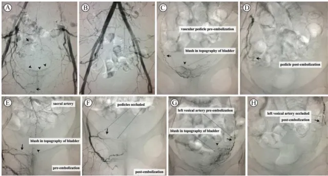
Hypervascularization was identiied in the pelvic area
and contrast leakage was seen in the topography of the bladder. Superselective angiographs demonstrated that the bleeding was not only related to the vesical arteries, but also originated from abnormal branches that had probably developed in response to radiotherapy.
All arterial pedicles identiied as involved in the
bleeding were catheterized superselectively with a 3F microcatheter and embolized with 500 to 700 µm embosphere-type microparticles (Medical). Control angiography demonstrated a considerable reduction in pelvic hypervascularization and an absence of contrast leakage in the topography of the bladder (Figure 1).
The patient returned to the ICU for postoperative recovery and was transferred to a private room 1 day after the operation. Hematuria intensity was reduced in the immediate postoperative period and continued to abate gradually. Bladder irrigation was resumed 2 days after the operation, by which point the patient had discrete hematuria, which was interpreted as residual, since hemometric
results did not drop off again. On the irst day after
embolization, the patient complained of mild to moderate pain in the lower abdomen, but palpation
of the area revealed no relevant indings. The pain
was interpreted as post-embolization syndrome and improved with symptomatic treatment. The patient remained in hospital for 5 days after the operation for monitoring to rule out the possibility of bladder necrosis and was discharged free from intercurrent conditions.
The patient has been in follow-up for 9 months. She has not needed further hospital admissions, blood transfusions or bladder irrigation, but she has developed urinary incontinence and reports discrete hematuria after situations that increase intra-abdominal pressure, such as defecatory effort. Despite chemotherapy for metastasis diagnosed in the lungs, the patient reports a marked improvement in quality of life.
DISCUSSION
While the technique for selective embolization of the vesical arteries was described more than 30 years ago,7 the few articles that have been published on the
subject2 are mostly reports of cases or small series.
Additionally, searches of the PubMed, Bireme and SciELO databases did not identify any Brazilian articles on the subject.
endoscopic urology procedures, severe hematuria is potentially lethal and constitutes a therapeutic challenge.1,2
In patients who are already weakened by intense bleeding and whose general health is compromised by advanced cancer, complex surgical procedures such as ligature of the hypogastric arteries, urinary shunts4
and radical cystectomy with or without construction of a neobladder, are associated with high morbidity and mortality rates.4,6
In cases in which the cause of bleeding has not yet
been identiied and pulsating hemorrhage is detected
during cystoscopy, angiotomography or arteriography can be used to facilitate diagnosis.5 Of the two,
arteriography provides more precise details of pelvic vascularization. Depending on the type of bleeding that
is identiied, the diagnostic angiographic procedure
can be converted into a therapeutic intervention. Before embolization, the patient should undergo rigorous intravenous hydration, the bladder should be irrigated, any clots should be removed and blood transfusion should be performed as necessary.2 While
this procedure can be conducted with local anesthesia, the choice of anesthetic technique should take into account the general condition of the patient and the need for their collaboration in terms of remaining
immobile during a procedure the duration of which is unpredictable because superselective catheterization of the vessels feeding the bleeding can be challenging.
Certain angiographic and technical aspects must be considered when vesical embolization is proposed. The inferior vesical artery is a branch of the anterior division of the hypogastric artery. This vessel supplies the fundus of the bladder, the seminal vesicles, the deferent duct and the prostate, and is analogous to the vaginal artery in females. Some authors consider that this artery exists in both sexes and, in this case it would be a branch of the vaginal artery in women. Alternative origins of the inferior vesical artery include from a common trunk with the superior gluteal and the pudendal arteries or emergence directly from the pudendal artery.2 In turn, the superior vesical artery
gives rise to branches that supply higher areas of the bladder and distal segments of the urethras.2
The large number of anatomic variants2 means that
vascular intervention is dependent on identiication of
signs that indicate which vessel should be occluded. It is more common to identify areas of hypervascularization than clear leakage of contrast;2 spiral or ectatic arteries
and retention of contrast in terminal branches also suggest a connection with the bleeding.
Every effort should be made to ensure that the bilateral embolization is as selective as possible.2
Hematuria secondary to actinic cystitis is often associated with bleeding fed by several different vessels, as in the case described here, but if it appears that only one of the vesical arteries is involved, contralateral embolization can be conducted at a later date to reduce the risk of necrosis of the bladder.4,6 Since there are
few reports in the literature, there is no consensus on the ideal interval between embolizations or on whether this strategy really does afford protection from necrosis of the bladder. Additionally, the majority of authors recommend simultaneous bilateral embolization for severe hematuria.2,6
A range of different catheters and microcatheters can be used at different stages of the procedure and
the choice is inluenced by the anatomy of arteries,
by availability and by the endovascular surgeon’s preference.
A great variety of embolization agents have been used for embolization of vesical arteries. In emergency situations and when non-absorbable materials are unavailable, absorbable hemostatic materials such as Gelfoam can be used for this purpose. There are also descriptions of the use of non-absorbable microparticles with diameters calibrated to 300-500 µm and 500-700 µm2 and of n-butyl cyanoacrylate surgical
glue mixed with lipiodol at a proportion of 1:3 to make it radiopaque.2
When superselective catheterization of the vesical arteries is not possible, proximal occlusion of arteries that are not feeding the bleeding can be achieved with coils before embolization with particulate material (the springs divert the particles in the direction of the target vessel and protect the distal bed of vessels that are not associated with the hemorrhage).1
There is no consensus on the inluence of the
type of embolization agent because the scarcity of published studies precludes conclusions, but the current preference of the majority of authors is for permanent particulate agents, such as the calibrated microspheres used in the case described here.2
Treatment success is achieved in 90% of cases,
when the vesical arteries can be identiied.2
In the literature, early clinical success is deined
as absence of macroscopic hematuria, no formation of intravesical clots, no fall in hemoglobin by more than 2.0 mg/dl and/or failure of more conservative measures for management of hematuria during the
irst month after embolization.1 Relapse of bleeding
is more common in cases of actinic cystitis than in cases of local neoplasm, primarily because of the greater life expectancy of the patients.2 In the case
described here the patient has been in follow-up for 9 months and is doing well.
Complications related to this method include post-embolization syndrome (self-limiting manifestations of pain, nausea, vomiting and fever), gluteal and perineal pain, Brown-Séquard syndrome caused by anastomoses between vesical arteries and sacral arteries (which should be screened for during arteriography), gluteal paresis and cutaneous necrosis; necrosis of the bladder is rare because of the organ’s rich vascularization.2,6
Necrosis of the bladder and other severe ischemic complications were reported with greater frequency in older samples, when bilateral occlusion of hypogastric arteries was performed.6 More modern studies using
the superselective embolization technique report complication rates of less than 10%.2
Frequent hospital admissions for blood transfusions and irrigation of the bladder are not practical and compromise the patient’s quality of life, while radical surgery can be high risk.1,2
This procedure offers immediate control of potentially fatal hemorrhage, improves palliative care and quality of life and reduces the need for frequent cystoscopy, irrigation of the bladder and blood transfusions.2,6
In the majority of cases, embolization is well-tolerated, reducing the need for surgery,2,6 and can be repeated if
necessary.6 If there is a need for surgical intervention,
the patient’s general health status will be better preserved and there tends to be a reduced need for intraoperative transfusions.6
REFERENCES
1. Delgal A, Cercueil JP, Koutlidis N, et al. Outcome of transcatheter arterial embolization for bladder and prostate hemorrhage. J Urol. 2010;183(5):1947-53. http://dx.doi.org/10.1016/j.juro.2010.01.003. PMid:20303518.
2. Loffroy R, Pottecher P, Cherblanc V, et al. Current role of transcatheter arterial embolization for bladder and prostate hemorrhage. Diagn Interv Imaging. 2014;95(11):1027-34. http://dx.doi.org/10.1016/j. diii.2014.03.008. PMid:24746761.
3. Han Y, Wu D, Sun A, et al. Selective embolization of the internal iliac arteries for the treatment of severe hemorrhagic cystitis following hematopoietic SCT. Bone Marrow Transplant. 2008;41(10):881-6. http://dx.doi.org/10.1038/bmt.2008.4. PMid:18246111.
4. Palandri F, Bonifazi F, Rossi C, et al. Successful treatment of severe hemorrhagic cystitis with selective vesical artery embolization. Bone Marrow Transplant. 2005;35(5):529-30. http://dx.doi.org/10.1038/ sj.bmt.1704818. PMid:15665843.
5. Zheng X, Lin Y, Chen B, et al. Severe hematuria after transurethral electrocoagulation in a patient with an arteriovesical fistula. BMC Urol. 2013;13(1):68-72. http://dx.doi.org/10.1186/1471-2490-13-68. PMid:24289138.
7. Kobayashi T, Kusano S, Matsubayashi T, Uchida T. Selective embolization of the vesical artery in the management of massive bladder hemorrhage. Radiology. 1980;136(2):345-8. http://dx.doi. org/10.1148/radiology.136.2.7403507. PMid:7403507.
*
Correspondence
Adenauer Marinho de Oliveira Góes Junior Rua Domingos Marreiros, 307/802 – Umarizal CEP 66055-210 – Belém (PA), Brazil E-mail: [email protected]
Author information
AMOGJ - Vascular surgeon certiied in angioradiology and endovascular surgery; MSc and PhD candidate from the Graduate Program in Interdisciplinary Surgical Sciences, Escola Paulista de Medicina (EPM), Universidade Federal de São Paulo (UNIFESP); Scientiic director, Sociedade Brasileira de Angiologia e de Cirurgia Vascular do Pará (SBACV-PA); Professor, Universidade Federal do Pará (UFPA) and Centro Universitário do Pará (CESUPA). SAHJ - Vascular surgeon; Professor, Universidade Federal do Pará (UFPA). MVBQ - Urologist; PhD in Urology from Universidade de São Paulo (USP), Universidade Federal do Pará (UFPA), and Centro Universitário do Pará (CESUPA).
Author contributions
Conception and design: AMOGJ Analysis and interpretation: AMOGJ, SAHJ, MVBQ Data collection: AMOGJ, MVBQ Writing the article: AMOGJ Critical revision of the article: AMOGJ Final approval of the article*: AMOGJ, SAHJ, MVBQ Statistical analysis: N/A. Overall responsibility: AMOGJ