CASE REPORT
Hydrothorax due to parenteral nutrition – a case report
Hidrotórax secundário à nutrição parenteral – relato de caso
José Henrique Silvah1, Cristiane Maria Mártires Lima1, Francisco Abaeté das Chagas-Neto2, Guilherme Teixeira Araújo1,
Fernando Bahdur Chueire3, Selma Freire de Carvalho Cunha4, Júlio Sérgio Marchini5
Introduction
Central vein catheterization for the administration of medications, chemotherapy drugs, hemoderivatives and parenteral nutrition is a routine procedure in hospitals, but it is not free of complications1. We report the case of
a patient who developed hydrothorax ater insertion of a long-term central venous catheter (Port-a-Cath®) for total
parenteral nutrition (TPN).
Case report
A 42-year-old female patient was admitted to the Nutrology Service of the Hospital das Clínicas de Ribeirão Preto in April, 2009, with short bowel syndrome (small bowel length of less than 15 cm) secondary to vascular pedicle tor-sion, plus a history of two previous abdominal operations.. She had been submitted to jejunoileal enterectomy from 15
cm distal to the ligament of Treiz to the terminal ileum, and to a gastrojejunal bypass, similar to a Roux-en-Y anastomosis.
Total parenteral nutrition (TPN) was administered through central venous catheter inserted by the Seldinger technique. Twenty days later, a Port-a-Cath® catheter was
placed through the right internal jugular vein for long-term parenteral nutrition. Adequate positioning of the catheter tip and blood low were conirmed by luoros-copy. he short-term catheter was removed. Nineteen days later, the Port-a-Cath® device was punctured with a
Huber needle (special needle, designed by Huber in 1946, that produces a non-coring puncture in the reservoir dia-phragm), in order to start TPN infusion1. Aspiration of
blood through the catheter was not performed. On the irst day of TPN, the patient complained of intense thorac-ic pain, tachypnea, tachycardia, abdominal pain referred to the right shoulder, decreased breath sounds in the right side and hemodynamic instability.
Abstract
Hydrothorax due to parenteral nutrition infusion is a rare, although increasingly common event. his report shows a short bowel patient who developed hemodynamic instability and respiratory failure few hours after parenteral nutrition infusion’s start. We also emphasize the maneuvers to avoid and treat such complication.
Keywords: hydrothorax; parenteral nutrition; short bowel syndrome.
Resumo
Hidrotórax secundário à infusão de nutrição parenteral é uma condição rara, embora se apresente cada vez mais comum. Neste relato de caso, uma paciente com síndrome do intestino curto desenvolveu instabilidade hemodinâmica e insuiciência respiratória algumas horas após o início da infusão de nutrição parenteral. Ressaltamos também as manobras para evitar e tratar tal complicação.
Palavras-chave: hidrotórax; nutrição parenteral; síndrome do intestino curto.
Study carried out at Hospital das Clínicas, Faculdade de Medicina de Ribeirão Preto, Universidade de São Paulo (USP) – Ribeirão Preto (SP), Brazil.
1 Resident of the Department of Internal Medicine and Nutrology at Faculdade de Medicina de Ribeirão Preto of USP – Ribeirão Preto (SP), Brazil. 2 Resident of the Department of Internal Medicine and Radiology at Faculdade de Medicina de Ribeirão Preto of USP – Ribeirão Preto (SP), Brazil. 3 Assistant Physician of Internal Medicine and Nutrology at Faculdade de Medicina de Ribeirão Preto of USP – Ribeirão Preto (SP), Brazil. 4 PhD; Professor of Internal Medicine and Nutrology at Faculdade de Medicina de Ribeirão Preto of USP – Ribeirão Preto (SP), Brazil. 5 Full Professor of Internal Medicine and Nutrology at Faculdade de Medicina de Ribeirão Preto of USP – Ribeirão Preto (SP), Brazil.
Hydrothorax due to parenteral nutrition - Silvah JH et al. J Vasc Bras 2011, Vol. 10, Nº 3
240
he patient was placed on non-invasive ventilatory support, and a double-lumen catheter was placed on the let jugular vein for the infusion of vasoactive drugs, luids and antibiotic therapy. Chest radiography showed pleural efusion at the right side (Figures 1 and 2). horacocentesis was performed at bedside and the drainage of 2,900 mL of a typical serous luid conirmed the suspicion of hy-drothorax from the TPNn infusion (Figure 3). he meth-ylene blue test which consists in the infusion of methmeth-ylene blue through the central venous catheter, with drainage of the dye through the chest tube, conirmed the diagnosis of hydrothorax (Figure 4). Fluoroscopy and chest radiog-raphy conirmed misplacement of the Port-a-Cath in the right side (Figure 5). Ater investigation and patient stabi-lization, the catheter was removed. he catheter for short-term infusion was maintained on the let jugular vein to continue the treatment that had been previously initiated to prevent a possible infection due to the patient’s hemo-dynamic instability.
Hemothorax, a possible complication after the re-moval of a misplaced long-term catheter was not ob-served. With the discontinuation of TPN infusion in the pleural space and after chest drainage, the patient re-covered well. Antibiotic therapy and ventilatory support were discontinued.
Discussion
he present paper reports a complication of central venous catheter insertion. Pneumothorax, hemorrhage, thrombosis, embolism, and vessel perforation, such as the pulmonary artery, are some of the complications reported
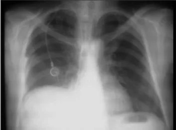
Figure 1. Chest radiography before total parenteral nutrition showing the central venous catheter at the right side, with its distal tip projected to the right atrium and right diaphragmatic cupule.
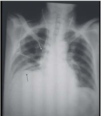
Figure 2. Chest radiography showing pleural efusion at the right side, mediastinal structures and central venous catheter (white arrow) devi-ated to the left; note the distal tip positioned at the right pleural cavity and the other venous catheter at the left jugular vein in adequate posi-tion (black arrow).
Figure 3. Pleural luid obtained by toracocentesis.
Hydrothorax due to parenteral nutrition - Silvah JH et al. J Vasc Bras 2011, Vol. 10, Nº 3 241
in literature1,3-6. In order to avoid such complications, even
when the procedure is performed by a trained physician and guided by luoroscopy, a check list for every stage of the procedure should be followed and no short cuts should be permitted. Routine aspiration of the catheter before starting the infusion is recommended, but it was not done in this case. Hydrothorax was attributed to misposition-ing of the long-term catheter.
Follow-up radiographies constitute an alternative when blood flow is absent at catheter aspiration, and should be performed in two incidences (antero-pos-terior and lateral). Fluoroscopy, on the other hand, is the most reliable method to confirm the catheter posi-tion at the moment of inserposi-tion, as recommended by the American College of Surgeons. Although there are not data to support the routine use of radiography to prevent complications, such measures could help to de-termine the catheter position7,8.
Anatomic landmarks have been used to estimate the adequate position of the distal tip of the catheter. For in-stance, the tip of catheters inserted in the subclavian or jugular vein should be positioned below the right bron-chus, which corresponds to the cavo-atrial junction8. If
there is any doubt as to the adequate position, the catheter should not be used.
Ultrasound-guided puncture, which was not performed in the patient in this case, may shorten procedure time and decrease the chance of immediate complications9.
Cases of vascular erosion that result in pleural effu-sion have been reported as a late complication (onset up to 11 days after the insertion of the catheter) that may lead to the patient’s death10. Our hypothesis is that this
was the cause of hydrothorax in our patient, but we could not confirm it.
Specific treatments for hydrothorax following TPN have been little studied in literature. However, measures such as ventilatory and hemodynamic support, along with immediate suspension of TPN and chest tube drainage, were effective to prevent a bad outcome in this case. Fortunately, this event is rare, even though re-ported with increasing frequency, and may lead to death if not properly diagnosed and treated11. We emphasize
the importance of confirming catheter position before its effective use. Radiographic examination after cath-eter insertion and the maneuver of blood reflux into the catheter are mandatory.
References
1. Pittiruti M, Hamilton H, Bii R, et al. ESPEN Guidelines on Parenteral Nutrition: central venous catheters (access, care, diagnosis and therapy of complications). Clin Nutr. 2009;28(4):365-77.
2. Haindl H, Müller H. An atraumatic needle for the puncture of ports and pumps. Klin Wochenschr. 1988;66(20):1006-9.
3. Lu WH, Huang TC, Pan JY, et al. A potentially fatal complication during subclavian vein catheterization in an infant with congenital heart disease-puncture to pulmonary artery directly: a case report. J Clin Anesth. 2008;20(3):225-7.
4. Duong MH, Jensen WA, Kirsch CM, et al. An unusual complica-tion during central venous catheter placement. J Clin Anesth. 2001;13(2):131-2.
5. Lee EK. An unexpected left hydrothorax after left internal jugular venous catheterisation for total parental nutrition and antibiot-ics. Ann Acad Med Singapore. 2006;35(10):742-4.
6. Machado JD, Suen VM, Figueiredo JF, et al. Bioilms, infection, and parenteral nutrition therapy. JPEN J Parenter Enteral Nutr. 2009;33(4):397-403.
7. College’s Committee on Perioperative Care. Statement on recom-mendations for use of real-time ultrasound guidance for placement of central venous catheters. Bull Am Coll Surg. 2008;93(9):35-6.
8. Askegard-Giesmann JR, Caniano DA, Kenney BD. Rare but seri-ous complications of central line insertion. Semin Pediatr Surg. 2009;18(2):73-83.
9. Turker G, Kaya FN, Gurbet A, et al. Internal jugular vein cannula-tion: an ultrasound-guided technique versus a landmark-guided technique. Clinics (Sao Paulo). 2009;64(10):989-92.
Hydrothorax due to parenteral nutrition - Silvah JH et al. J Vasc Bras 2011, Vol. 10, Nº 3
242
10. Duntley P, Siever J, Korwes ML, et al. Vascular erosion by cen-tral venous catheters. Clinical features and outcome. Chest. 1992;101(6):1633-8.
11. Westermann SA, Pahlplatz PV, Brouwers MA. Timeline of celluli-tis and late development of hydrothorax induced by a right-sided central venous catheter: report of a case. JPEN J Parenter Enteral Nutr. 2010;34(3):341-3.
Correspondence José Henrique Silvah Departamento de Clínica Médica – Hospital das Clínicas Av. Bandeirantes, 3.900, 6° andar CEP 14048-900 - Ribeirão Preto (SP), Brazil. E-mail: [email protected]