CLINICAL SCIENCE
Effects of thyroxine replacement on endothelial
function and carotid artery intima-media thickness in
female patients with mild subclinical hypothyroidism
Monica Dias Cabral,IPatricia Teixeira,IDebora Soares,ISandra Leite,II Elizabeth Salles,IMario WaismanI IUniversidade Federal do Rio de Janeiro, Rio de Janeiro/RJ, Brazil.IICentro de Estudos e Pesquisas da Mulher, Rio de Janeiro/RJ, Brazil.BACKGROUND: Previous studies have suggested an association between subclinical hypothyroidism and coronary artery disease that could be related to changes in serum lipids or endothelial dysfunction.
METHODS: Thirty-two female subclinical hypothyroidism patients were randomly assigned to 12 months of L-thyroxine replacement or no treatment. Endothelial function was measured by the flow-mediated vasodilatation of the brachial artery, as well as mean carotid artery intima-media thickness, and lipid profiles were studied at baseline and after 12 months of follow-up.
RESULTS:The mean (¡SD) serum thyroid-stimulating hormone levels in the L-thyroxine replacement and control groups were 6.09¡1.32 and 6.27¡1.39mUI/ml, respectively. No relationship between carotid artery intima-media
thickness or brachial flow-mediated vasodilatation and free T4 and serum thyroid-stimulating hormone was found. The median L-T4 dose was 44.23¡18.13mg/day. After 12 months, there was a significant decrease in the
flow-mediated vasodilatation in the subclinical hypothyroidism control group (before: 17.33¡7.88 to after: 13.1¡4.75%, p = 0.03), but there were no significant differences in flow-mediated vasodilatation in the L-thyroxine treated group (before: 16.81¡7.0 to after: 18.52¡7.44%, p = 0.39). We did not find any significant change in mean carotid intima-media thickness after 12 months of L-thyroxine treatment.
CONCLUSION: Replacement therapy prevents a decline in flow-mediated vasodilatation with continuation of the subclinical hypothyroidism state. Large prospective multicenter placebo-controlled trials are necessary to investigate endothelial physiology further in subclinical hypothyroidism patients and to define the role of L-thyroxine therapy in improving endothelial function in these patients.
KEYWORDS: Subclinical hypothyroidism; Endothelial function; Flow-mediated vasodilatation; Carotid artery intima-media thickness; Levothyroxine treatment.
Cabral MD, Teixeira P, Soares D, Leite S, Salles E, Waisman M. Effects of thyroxine replacement on endothelial function and carotid artery intima-media thickness in female patients with mild subclinical hypothyroidism. Clinics. 2011;66(8):1321-1327.
Received for publication onJanuary 6, 2011;First review completed onJanuary 26, 2011;Accepted for publication onApril 19, 2011
E-mail: [email protected]
Tel.: 55 21 32825071
INTRODUCTION
Subclinical hypothyroidism (SHT) is a disorder character-ized by elevated serum thyroid-stimulating hormone (TSH) levels despite normal free thyroid hormone values.1 The prevalence of SHT ranges from 5 to 10%, and the condition affects 6 to 10% of women (approximately 15% of women over 60 years of age) and 2.4 to 3% of the male population.2,3 A recent meta-analysis demonstrated that SHT can lead to an increased risk of developing cardiovascular disease and, consequently, elevate cardiovascular-associated mortality, especially in patients under 65 years of age.4-6 However, different authors propose that this higher risk does not
apply to all patients with SHT, especially when the serum TSH is only slightly elevated and there are no other associated cardiovascular risks.7 It is unclear whether the association between SHT and coronary artery disease is related to SHT-induced changes in serum lipid levels.8-10In a population-based survey, SHT emerged as a significant risk factor for aortic atherosclerosis and myocardial infarc-tion in elderly women independently of serum cholesterol level.11Therefore, further studies should clarify the role of SHT in cardiovascular disease.
The endothelium plays a major role in the maintenance of vascular function and integrity through the production of vasodilator and vasoconstrictor substances.12,13Endothelial dysfunction that results in reduced availability of nitric oxide is an early marker of atherosclerosis and can be used to predict coronary artery disease before the development of atherosclerotic changes.14 Recent studies have shown that hypothyroidism and SHT may have adverse effects on endothelial function independently of other well-known
Copyrightß2011CLINICS– This is an Open Access article distributed under
atherosclerotic risk factors.15-22 Considerable controversy surrounds the hypothesis that SHT has a significant effect on the risk profile of cardiovascular disease. Boekholdt et al.23provided important information that addressed this issue. Although higher elevated LDL cholesterol levels were found in the SHT group, thyroid dysfunction was not linked with a significantly increased risk of coronary heart disease. Despite the association between thyroid hormone levels and cardiovascular risk factors, thyroid status was not statisti-cally significantly associated with the risk of future coronary heart disease or all-cause mortality in that large cohort over a follow-up of 10.6 years. The endothelial physiology in SHT and the role of levothyroxine (L-T4) therapy in improving endothelial function are still uncertain.
We previously reported no differences in flow-mediated vasodilatation (FMD) of the brachial artery and carotid artery intima-media thickness (IMT) in a group of women with mild SHT without previously diagnosed cardiovascu-lar disease compared with euthyroid subjects.24,25
The aims of the present study were to assess the effect of L-T4 replacement on endothelial function measured by FMD of the brachial artery, as well as carotid artery IMT in a group of female patients with mild SHT.
PATIENTS AND METHODS
The study was a randomized open-label prospective trial comparing the use of L-T4 in female patients with mild SHT. Thirty-two women with mild SHT were recruited from the Outpatient Clinic of the Clementino Fraga Filho University Hospital (HUCFF), Federal University of Rio de Janeiro (UFRJ). The inclusion criteria were at least two documented laboratory determinations of SHT conducted at least six weeks apart, as defined by both elevated TSH (.4.0mIU/ml) and normal free thyroxine (FT4) levels. The
patients had no previous history of thyroid disease. The maximum TSH value accepted was 12mIU/ml. This cutoff
represents three times the superior acceptable limit of the TSH level, which is 4mIU/ml. Individuals were excluded
from the study if they had a history of alcohol use or were suffering from any cardiovascular disease or concomitant non-thyroid illnesses (e.g., obesity, diabetes mellitus, arterial hypertension, liver, or renal diseases). We examined their personal histories, physical examinations, and laboratory tests. The routine laboratory test included kidney function tests (urea, creatinine), liver function tests (ALT, AST, and GGT), and mean glycemia.
The routine laboratory chemistry was normal in all patients, and no patient was taking any drug that could interfere with thyroid, lipoprotein, or endothelial function. The drugs that could interfere included amiodarone, corticosteroids, estrogens, lithium, statins, diuretics, and fibrates; additional drugs were used to treat diabetes, arterial hypertension, or obesity. The patients had no history of recent hospitalization. Patients with SHT were randomly allocated to treatment with L-T4 (levothyroxine group) or no treatment (observation group). Fixed-block numbers were used to generate the random allocation sequence of the treatment group. Each patient received a number in a block of four or six random numbers. Each number corresponded to one of the follow-up groups (levothyroxine group or observation group). Blocks of six and four numbers were continuously distributed, and each block had half of the numbers corresponding to treatment and
another half corresponding to observation. For those patients who were treated, the initial L-T4 dose was 0.75mg/kg/day. Patients were re-evaluated every four
weeks based on the clinical evaluation (including diet habits and physical activity) and TSH and FT4 levels to adjust the dose. The dose that achieved a normal serum TSH was subsequently maintained. After a normal TSH level was obtained, patients were seen every three months to assess the presence of adverse effects, disease progression, and whether the L-T4 dose needed adjustment. The patients in the observation group (OG) were re-evaluated every four weeks, and blood tests were used to measure TSH and FT4 levels.
All patients were instructed to reduce the fat in their daily diet. The patients received general recommendations of a healthy diet, including the following: consume fish, espe-cially oily fish, at least twice a week; limit the intake of saturated fat and trans fat by choosing lean meats and vegetable alternatives, as well as fat-free or low-fat dairy products and minimizing the intake of partially hydro-genated fats; minimize the intake of beverages and foods with added sugars; and choose and prepare foods with little or no salt. These recommendations were written and provided to all patients.
A re-evaluation of serum lipids, FMD and carotid IMT was performed one year after the restoration of the euthyroidism state or one year after inclusion in the group of patients with SHT who did not receive L-T4 treatment. The subjects did not use any lipid-lowering medications during the study.
The study was approved by the UFRJ Institutional Ethics Committee, and all subjects provided written informed consent to participate. The number of the protocol for the Ethics Committee in Research was 12-01.
A general physical examination was performed, including assessment of height (without shoes), weight, and waist circumference (the minimum value between the iliac crest and the lateral costal margin). BMI was calculated as the weight (kg) divided by the height squared (m2). Systolic and diastolic blood pressures were measured from the right brachial artery of the subjects in a supine position after 10 min of rest using a pneumatic sphygmomanometer.
Venous blood samples were drawn between 8:00 and 9:00 am after a 12-hour overnight fast. Serum was centrifuged and stored at -80˚C until assayed. Serum TSH, FT4 and thyroid peroxidase antibody (TPO-Ab), total cholesterol (TC), high-density lipoprotein cholesterol (HDL-c), trigly-ceride (TG), apoprotein B (apoB), apoprotein A (apoA), and lipoprotein a [Lp(a)] levels were determined in both groups. Serum TSH, FT4 and TPO-Ab were measured by immu-nochemiluminescence (Immulite 2000; DPC, Diagnostic Products Corporation, USA). Reference ranges for TSH and FT4 were 0.4-4.0mIU/ml and 0.8-1.9 ng/dl, respectively.
Education Program (NCEP).26The Lp(a) concentration was determined by an immunoradiometric assay (Diagnosis System International, Novatech, USA) and apoA and apoB were measured by a rate nephelometric immunoassay (Beckman Coulter, USA), and their reference ranges were lower than 30, 90-200, and 30-100 mg/dl, respectively.
The subjects were investigated by high-resolution color-Doppler ultrasound imaging (Toshiba Nemium, Japan; 14-mHz linear probe) of the brachial artery in the dominant arm. The study was performed in a temperature-controlled room (25˚C) with subjects resting in the supine position. The blood pressure and heart rate were recorded on the opposite arm every three min using an automatic sphygmoman-ometer. The subjects’ dominant arm was comfortably immobilized in the extended position to allow consistent access to the brachial artery. Doppler ultrasound measure-ments were performed before and 60 seconds after reactive hyperemia (RH). To avoid interobserver variability, all measurements were performed by the same examiner, who was blinded to the subjects’ clinical status. Brachial artery vasodilation in response to RH was determined by a previously validated technique.27,28The intraclass correla-tion coefficient of this technique has been reported previously by our laboratory and ranges from R = 0.7001 to R = 0.8420 (p,0.05).29,30The scans were recorded on S-VHS videotape. The internal diameter of the brachial artery was assessed at the end of diastole, and the arterial flow was measured using the pulse Doppler sample volume at an angle of 60˚or less in the center of the artery. For each subject, optimal brachial artery images were obtained approximately 5 cm above the antecubital fossa. Arm pressure was generating by inflating a pneumatic arm band above the elbow cuff to a pressure up to 30 mmHg higher than the subject’s systolic arterial pressure for 5 min. The cuff was then deflated, the arterial flow was immediately recorded and the diameter was measured 60 to 90 seconds after deflation. For both diameters, one measurement was recorded. FMD was calculated according to the following formula: FMD = (post-occlusion diameter - baseline diameter)6100/baseline diameter.
The carotid images were obtained with the patient in the supine position with the neck mildly extended and the head rotated contralaterally to the side. The imaging protocol involved obtaining a minimum of four longitudinal B-mode images of the distal 10 mm of the right and left common arteries and the carotid bifurcation. The whole image session was recorded on videotape. The IMT of the common carotid artery (CCA) and carotid bifurcation were calculated by the same examiner with high-resolution ultrasound imaging (Acuson Aspen Advanced, 10 mHz linear probe, USA), as previously described.31 The final analysis was performed with IMT images of both the near and far wall of the right and left common artery and the carotid bifurcation. The recordings were evaluated by the same reader, who was blinded to the clinical data. The diastolic frames of each carotid segment were digitized. Lines were drawn along the lumen-intimal and medial adventitial interfaces in all analyzed segments, and the IMT of each segment was automatically computed as an average of several measure-ments. For statistical analysis, IMT readings in all the carotid segments were averaged (mean IMT). Plaque was considered as a focal structure if it encroached into the arterial lumen by at least 0.5 mm or 50% of the surrounding IMT value or demonstrated a thickness .2.0 mm, as
measured from the media-adventitia interface to the intima-lumen interface. The reproducibility of the IMT measurement was acceptable, as demonstrated by coeffi-cients of variation of 7.7¡4.3%.31
Statistical analysis
A statistical analysis was performed using SPSS for Windows, version 10.0. The mean values of the numerical variables were compared between groups by the Mann-Whitney test if they were not normally distributed, as detected by the Kolmogorov-Smirnov Liliefors test. Parametric variables were compared by the Student t-test. Data were expressed as means ¡ SD values unless
otherwise stated. We also determined the frequencies of qualitative variables. For multi-group comparisons, one-way analysis of variance (ANOVA) or the Kruskal-Wallis test was applied, as appropriate. Analysis of frequencies was performed with the use of contingency tables (chi-square test). Correlations between parameters were tested by Pearson or Spearman correlation coefficients. The variation in the studied variables for each SHT patient who completed the follow-up protocol was calculated. The effect of LT-4 was assessed by comparing the mean variations between the two SHT groups (treated and observational), based on a per-protocol approach. The different effects of L-T4 replacement were also assessed by comparing the means before and after intervention by the Wilcoxon Signed Rank Test or Student’s t-test. A p value of ,0.05 was considered to be significant.
RESULTS
Thirty-two female patients with mild SHT without previously treated thyroid or vascular disease were ran-domly assigned to 12 months of L-T4 replacement or no treatment and were evaluated. 14 patients were randomly assigned to the L-T4 treatment group (LtG), and 18 patients were randomly assigned to the observational group (OG). During the study, no patient was excluded. Both groups were similar with respect to age, BMI, waist circumference, activity level, smoking behavior, menopausal status, and laboratory and vascular parameters.
Baseline data:
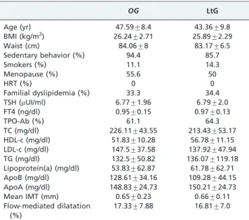
Table 1 shows the baseline information of the patients in the different studied groups. All patients had spontaneous and primary hypothyroidism, and the majority had positive circulating TPOAb (Table 1). No relationship between carotid IMT or brachial FMD and free T4 and TSH was found.
Follow-up data:
TSH and FT4 levels at the end of the study (12 months) are shown in Table 2. No significant changes were observed in BMI and waist circumference (data not shown) or in other laboratory variables (Table 2). The median dose of L-T4 was 44.23¡18.13mg/day.
When comparing the mean changes during the study in the two TPO-Ab patients within the prospective SHT groups, no significant differences in brachial artery FMD were found (LtG, 1.13¡9.5; OG -5.58¡9.59;p= 0.12).
When the patients in each group were subdivided into low ($4 but,8mIU/ml) or high ($8 but,12mIU/ml) TSH
levels, no differences were observed in metabolic or vascular parameters (data not shown).
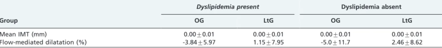
A stratified analysis that considered the presence of any type of dyslipidemia based on NCEP criteria was per-formed. Dyslipidemia was defined as elevated TC, elevated
triglycerides, elevated LDL-c and/or decreased HDL-c. There was a non-significant decrease in FMD in the OG in both subgroups (dyslipidemia present or absent) and a non-significant increase in FMD in the LtG in both subgroups. Changes in brachial FMD and carotid artery IMT after one year of follow-up in the LtG and OG based on the presence or absence of dyslipidemia are shown in Table 3.
DISCUSSION
LT-4 treatment did not significantly improve endothelial function or reduce the carotid IMT in subjects with mild SHT who were similar with respect to age, BMI, smoking, menopausal status, and endothelial function modifiers. After one year, there was a significant decrease in brachial artery FMD in the OG and no difference in LtG, which suggests that thyroid hormone replacement could prevent a decline in FMD in patients with SHT. The exclusion of participants with concomitant hormonal, renal, or liver diseases allowed us to control our analysis for confounding variables and yielded a homogeneous population. In addition, by excluding co-morbid conditions, the mean age was reduced, allowing a smaller sample size for the study.
Few studies have evaluated carotid IMT and the effect of L-T4 replacement in subclinical thyroid dysfunction. Monzani et al.15 were the first to show higher mean IMT values in SHT patients compared with age- and sex-matched controls. The maximal thickness in any particular segment was also measured (maximal IMT) and was higher in the SHT group. We previously reported no significant differences in the mean carotid IMT in a group of subclinical hypothyroid patients compared with a euthyroid group, which suggests that mild SHT is not associated with an increase in cardiovascular risk when assessed by carotid IMT.24,25Monzani et al.25were also the first to evaluate the benefit of six months of L-T4 replacement therapy on IMT in patients with SHT. L-T4 significantly reduced both LDL-c and the mean IMT, suggesting that lipid infiltration of the arterial wall may represent the main mechanism underlying the increase in IMT in patients with SHT. In the present work, we did not find any association between carotid IMT
Table 1 -Baseline data of levothyroxine-treated and observation groups.
OG LtG
Age (yr) 47.59¡8.4 43.36¡9.8
BMI (kg/m2) 26.24¡2.71 25.89¡2.29
Waist (cm) 84.06¡8 83.17¡6.5
Sedentary behavior (%) 94.4 85.7
Smokers (%) 11.1 14.3
Menopause (%) 55.6 50
HRT (%) 0 0
Familial dyslipidemia (%) 33.3 34.4
TSH (mUI/ml) 6.77¡1.96 6.79¡2.0
FT4 (ng/dl) 0.95¡0.15 0.97¡0.13
TPO-Ab (%) 61.1 64.3
TC (mg/dl) 226.11¡43.55 213.43¡53.17
HDL-c (mg/dl) 51.83¡10.28 56.78¡11.15
LDL-c (mg/dl) 147.5¡37.58 137.92¡47.94
TG (mg/dl) 132.5¡50.82 136.07¡119.18
Lipoprotein(a) (mg/dl) 53.83¡62.87 61.78¡62.71
ApoB (mg/dl) 128.61¡34.16 109.28¡44.15
ApoA (mg/dl) 148.83¡24.73 150.21¡24.73
Mean IMT (mm) 0.65¡0.23 0.66¡0.11
Flow-mediated dilatation (%)
17.33¡7.88 16.81¡7.0
Notes: Values are expressed as means¡SD or frequency. The p values were.0.05.
OG, observation group; LtG, levothyroxine group; HRT, hormone replacement treatment; TSH, thyroid-stimulating hormone; FT4, free thyroxine; TPO-Ab, antiperoxidase antibody; TG, triglycerides; HDL-c, high-density lipoprotein cholesterol; TC, total cholesterol; LDL-c, low-density lipoprotein cholesterol; apoA, apoprotein A; apo B, apoprotein B; IMT, intima-media thickness; FMD, flow-mediated vasodilatation.
Table 2 -Initial (baseline) and final (after one year) results of the levothyroxine-treated and observation groups.
Observation group Levothyroxine Group
Baseline 1 year Baseline 1 year
TSH (mUI/ml) 6.77¡1.96b
(4.2-11.0)
6.69¡3,51b,
(3.27-13.5)
6.79¡2.0a,b
(4.04-10.65)
3.02¡0.89a,b
(1.54-4.0)
FT4 (ng/dl) 0.95¡0.15 1.06¡0.15 0.97¡0.13 1.16¡0.18
TC (mg/dl) 226.11¡43.55 227.6¡36.9 213.43¡53.1 208.4¡36.7
HDL-c (mg/dl) 51.83¡10.28 49.61¡9.74 56.78¡11.15 54.36¡12.3
LDL-c (mg/dl) 147.5¡37.58 150.6¡34.74 137.92¡47.94 132.8¡37.5
TG (mg/dl) 132.5¡50.82 137.0¡56.06 136.07¡119.18 106.0¡36.7
Lipoprotein (a) (mg/dl) 53.83¡62.87 48.21¡47.22 61.78¡62.71 58.87¡64.09
ApoB (mg/dl) 128.61¡34.16 125.94¡27.47 109.28¡44.15 105.1¡27.17
ApoA (mg/dl) 148.83¡24.73 136.78¡24.04 150.21¡24.73 144.14¡25.57
Mean IMT (mm) 0.65¡0.23 0,67¡0,18 0.66¡0.11 0.66¡0.15
FMD (%) 17.33¡7.88a,b 13.10¡4.75a,b 16.81¡7.0b 18.52¡7.44b
Values are expressed as means¡SD; TSH values are expressed as the mean¡SD (interquartile range).
a
p,0.05 comparing before and after values within each group.
b
p,0.05 comparing the mean changes between the two prospective SHT groups during the study. For all other values,p.0.05.
and the variables studied. Furthermore, no reduction of carotid IMT was appreciated after one year of L-T4 therapy. The cardiovascular system is a specific target of thyroid hormones (THs); thus, thyroid dysfunction is accompanied by profound changes in cardiovascular hemodynamics.26,27 Both the vasculature and the endothelium play pivotal roles in modulating vascular tone, and both are potential targets of THs.28 Several studies have demonstrated the presence of thyroid receptors in endothelial cells.32,33Some studies have suggested a role for vascular smooth muscle (VSM) cells in mediating the vasodilating effect of T3 infusion in the endothelium in human euthyroid subjects.34,35
Little is known about the interaction of TSH with endothelial cells. The TSH receptor is present in the thyroid gland and, also, in many other sites, such as VSM cells.34 Dardano et al. demonstrated that an acute systemic increase of serum TSH levels by rhTSH injection induced a significant impairment of endothelial vasodilatation in conduit arteries. Few studies have evaluated the relationship between SHT and endothelial dysfunction and the effect of L-T4 replace-ment in the endothelial function of subclinical hypothyroid patients. Lekakis et al.17were the first to describe the negative association between borderline and mild hypothyroidism and FMD. In their study, cholesterol did not differ significantly among groups but tended to be higher in the hypothyroidism and SHT groups, and the authors concluded
that higher cholesterol levels may be associated with endothelial dysfunction. Dagre et al.36 noninvasively assessed the NO-dependent endothelial function of resis-tance arteries in subjects with hypothyroidism of varying severity by measuring the forearm blood flow response during reactive hyperemia via utilizing venous occlusion strain-gauge plethysmography. Endothelial dysfunction was only detected in the microvasculature of patients with overt hypothyroidism. We previously reported no significant differences in brachial FMD in a group of subclinical hypothyroid patients compared with a euthyroid group, which suggests that mild thyroid dysfunction had no adverse effects on endothelial function in the population studied.24 Taddei et al.22showed in a group of subclinical hypothyroid patients that 6 months of euthyroidism by L-T4 replacement increased acetylcholine-induced vasodilation. They also observed a significant correlation between the maximal response to acetylcholine and changes in LDL-c. In a randomized, double-blind, crossover study of L-T4 or placebo for 12 weeks, brachial FMD was significantly improved in the group of patients who took a daily dose of 100mg of L-T4 compared with the placebo group.
Multivariate analysis showed that increased serum FT4 levels were the most significant variable predicting a reduction in TC or improvement in FMD.23 Xiang et al. showed that TSH, FT3 (free T3), LDL-c and Lp(a) levels were
Table 3 -Changes in brachial FMD and mean EIM after one year of follow-up in the L-T4 group and the observation group based on the presence or absence of dyslipidemia.
Dyslipidemia present Dyslipidemia absent
Group OG LtG OG LtG
Mean IMT (mm) 0.00¡0.01 0.00¡0.01 0.00¡0.01 0.00¡0.01
Flow-mediated dilatation (%) -3.84¡5.97 1.15¡7.95 -5.0¡11.7 2.46¡8.62
Values are expressed as the mean changes between the two prospective SHT groups through the study. All otherp.0.05. OG, observation group; LtG, levothyroxine group; FMD, flow-mediated vasodilatation; IMT, intima-media thickness.
significant factors associated with endothelium-dependent arterial dilation in Hashimoto’s thyroiditis patients with euthyroidism.35The lack of FT3 measurement is a limitation of our study and is also important for ruling out the possible inclusion of subjects with non-thyroidal illness syndrome.
The interpretation of the results strongly depends on the effectiveness of substitutive treatment. According to the literature,7in this case, the optimal values for TSH in L-T4 treated patients are unclear. As shown in some studies, it is not uncommon for patients in clinical trials to be outside of the therapeutic target at the end of the study. Ravzi et al.6 demonstrated a significant improvement in cardiovascular risk factors in a group of patients with SHT treated by LT-4, and 27 patients had discordant thyroid function tests during the course of the study.
This study was a randomized open-labeled prospective trial. A double-blind, placebo-controlled study would be a more suitable approach. Another limitation of this study was the small sample size, which makes it difficult to obtain a clear decision about the benefit of L-T4 treatment in SHT patients. More studies with larger patient populations are needed to elucidate this subject more fully.
We did not measure the maximal IMT during the internal carotid artery measurements, which could be considered a limitation of the study. An important issue is which IMT measure (e.g., mean IMT value; maximal IMT value; internal carotid artery from the anterior, lateral, and posterior angle; and composite measures from the left and right side or from different arterial sides) should be used. There is no definite answer to this question, but, for study purposes, all segments should be analyzed.
The brachial artery vasodilation in response to RH was determined by FMD of the brachial artery. FMD is an indirect way of measuring the endothelial function using vascular reactivity. We did not assess endothelium-independent vasodilatation, which could be assessed with a dose-response curve to sodium nitroprusside. While it is important to perform nitrate dilatation to confirm that the findings were not due to media disease, as opposed to endothelial dysfunction, we were unable to perform this evaluation and suggest that it be performed in future works. Some studies that assessed the response to sodium nitroprusside (which specifically acts on smooth muscle cells) have shown similar responses in both groups (control and SHT groups) and a reduced vasodilatory effect of acetylcholine in SHT patients; these results indicate that SHT is characterized by the presence of endothelial dysfunction.17,22
Another limitation of this study is that we did not measure estradiol levels or the duration of menopause. This finding is important because abnormal FMD values can be obtained in older women and women who experienced a long menopausal period. Although both groups were similar in relation to age, the control group was more than four years older than the treated one; thus, the changes in FMD could be related to the development of climacterium. In the present study, the reported mean values for TSH after treatment were in the upper limit of the normal range. Some patients were under-treated, and this finding could explain why we did not find a significant benefit after LT-4 treatment.
CONCLUSION
Our data suggest that thyroid hormone replacement therapy could prevent a decline in FMD with the continuation
of the SHT state in comparison with untreated subjects. Large, prospective, multicenter placebo-controlled trials are neces-sary to further investigate the endothelial physiology of patients with SHT and to define the role of L-T4 therapy in improving endothelial function in patients with SHT.
REFERENCES
1. Ayala A, Wartofsky L. Minimally symptomatic (subclinical) hypothyr-oidism. The Endocrinologist. 1997;7:44-50.
2. Canaris GJ, Manowitz NR, Mayor G, Ridgway EC. The Colorado thyroid disease prevalence study. Arch Intern Med. 2000;160:526-34.
3. Cooper DS. Clinical practice. Subclinical hypothyroidism. N Engl J Med. 2001;345:260-5.
4. Rodondi NR, Aujesky D, Vittinghoff A, Cornus J, Bauer D. Subclinical hypothyroidism and the risk of coronary heart disease: a meta-analysis. Am J Med. 2006;119:541-51
5. Ochs N, Auer R, Bauer D, Nanchen D, Gussekloo J, Cornuz J, et al. Meta-analysis: subclinical thyroid dysfunction and the risk for coronary heart disease and mortality. Ann Intern Med. 2008;148:832-45.
6. Razvi S, Ingle L, Keeka G, Oates C. The beneficial effect of L-thyroxine on cardiovascular risk factors, endothelial function, and quality of life in subclinical hypothyroidism: randomized, crossover trial. The Journal of Clin Endoc Metab. 2007;92:1715-23.
7. Biondi B, Cooper D. The clinical significance of subclinical thyroid dysfunction. Endoc Rev. 2008;29:76-131.
8. Caraccio N, Ferrannini E, Monzani F. Lipoprotein profile in subclinical hypothyroidism: response to levothyroxine replacement, a randomized placebo-controlled study. The Journal of Clin Endoc Metab. 2002;87: 1533-8.
9. Teixeira PF, Reuters VS, Ferreira MM, Almeida CP, Reis FA, Melo BA, et al. Treatment of subclinical hypothyroidism reduces atherogenic lipid levels in a placebo-controlled double-blind clinical trial. Horm Metab Res 2008;40:50-5.
10. Rodondi N, Newman AB, Vittinghoff E, Rekeneire N, Satterfield S, Harris T, et al. Subclinical hypothyroidism and risk of heart failure, other cardiovascular events and death. Arch Inter Med. 2005;165:2460-6. 11. Hak AE, Pols HA, Visser TJ, Drexhage H, Hofman A, and Witteman
JCM. Subclinical hypothyroidism is an independent risk factor for atherosclerosis and myocardial infarction in elderly women: the Rotterdam Study. Ann Intern Med. 2000;132:270-8.
12. Vane JR, Anggard EE, Botting RM. Regulatory functions of the vascular endothelium. N Engl J Med. 1990;323:27-36.
13. Healy B. Endothelial cell dysfunction: an emerging endocrinopathy linked to coronary disease. J Am Coll Cardiol. 1990;16:357-8.
14. Bonetti PO, Lerman LO, Lerman A. Endothelial dysfunction: a marker of atherosclerotic risk. Arterioscler Thromb Vasc Biol. 2003;23:168-75. 15. Monzani F, Caraccio N, Kozakowa M, Dardano F, Vittone A, Virdis S,
et al. Effect of levothyroxine replacement on lipid profile and intima-media thickness in subclinical hypothyroidism: a doubleblind, placebo-controlled study. The Journal of Clin Endoc Metab. 2004;89:2099-106. 16. Nagasaki T, Inaba M, Henmi Y, Kumeda Y, Ueda M, Tahara H, et al.
Decrease in carotid intima-media thickness in hypothyroid patients after normalization of thyroid function. Clin Endocrinol. 2003;59:607-12. 17. Lekakis J, Papamichael C, Alevizaki M, Piperingos G, Marafelia P,
Mantzos J, et al. Flow-mediated, endotheliumdependent vasodilation is impaired in subjects with hypothyroidism, borderline hypothyroidism, and high-normal serum thyrotropin (TSH) values. Thyroid. 1997;7:411-4. 18. Papaioannou GI, Lagasse M, Mather JF, Thompson PD. Treating hypothyroidism improves endothelial function. Metabolism. 2004;53: 278-9. 19. Cikim AS, Oflaz H, Ozbey N, Cikim K, Umman S, Meric M, et al. Evaluation of endothelial function in subclinical hypothyroidism and subclinical hyperthyroidism. Thyroid. 2004;14:605-9.
20. Volzke H, Robinson DM, Schminke U, Lu¨demann J, Rettig R, Felix S, et al. Thyroid function and carotid wall thickness. The Journal of Clin Endoc Metab. 2004;89:2145-9.
21. Erbil Y, Ozbey N, Giris M, Salmaslıog˘lu S, O¨ zarmag˘an S, Tezelman S. Effects of thyroxine replacement on lipid profile and endothelial function after thyroidectomy. Br J Surg. 2007;94:1485-90.
22. Taddei S, Caraccio N, Virdis A, Dardano A, Versari D, Ghiadoni L, et al. Impaired endothelium-dependent vasodilatation in subclinical hypothyroidism: beneficial effect of levothyroxine therapy. The Journal of Clin Endoc Metab. 2003;88:3731-7.
23. Boekholdt MS, Titan SM, Wiersinga WM. Initial thyroid status and cardiovascular risk factors: The EPIC-Norfolk prospective population study. Clin Endocrinol (Oxf). 2009;21:6-9.
24. Cabral MD, Teixeira PFT, Silva NA, Morais FC, Soares DV, Salles E, et al. Normal flow-mediated vasodilation of the brachial artery and carotid artery intima-media thickness in subclinical hypothyroidism. Braz J Med Biol Res. 2009;42:426-33.
in patients with subclinical hypothyroidism. Arq Bras Endocrinol Metabol. 2007;51:472-7.
26. Klein I, Ojaama K. Thyroid hormone and the cardiovascular system: from theory to practice. The Journal of Clin Endoc Metab. 1994; 78:1026-7. 27. Kahaly GJ, Dillmann WH. Thyroid hormone action in the heart. Endocr
Rev. 2005;26:704-28.
28. Corretti MC, Anderson TJ, Benjamin EJ, Celermajer D, Charbonneau F, Creager MA, et al. Guidelines for the ultrasound assessment of endothelial-dependent flow-mediated vasodilation of the brachial artery: a report of the International Brachial Artery Reactivity Task Force. J Am Coll Cardiol. 2002;39:257-65.
29. Meirelles CM, Leite SP, Montenegro CA, Gomes PS. Reliability of brachial artery flow-mediated dilatation measurement using ultrasound. Arq Bras Cardiol. 2007;89:180-3.
30. Garrido KU, Leite S, Montenegro C, Koch H, Soares A. Artery flow-mediated dilatation measurement: study of endothelial function in women in menopause. Rev Bras Ecocardiog. 2008;21:22-6.
31. Soares DV, Spina LD, Brasil RR, Silva EC, Lobo PM, Salles E, et al. Carotid artery intimamedia thickness and lipid profile in adults with
growth hormone deficiency after longterm growth hormone replace-ment. Metabolism. 2005;54:321-9.
32. Dietrich JB, Kuchler-Bopp S, Boutillier S. Expression of thyroid hormone receptors alpha and beta-1 messengers RNA in human endothelial cells. The T3 hormone stimulates the synthesis of the messenger RNA of the intercellular adhesion molecule-1. Cellular and Molecular Biology. 1997;43:1205-12.
33. Baumgartner-Parzer SM, Wagner L, Reining G. Increase by tri-iodothyronine of endothelin-1, fibrobectin and von Willebrand factor in cultured endothelial cells. The Journal of Endocrinology. 1997;154: 231-9.
34. Ojamara K, Klemperer JD, Klein I. Acute effects of thyroid hormone on vascular smooth muscle. Thyroid. 1996;6:505-12.
35. Yoneda K, Takasa N, Hija S. Direct effects of thyroid hormones on rat coronary artery: nongenomic effects of triiodothyronine and thyroxine. Thyroid. 1998;8:609-13.