Gonçalo Alexandre dos Santos Marcelo
Licenciado em Ciências da Engenharia Química
e Bioquímica
Synthetic Optimization Process of
Mesoporous Nanoparticles for Protein
Extraction in Biological Samples
Dissertação para a obtenção do Grau de Mestre em Engenharia
Química e Bioquímica
Orientador: Dr. Elisabete Oliveira (FCT/UNL)
Co-orientador: Dr. José Paulo Mota (FCT/UNL)
Composição do Júri:
Presidente de mesa
Prof. Doutor Mário Eusébio (FCT/UNL)
Orientador
Doutora Elisabete Oliveira (FCT/UNL)
Arguente
Prof. Doutor Jorge Parola (FCT/UNL)
Setembro 2017
Licenciatura em Ciências da Engenharia Química
e Bioquímica
Synthetic Optimization Process of
Mesoporous Nanoparticles for Protein
Extraction in Biological Samples
Dissertação para obtenção do Grau de Mestre em Engenharia
Química e Bioquímica
Orientador: Dr. Elisabete Oliveira (FCT/UNL)
Co-orientador: Dr. José Paulo Mota (FCT/UNL)
Composição do Júri:
Presidente de mesa
Prof. Doutor Mário Eusébio (FCT/UNL)
Orientador
Doutora Elisabete Oliveira (FCT/UNL)
Arguente
Prof. Doutor Jorge Parola (FCT/UNL)
Copyright © Gonçalo Alexandre dos Santos Marcelo, Faculdade de Ciências e Tecnologia, Universidade Nova de Lisboa
A Faculdade de Ciências e Tecnologia e a Universidade Nova de Lisboa têm o direito, perpétuo e sem limites geográficos, de arquivar e publicar esta dissertação através de exemplares impressos reproduzidos em papel ou de forma digital, ou por qualquer outro meio conhecido ou que venha a ser inventado, e de a divulgar através de repositórios científicos e de admitir a sua cópia e distribuição com objetivos educacionais ou de investigação, não comerciais, desde que seja dado crédito ao autor e editor.
‘ It is about the fact that our lives are absurd. There is no God there is no
Morality. Society invents rules to keep us from happiness, but every
minute of every day we are free to decide who to be and how to live. ‘
Tom Spezialy
I would like to express my sincere gratitude to my supervisor Dr. Elisabete Oliveira for the continuous support on my MSc study and research, for her patience, motivation, enthusiasm, laugh and sincerity. Her guidance and insight were essential for the development and finishing of this work. My deepest thanks also go to Prof. José Mota, who co-supervised this work with always a cheering prospect and enthusiasm.
My sincere thanks also go to Prof. Carlos Lodeiro, Prof. José Luís Capelo and Dr. Hugo Santos for their constant advices and wise words. I thank my fellow lab mates from the Bioscope Group for the ever-present stimulating discussions, for their uncountable teachings and for all the fun we have had. I also thank the group for their investment and trust on my abilities and for their continuous care for my growth within the scientific world.
Last but not the least, I would like to thank my family and close friends for their constant support and counselling on the most tougher decisions and situations throughout this stage; especially to my parents Paula Rocha and João Marcelo, for being the wardens of my motivation, and my friends Ana Patricia, Inês Ferreira, Gonçalo Martins and Guilherme Nunes, for making every day easier, happier and for being the source of the most maddening and inspiring ideas.
O trabalho aqui apresentado serve como “prova de conceito” a um sistema capaz de extrair proteínas de meios biológicos, com recurso a nanoparticulas de sílica funcionalizadas.
Apostando num desenvolvimento de raiz, foi tido como primeiro passo a síntese de nanoparticulas de sílica mesoporosas, seguido de uma caracterização física através de análises espectroscópicas de infravermelhos e de difração de raios X, bem como de isotérmicas de azoto a 77 K. Visou-se ainda a modelação do tamanho do poro e da interação partícula-partícula, através do uso respetivo de agentes expansores e de açúcares, de onde resultaram poros de 4 e 5 nm com SBET de 343 e 534 cm2/g e um rácio ótimo de açúcar : partícula de 1:1. Os poros foram funcionalizados com um derivado de rodamina B (RhNHS), com afinidade para com aminas primárias presentes em proteínas, e um máximo de encapsulação de 44 mg/g obtido. Consequentes testes de libertação em diferentes pH demonstraram que uma maior libertação é favorecida por meios ácidos.
Os testes de extração de proteínas foram levados a cabo em condições fisiológicas, para tempos de incubação de 30 minutos e 2 horas. A quantidade incubada (% L) e a capacidade de extração do sistema (mg/g) foram calculados após uma quantificação, por teste de Bradford, dos sobrenadantes. O aumento do rácio proteína : partícula em conjunto com o uso de RhNHS, levam a um aumento na quantidade de proteína adsorvida (464-519 mg/g), bem como na uma seletividade para proteínas ricas em grupos NH2 (i.e. ovalbumina).
Por fim, o sistema foi ainda testado na extração e deteção a olho nu de iões metálicos tóxicos (Hg2+, Cu2+ e Pb2+) de meios aquosos contaminados. A incorporação de uma porfirina e/ou de RhNHS permitiram ao sistema devolver uma resposta colorimétrica aquando a deteção destes iões, com sensibilidade até concentrações de 5 ppm. A sua capacidade de extração foi ainda avaliada por ICP, bem como a sua reversibilidade e reutilização.
Palavras-Chave: nanoparticulas mesoporosas de sílica; RhNHS; tamanho de poro; proteínas;
In this present work, it is proposed the development of a proof of concept for a solid-state device capable of extracting proteins from either artificial or biological media, based on functionalised silica mesoporous nanoparticles.
Following a “from scratch’ approach, cubic-phased SBA-16 silica nanoparticles were firstly synthesised. Their characterisation was achieved by infrared and X-ray diffraction spectroscopic techniques, along with N2 physio sorption isotherms, at 77 K. Pore modulation and dispersion behaviour were controlled using expanding agents and sugar coatings, respectively. Studied pore widths were of 4 and 5 nm, with SBET of 343 and 534 cm2/g, and an optimal sugar : protein ratio for minimal agglomeration was of 1:1 (wt.). Pore functionalisation was attained through the adsorption of a NHS-based rhodamine B derivative (RhNHS), with affinity towards amine groups in proteins. A pore width-independent loading maximum of 44 mg/g was achieved and release tests were performed for different pH values, with acidic conditions favouring a stronger release.
Protein extraction assays were run on PBS 7.4 to mimic physiological conditions, with incubation times of 30 minutes and 2 hours. Loading yields (% L) and capacities (mg/g) were obtained after supernatants quantification by Bradford assay. An increase on the protein : particles ratio (i.e 1:1), as well as the use of RhNHS, had a general positive effect on the total amount of loaded protein (464-519 mg/g) and a selectivity towards NH2 residue-rich proteins (i.e ovalbumin) was also observed.
Lastly, an environmental extension of the work aimed for a colorimetric detection and the extraction of toxic metal ions (Hg2+, Cu2+ and Pb2+), from aqueous and biological media (urine), with porphyrin and RhNHS doped silica nanoparticles successfully working, at naked-eye, down to 5 ppm concentrations. Extracted quantities were quantified by an induced coupled plasma technique. Reversibility and reusability were also tested for both systems.
Keywords: SBA-16 mesopores silica nanoparticles; RhNHS; pore size; protein; porphyrin; toxic metal
G. Marcelo, E. Oliveira, H. M. Santos, José Paulo Mota, J.L. Capelo and C. Lodeiro
“Synthetic optimization process of mesoporous nanoparticles for protein extraction in biological samples”
XXV Encontro Nacional da Sociedade Portuguesa de Química; Lisboa, Portugal – 12-15th July 2017 Type of Contribution: Poster Communication - http://xxvenspq.eventos.chemistry.pt/
Gonçalo Marcelo, Elisabete Oliveira, Sónia Pires, M. Graça P. M. S. Neves, José Paulo Mota, José Luís Capelo, Carlos Lodeiro
“Mesoporous Silica Nanoparticles functionalized with Porphyrins as a reusable solid-state system for metal detection via emission and colorimetric effects “
Jena Symposium on Remediation 2017; Friedrich-Schiller University of Jena, Germany – 5th October 2017
Type of Contribution: Poster Communication
G. Marcelo, E. Oliveira, H. M. Santos, José Paulo Mota, J.L. Capelo and C. Lodeiro
“Development of reusable solid-state chemosensors based on fluorophores for Hg2+ detection in aquatic media”
2nd International Caparica Conference on Pollutant Toxic Ions and Molecules (PTIM); Costa da Caparica, Portugal – 6-9th November 2017
Type of Contribution: Poster and Shot-gun Communication - http://www.ptim2017.com Awards: Excellent Shotgun Communication Award
G. Marcelo, E. Oliveira, H. M. Santos, José Paulo Mota, J.L. Capelo and C. Lodeiro
“Synthetic optimization of mesoporous silica-based nanosystems for protein extraction in biological samples”
2nd International Caparica Christmas Congress on Translational Chemistry (IC3TC); Costa da Caparica, Portugal – 4-7th December 2017
Type of Communication: Poster and Shot-gun Communication - http://www.ic3tc2017.com/
Scientific Paper
Gonçalo Marcelo, Elisabete Oliveira, M. Graça P. M. S. Neves, Sónia Pires, José Paulo Mota, Carlos Lodeiro, José Luís Capelo
“Mesoporous SBA-16 nanoparticles with dual Porphyrin and Rhodamine cargos as new colorimetric probes for toxic metal ions detection / extraction”
i
1
Introduction ... 1
1.1
Motivation ... 1
1.1.1
Mesoporous Silica Nanoparticles ... 1
1.1.2
SBA-16 and Applications ... 6
1.1.3
Mesoporous nanoparticles for protein extraction ... 7
1.1.4
Mesoporous nanoparticles for metal detection ... 9
1.2
Synthetic pathways ... 10
1.2.1
Sol-Gel Method ... 10
1.2.2
Hydrothermal Method ... 13
1.3
Characterisation Techniques ... 14
1.3.1
X-Ray Diffraction ... 14
1.3.2
(Scanning & Transmission) Electron Microscopy ... 15
1.3.3
Dynamic Light Scattering and Zeta Potential ... 15
1.3.4
Fourier Transform Infrared Spectroscopy... 16
1.3.5
UV-Visible Spectroscopy ... 16
1.3.6
1H /
13C Nuclear Magnetic Resonance Spectroscopy ... 16
1.3.7
BET (Brunauer-Emmett-Teller) / BJH (Barrett-Joyner-Halend) Theory ... 17
1.3.8
Bradford Protein Assay... 19
1.3.9
1D PAGE Electrophoresis ... 20
2
Objectives ... 23
2.1
Focus ... 23
2.2
Structure ... 26
3
Synthesis and characterisation of pluronic (SBA-16) mesoporous nanoparticles ... 27
3.1
Synthesis ... 27
3.2
Pore Modulation ... 32
3.3
Additional Characterisations ... 34
ii
4
Synthesis and characterisation of NHS-Rhodamine-based derivative. Encapsulation
studies of luminescent chromophores into mesoporous nanoparticles ... 41
4.1
Synthesis ... 41
4.2
Loading of Rhodamine B-Based Chromophores... 43
4.3
Release Trials ... 47
4.4
Loading of L5 Porphyrin ... 49
4.5
Concluding Remarks ... 50
5
Application of dye-doped mesoporous nanoparticles for extraction of proteins. ... 51
5.1
Proteins Extraction Quantification ... 51
5.2
Proteins Influence over SBA systems stability... 55
5.3
Particle-Protein Interaction Validation ... 55
5.4
Concluding Remarks ... 57
6
Use of dye-doped mesoporous nanoparticles for pollutant detection. Assays in real tap
water and urine. ... 59
6.1
Detection and Extraction Trials with L5@SBA ... 59
6.2
Detection and Extraction Trials with RhNHS@SBA ... 64
6.3
Concluding Remarks ... 65
7
Experimental Procedures ... 67
7.1
Chemicals and Starting Materials ... 67
7.2
Instrumentation ... 68
7.3
Synthesis, Functionalisation and Protein Assays ... 69
7.3.1
SBA-16 Synthesis & Characterisation... 69
7.3.2
SBA-16 Surface Functionalisation ... 70
7.3.3
RhNHS Synthesis & Purification ... 70
7.3.4
RhNHS Doping of SBA-16 Particles ... 71
7.3.5
RhNHS Release from SBA-16 Particles ... 72
7.3.6
Proteins Adsorption ... 72
iii
7.4
Fluorescence Porphyrin-Based SBA-16 nanoparticles for metal sensing ... 76
7.4.1
Metallic Ions Detection/Extraction in DI Water ... 77
7.4.2
Hg 2+ Spikes Detection/Extraction in Urine ... 78
8
General Conclusions & Future Prospects ... 81
v
Figure 1.1 | Spatial arrangement of siloxane (Si-O-Si) and silanol (Si-OH) groups within silica materials matrixes. ... 1 Figure 1.2 | M41S family of mesoporous silica structures, with its members named after their conformation. Adapted from [21]. ... 2 Figure 1.3 | Synthetic procedure for silica nanoparticles, following the LCT mechanism: I - self-assembly and II - cooperative paths. Adapted from [25]. ... 3 Figure 1.4 | Structures of pluronic P123 (EO20PO70EO20) and F127 (EO106PO70EO106) directing agents and some of the resulting mesoporous structures: a) laminar, b) hexagonal and c) cubic arrangements, depending on EO:PO ratios and pluronic concentrations. Adapted from [31]. ... 4 Figure 1.5 | Close view of SBA mesoporous silicas of SBA-15 (left), with microporous connecting its macroporous channels, and SBA-16 (right) side view of its intricate matrix of macroporous channels. Adapted from [31]. ... 5 Figure 1.6 | Pluronic EOxPOyEOx arrangement inside cubic-structured silica material, where x > y. .... 6
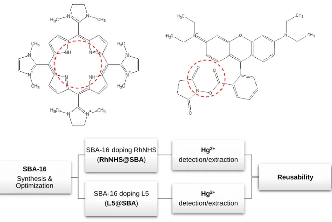
Figure 1.7 | Schematic summary of sol-gel process and its many final products. Uniform particles formation along a precipitation pathway is evident. Adapted from [93-106]. ... 11 Figure 1.8 | Polymerisation pathways and its final products, from 3D reticulated grids to sols. Adapted from [107]. ... 12 Figure 1.9 | XRD schematics. Single λ radiation incidence over a crystalline structure of defined interplanar distance (d), at a defined incidence angle θ. ... 14 Figure 1.10 | Representation of (A) all six types of isotherms and (B) types of hysteresis. Adapted from [107] ... 18 Figure 1.11 | Coomassie Brilliant Blue G-250 dye native form. ... 19 Figure 2.1 | Schematic representation of the here in proposed SBA-16 system for proteins extraction from biological media, using an NHS-modified chromophore. ... 24 Figure 2.2 | Brief schematics of the synthetic path of the here in proposed system. ... 24 Figure 2.3 | Brief schematics of the here in proposed device, from its synthesis to doping - with L5 porphyrin (upper left) and RhNHS (upper right) - and application. Coordination centres signalled by red dotted line. ... 25 Figure 3.1 | FT-IR spectra of SBA-16 M2 T1 sample after (a) and before (b) template removal. A diminish in transmittance % (%T) is highlighted for F127 characteristic 1342 cm-1 and 1279 cm-1 bands; and for CTAB 2919 cm-1 and 2835 cm-1 typical bands. ... 29
vi
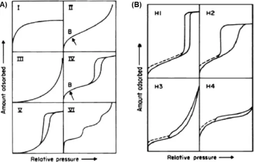
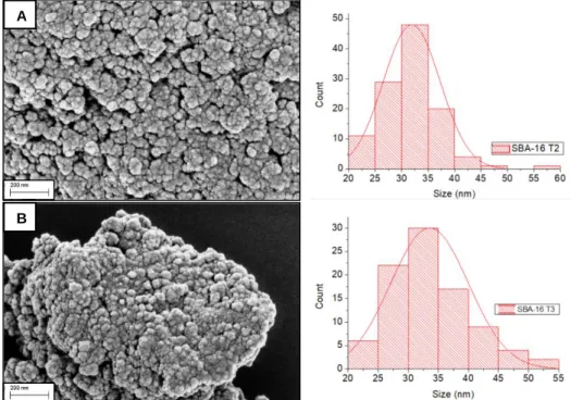
Figure 3.3 | XRD patterns of both T2 (grey) and T3 (black) samples. ... 35 Figure 3.4 | SEM images and particle size distributions of both (A) SBA-16 T2 and (B) SBA-16 T3 samples. ... 35 Figure 3.5 | SEM micrographs of sucrose functionalised SBA-16 nanoparticles (2:1 ratio) with clear (A) disruption of aggregates and (B) occurrence of vesicles between nanoparticles... 38 Figure 3.6 | Brief schematics of the whole synthesis procedure, from particles formation to its surface functionalization, with (a) 1:1, (b) 2:1 and (c) 3:1 sugar : SBA ratios. ... 39 Figure 4.1 | Step-by-step reaction pathway of a DCC catalysed Steglish esterification for the synthesis of RhNHS (right), with the co-production of DCU (left). ... 42 Figure 4.2 | A) Post DCU precipitation RhNHS product solution (in acetone) with its experimental 1H NMR spectrum (A.1), in CDCl3, vs RhNHS theoretical spectrum (A.2). 2.7 ppm NHS peak is highlighted, in red, and DCU interfering peaks, in blue. ... 43 Figure 4.3 | Rhodamine B Isothiocyanate structure. ... 44 Figure 4.4 | Progression on pellet recovery with consecutive washes with MetOH, or MeOH containing solvents ... 44 Figure 4.5 | Time dependent (left) and pH dependent (right) progression of % L RhNHS release profiles in different pH buffers. Trials run on 4.5 PBS, 4.7 PBS and Tris base 8.5 buffers. ... 48 Figure 5.1 | % L (left) and loading capacities (right) of all 30 min incubations on (A) SBA-16 T2 blank, (B) RhNHS@SBA T2 and (C) RhISO@SBA T2 samples, for Protein : Particle wt. ratios of 1:4, 1:2 and 1:1; after Bradford assay quantification. ... 53 Figure 5.2 | % L (left) and loading capacities (right) of all 30 min incubations on (A) SBA-16 T2 blank, (B) RhNHS@SBA T3 and (C) RhISO@SBA T3 samples, for Protein : Particle wt. ratios of 1:4, 1:2 and 1:1; after Bradford assay quantification. ... 54 Figure 5.3 | A) Pellets (+20% lightning & +40% contract) and B) supernatants (+20% lightning & +20% contract) SDS 1D-PAGE electrophoresis digitalized gels, with 30 min (green) and 2h (orange) 1:4 ratio samples. Where, A = BSA, B = LYS, C = Cyt C, D = Myo, E = Hb, F = CA, G = OVA. ... 56 Figure 6.1 | Colorimetric gradient of L5-doped SBA-16 with growing concentrations of Hg2+ aqueous solutions. ... 60 Figure 6.2 | (a) Solid-state emission spectra of L5@SBA upon addition of increasing Hg2+concentrations, at 579 nm; All intensities are normalized for a blank maximum of 5.6x10⁶ csi and presented in a logarithmic scale. (b) Photos of L5@SBA in the presence of different Hg2+ concentrations, under a 365 nm UV lamp. ... 60
vii
365 nm UV lamp. ... 61 Figure 6.4 | Solid-state fluorescence spectra of L5@SBA upon addition of increasing Hg2+concentrations (5, 15, 20, 25, 35, 40, 50 ppm), under excitation at 579 nm; All intensities are normalized for a L5 blank maximum of 5.6x10⁶ csi. ... 62
Figure 6.5 | Solid-state emission spectra of L5@SBA upon addition of increasing (A) Pb2+ (B) Cu2+ or (C) Hg2+ concentrations (5, 15, 20, 25, 35, 40, 50 ppm), under excitation at 579 nm with a 2 nm slit. Superimposed spectra after 10-5 M solutions addition, of all metals. All intensities are normalized for a L5 blank maximum of 5.6x10⁶ csi. ... 62
Figure 6.6| Colorimetric (below) and fluorescent (above) calibration curves of L5@SBA in the presence of different Pb2+ concentrations, ranging from 5 ppm to 50 ppm. Fluorescence photos acquired under a 365 nm UV lamp. ... 63 Figure 6.7 | Colorimetric (below) and fluorescent (above) calibration curves of L5@SBA in the presence of different Cu2+ concentrations, ranging from 5 ppm to 50 ppm. Fluorescence photos acquired under a 365 nm UV lamp. ... 63 Figure 6.8 | Fluorescence spectra and under UV calibration curves of RhNHS@SBA upon addition of increasing Hg2+ concentrations, (A) in water and (B) in urine. Excitation at 556 nm, for a 3 nm slit, and photos under a 365 nm lamp. All intensities are normalized for a maximum of 1x10⁷ csi. ... 64
Figure 6.9 | RhNHS colour shift with pH. ... 65 Figure 7.1 | Experimental procedure for proteins adsorption experiments sample preparation. ... 73 Figure 7.2 | Loading schematics of supernatant or pellet samples on an electrophoresis gel. ... 75 Figure 7.3 | Range of analysed concentrations (in mol/L = M) per type of ionic specie: Hg - Hg (II), Pb - Pb (II) and Cu - Cu (II); and doped chromophore ligand: RhNHS, RhISO and L5. ... 77
ix
Table 1.1 | Main properties of most used silica mesoporous nanoparticles [3, 4, 14, 19, 26, 36, 37]. ... 5 Table 3.1 | Reaction yields, in % (wt.), based on initial weighted silica precursors, and corresponding SBA-16 mass. ... 28 Table 3.2 | Template removal yields, in % (wt.), based on initial as synthesised dry-powder, and corresponding SBA-16 mass. ... 29 Table 3.3 | Zeta potentials, after and before template removal, obtained by dynamic light scattering, in DMSO. ... 30 Table 3.4 | Hydrodynamic average pore diameters, after and before template removal, obtained by dynamic light scattering, in DMSO. ... 31 Table 3.5 | Comparison table between main parameters of SBA-16 M2 T1, T2 and T4 synthesis. No hydrodynamic diameter was included, since PdI values were of approximately 1 and obtained diameters above the range of measurement of the apparatus. ... 32 Table 3.6 | Comparison table between main parameters of SBA-16 M2 T1, T3 and T5 synthesis. No hydrodynamic diameter was included for T5 samples, since PdI values were of approximately 1 and obtained diameters above the range of measurement of the apparatus. ... 33 Table 3.7 | Obtained average hydrodynamic diameters, zeta potentials and corresponding PdI for all T2 samples surface functionalisation, in water, with sugar molecules, at different sugar : SBA ratios of 1:1, 2:1 and 3:1. Sucrose, fructose and trehalose were used as sugar sources. ... 36 Table 3.8 | Summary of all collected data from N2 physio absorption assays, for T1, T2 and T3 SBA-16 samples. Reported data from literature is also display as a comparison element. ... 38 Table 4.1 | Zeta potential of doped SBA-16 M2 T1 samples, with RhISO and RhNHS, in triplicates. . 45 Table 4.2 | Loading yield (% L) and capacity (LC, mg/g) obtained for RhNHS and RhISO doping trials in SBA-16 M2 T2 and T3 samples. Correspondent average (avg.) and standard deviation (σ) represented along the respective test. Chromophore and SBA-16 used masses are detailed, between brackets (in mg), bellow its respective LC. ... 46 Table 4.3 | Obtained loading yields (% L) and capacities (LC, mg/g), for all L5 doping trials in SBA-16 M2 T3 samples. Correspondent average (avg.) and standard deviation (σ) represented along the respective test. Used SBA-16 and L5 masses are also detailed. ... 49 Table 5.1 | Chosen set of proteins for further extraction trials and their correspondent properties. ... 52 Table 7.1 | Weighted proteins and pipetted buffer volume for each stock solution, according to each Protein : RhX@SBA ratio and initial protein concentration (with, X = NHS or ISO). ... 73 Table 7.2 | BSA calibration curve. ... 75
x
incubation sample, [ ] dil., protein the aimed diluted concentration for each analysed sample, V protein the volume to pipette from each protein’s incubation supernatant and V H20 the volume of H2O to pipette. ... 76 Table 7.4 | Weighted amount (in mg) of each metallic triflate specie according to aimed concentration and total volume of stock solutions. ... 77 Table 7.5 | Pipetted volumes for the preparation of all 10-2 to 10-6 M dilutions. ... 77
Table 7.6 | Pipetted volumes (V i, Metal.Sol), from any 10-3 M metallic solution (e.g. Hg2+, Pb2+ or Cu2+), for the preparation of all 10-5 M to 10-4 M (5 ppm to 50 ppm) dilutions. ... 78
xi
CTAB Cetyltrimethylammonium bromide EO Ethylene oxide
EDTA Ethylenediaminetetraacetic acid
EtOH Ethanol
FTIR Fourier Transform Infrared Spectroscopy ICP Induced Coupled Plasma
LCT Liquid Crystal Templating L5 Porphyrin chromophore
L5@SBA Porphyrin chromophore doped SBA silica nanoparticles
MeOH Methanol
MCM Mobil Composition of Matter
NHS N-hydroxysuccinimide PO Propylene oxide RhB Rhodamine B
RhNHS NHS-modified rhodamine-based chromophore
RhNHS@SBA NHS-modified rhodamine-based chromophore doped SBA silica nanoparticles RhISO Rhodamine B isothiocyanate chromophoro
RhISO@SBA Rhodamine B isothiocyanate doped SBA silica nanoparticles SBET Specific BET surface area
SBA Santa Barbara Amorphous SEM Scanning Electron Microscopy TBOS Tetrabutylorthosilicate
TEOS Tetraethylorthosilicate
TEM Transmission Electron Microscopy TMOS Tetrametylorthosilicate
Introduction | 1
1
Introduction
1.1
Motivation
Since early 1970’s the discovery and development of easy-to-control and tuneable inorganic material framework [1-4] has gained its place among most of the top industries and study areas around the world. Namely, the synthesis of silica particles has been one of the most thoroughly sought technique for the creation of many catalytic supports, [2, 5] drug delivery systems, [6] bioimaging probes [7, 8] or sorption devices, just to name a few examples. As it is currently proven and seen, even after 40 years this family of materials finds itself among the most requested building blocks for novel and innovative ideas [9, 10]; thus, vindicating its usage and improvement.
1.1.1 Mesoporous Silica Nanoparticles
Silica materials are known for being nontoxic, thermally / hydrothermally resistant inert materials [3] formed of repetitive tetrahedral SiO4 units. The assemble these ‘monomeric’ units leads to the formation of either crystalline or amorphous structures that can grow from tens of nanometres up to hundreds of millimetres, in diameter [4]. Whilst crystalline structures present a well-defined repetitive arrangement, and thus low available surface-area, amorphous ones are made up of a complex matrix of porous (acting as sponges) that give them large surface-areas of 100 to 1000 m2g-1 [3].
From an structural point a view, the above mentioned ceramic oxide ‘monomer’ (SiO4) promotes the formation of siloxane groups (R-Si-O-Si-R) that are responsible for the growth of silica materials, and of silanol surface groups (R-Si-OH) that control their interaction with the surrounding media, as well as its reactivity (Figure 1.1) [11].
Figure 1.1 | Spatial arrangement of siloxane (Si-O-Si) and silanol (Si-OH) groups within silica materials matrixes.
Introduction | 2
The conditions of growth of these materials along with the right choice of reagents, has allowed for the development of easy and reproducible methodologies [12-14] to tune this arrangement, and consequently its mean pore-size; ranging from microporous (d < 20 Å) [2, 4], to mesoporous (20 Å < d < 500 Å) [3, 15, 16] and up to macroporous (d > 200 nm) [17]. It is then of utter importance to, depending on the material’s application (e.g. biological molecules internalisation), find the best approach to synthesize the right structure.
In 1992, researchers from Mobil Oil Research and Development [2, 18] found a way to synthesize what is now one of the most commonly used mesoporous silica structures, the MCM-41. This one belongs to the M41S family of mesoporous silica materials which have large surface-areas and highly ordered mesoporous molecular sieves [19, 20]. A welcoming addition, at the time, to classical zeolites and still one of the most pertinent materials in industry. M41S family members have had their denomination after their pore spatial arrangement and are briefly described below (Figure 1.2).
MCM-41
Unidimensional hexagonal pore display
MCM-48
Three-dimensional cubic pore display
MCM-50
Parenthetic display of layers of silica and surfactant
Figure 1.2 | M41S family of mesoporous silica structures, with its members named after their conformation. Adapted from [21].
The development of these porous structures within silica nanoparticles (10 nm up to 1000 nm, in diameter) is based on a micellar approach and uses as structuring agents cationic or anionic surfactants (e.g. CTA+), in basic media [4, 18-20, 22]. Besides, surfactants hydrophobic chains size is also an important factor to take in consideration, for it preordains the display of our porous structures (MCM-41, MCM-48 & MCM-50); with long chains favouring layered silica structures [22].
Several models are thought to describe the assembly mechanisms of mesoporous silica nanoparticles, namely the co-condensation (or self-assembly) of opposite charged species of silica precursors and surfactants (S+ I- or S- I+), and the counter-ions mediated mechanism, which depends on ions of opposite charge of that of the head of the surfactant (S+ X- I+) [3, 4, 22] Nonetheless, all of them are based on the principle that surfactants are responsible for the formation of this final porous structure.
Being made of both an hydrophilic end (head) and an hydrophobic tail, when in aqueous solution, surfactants tend to aggregate and form micellar structures, whose form is mediated not only by its concentration but also by extrinsic factors as temperature, pH, presence of electrolytes and solvents [2, 4, 5, 12, 19, 20, 23].
Introduction | 3
It is over such structures that silica precursors will grow and form the rigid silica skeleton of nanoparticles. Thus, it has been proposed by Mobil researchers, that the growing mechanism follows what is called a Liquid Crystal Templating (LCT) [2], where an initial liquid crystal phase composed of semi-ordered structures of surfactant is fixed (and ordered) by the addition of silica precursors species. In detail, when in optimal conditions of pH and temperature, and in the presence of highly concentrated surfactant solutions, this liquid crystal phase self assembles and serves as base for silica aggregation, growth and pore walls formation; however, when in low concentrations of surfactant, silica precursors (Tetraethylorthosilicate – TEOS – or tetramethylorthosilicate – TMOS) also act as directing and structuring agents helping the formation of micelles. To both approaches we call, self-assembly and cooperative pathways, accordingly (Figure 1.3) [24].
Figure 1.3 | Synthetic procedure for silica nanoparticles, following the LCT mechanism: I - self-assembly and II - cooperative paths. Adapted from [25].
In a last step, the as-synthesised nanoparticles are then submitted to template (surfactant) removal techniques, be them calcination at temperatures near 500 ºC or chemical removal with solvents, to obtain empty pores. Even though calcination is known for being one the most used methodology to completely remove templating molecules, it is also taken to be a very sensible heating technique where a strict control over a rising temperature is needed in order to avoid overheating and consequent higher cross-linking degrees between mesoporous materials, as well as lower surface areas, pore volumes and surface available hydroxyl groups. Conversely, solvent-based extraction is a mild and effective surfactant removal technique that is able to not only promote a full recovery of these, but also assure the attainment of homogeneous pore structures along the materials [26]. Fortunately, since its discovery many others mechanisms have been developed and optimised, namely acidic and microwave assisted routes [27, 28].
Summarizing, due to their unique organization, large surface-areas (of approx. 1000 m2g-1), and high porous volumes, this group of materials, MCM-41 included, has found a large spectrum of applications through various research and industrial fields. However, production of pores of 1 – 10 nm width [3, 4,
Introduction | 4
18], as is the case of MCM-41 structures, find no real application in biological / biochemical fields for most molecules of interest (proteins, antibodies, drugs) are themselves some nanometres long. Therefore, along with exhaustive studies developed over this kind of materials at the time, a new family of mesoporous silica nanoparticles was discovered – Santa Barbara Amorphous (SBA) type material. In 1998, large surface-area (600-1000 m2g-1) hexagonal Micelle-Templated Silicas (MTS) were reported by Zhao et al., with pore sizes between 5 - 30 nm [3, 4]. This new kind of silica nanoparticles, named after the institute where they were synthesised (SBA-15), not only showed larger pores, and higher thermal and hydrothermal stability, due to their thick pore walls (with 3.5 – 6.4 nm), but also made use of a non-ionic amphiphilic surfactant – Pluronic P123 (EO20PO70EO20). Besides, pore size and wall thickness can both be tuned by varying reaction temperature and time (from 35 ºC to 140 ºC and 11 to 72 h, for SBA-15 case) [3, 4, 16]. This surfactant, a triblock copolymer with a central Ethylene Oxide (EO) hydrophobic block and two Propylene Oxide (PO) hydrophilic ends [29, 30], is to react with silica precursors through the assistance of a counter-ion pair species – usually denominated as co-surfactant. In detail, it has been proposed that the surfactant S0 (non-ionic) and the positively charged silica precursor I+ interact in acidic medium through an ionic pair S0 H+ X- I+, forming relatively week hydrogen bonds [16]. This leads to the formation of the most various forms of nanoparticles, which might be and not be a good asset.
It has been confirmed that the addition of a salt to the reaction mixture (e.g. cetyltrimethylammonium bromide – CTAB – or NH4F) is a crucial step when chasing sharper shapes and well-defined nanoparticles sizes [16, 31] . Whether in its absence, only undefined shapes and a wide range of sizes are obtained. This corroborates previous statements, that particles morphology is reigned mostly by the S0 X- I+ interaction [32].
Similar to M41S materials, the size and shape of the chosen surfactant is one of the most important factors when designing SBA mesoporous silicas. It has been observed that the EO:PO ratio plays a decisive role, resulting in hexagonal or lamellar dispositions at low ratios (for concentrations of 0.5-1 % (w/w) and 2-5 % (w/w), accordingly) and in cubic conformations at higher ratios (Figure 1.4) [3].
Figure 1.4 | Structures of pluronic P123 (EO20PO70EO20) and F127 (EO106PO70EO106) directing agents and some of the resulting mesoporous structures: a) laminar, b) hexagonal and c) cubic arrangements, depending on EO:PO ratios and pluronic concentrations. Adapted from [31].
P123 F127
Introduction | 5
A most immediate example of this influence is the synthesis of SBA-16 silica mesostructures that, using a more hydrophilic pluronic - F127 - as structuring agent, promotes the formation of body-centered nanocages with cubic arrangement [33] . While SBA-15 particles display well-ordered parallel hexagonal arrangements of pores that allies micro and mesoporosity with thick walls (Figure 1.5), SBA-16 creates a complex matrix of mesoporous channels (with each hollow sphere connected to eight other hollow spheres) that favours the internalisation of biomolecules [26, 31, 34].
Figure 1.5 | Close view of SBA mesoporous silicas of SBA-15 (left), with microporous connecting its macroporous channels, and SBA-16 (right) side view of its intricate matrix of macroporous channels. Adapted from [31].
It is important to recall that this family of materials is also dependent of external conditions such as temperature and pH, which are theorised to interfere directly with the protonation and hydrophilicity of EO blocks [3], as well as silica isoelectric point and condensation rate [35] . With amorphous materials favoured by neutral pHs (~ 7) and crystal ones by strong acidic media (pH ~ 2).
Another improvement that has been reported, is the reusability of pluronic surfactants once template removal is achieved. Washing the as-synthesised nanoparticles with ethanol at 78 ºC allows the organic copolymer to completely dissolve in the solvent and be reused in future synthesis [3] .
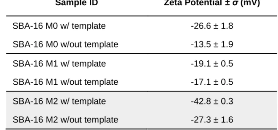
Table 1.1 | Main properties of most used silica mesoporous nanoparticles [3, 4, 14, 19, 26, 36, 37].
MCM-41 SBA-15 SBA-16
Surfactant type Cationic / Anionic Pluronic P123 (ion assisted)
Pluronic F127 (ion assisted)
Medium Basic Acidic Acidic
Pore-size 2-10 nm 5-30 nm 5-30 nm
Pore wall
thickness ~ 1 nm 3.5 – 6.4 nm 3.5 – 6.4 nm Stability Thermal Thermal & Hydrothermal Thermal & Hydrothermal
Mesophase
structures 2D hexagonal (p6mm) 2D hexagonal (p6mm) 3D cubic (lm3n) Main application Zeolites, Catalysts,
Delivery systems
Zeolites, Catalysts, Delivery systems
Zeolites, Catalysts, Delivery & Extraction systems
Introduction | 6
In conclusion, although M41S mesoporous synthesis methodologies are easily reproducible and less time-consuming than those of SBA mesoporous material, these last are able to overcome some of the problems that come with the synthesis of MCM-41; namely, small mean-pore diameters (up to 10 nm), thin pore walls (translating into less thermal or no hydrothermal stability) and low capability of internalisation of biomolecules (Table 1.1).
Therefore, allying large surface-areas, three-dimensional ordered mesoporous structures, thicker framework walls, high thermal and hydrothermal stability, super-large cages, complementary textural porosity with a more favourable mass transfer than that of unidirectional pore systems (due to their three-dimensional channel connectivity) [26] makes SBA-16 structures one of the best candidates for proteins, or other biochemical active species, loading and extraction from biological media.
1.1.2 SBA-16 and Applications
SBA-16 silica nanostructures are considered one of the most interesting mesostructures due to their unique 3D cubic arrangement of mesoporous (lm3m spacing) and the presence of large spherical cages that promote an optimal environment for catalytic reactions (acting as nano-reactors), molecules adsorption and metals/dyes extraction [31, 38-41], to name a few applications.
However, a limited number of SBA-16 synthesis methods have been so far reported, mostly due the narrow range of conditions that these cage-like structures can be obtained [3, 31, 33, 38, 42].
In 1988, along with SBA-15 discovery, it was reported that the existence of long hydrophilic EO chains within the triblock copolymer was crucial for the formation and development of cubic-phased SBA materials (Figure 1.6) [3].
However, improvements on its synthesis only appeared later in 2002, when single crystal particles (with a mean size ∼1 μm) were synthesised, under static conditions, using triblock copolymer F108 (EO133PO49EO133) as structuring agent, assisted by K2SO4 and HCl [42]. Concluding that crystal structures are favoured by highly charged counter-ions, whilst heterogeneous morphologies were attained for single charged ions (e.g. Cl-). It has also been reported by Kleitz et al. the synthesis of SBA-16 using F127 copolymer in a mixture of butanol-water, at low HCl concentrations [43]; where a relationship between silica precursor, co-solvent and cubic lm3m predominant phase is demonstrated. Additionally, the presence of acidic species promotes the already mentioned S0 X- I+ interactions, enhancing tunability of shape and pore as well as ensuring some reproducibility to the process [26].
Introduction | 7
Hence, it was proposed that both morphology and particle size are strongly affected by the regularity of the poly-condensation degree of silica species over surfactant micelles, which in turn is mainly determined by media temperature and pH [32, 36, 44]. A solution that helps regulate shape and easily control morphology was attained when synthesising SBA-16 with the help of ionic co-surfactant CTAB [16, 32, 36].
Unfortunately, a significant issue that arises from the synthesis and strongly hinders SBA-16 large scale applications is its time-consuming reaction (approx. 72h). The use of SDS or CTA+ co-surfactants is thought to shorten micellation time of pluronic species, acting as micellation promoters [33], and has already been demonstrated that it can produce SBA-16 nanoparticles within one hour of reaction [45]. Henceforth, although cubic-phase materials were already a current occurrence in mesoporous structures (MCM-48), the discovery of SBA-16 has brought a new solution to previous hitches of relatively week stability, small pore sizes and low mass transfer capacity. Still a scarcely studied mesoporous structured it has showed so far a very prominent material with a broadening spectrum of applications; from a ‘ship-in-a-bottle’ nanoparticle synthesis catalyst [40], FCC Diesel hydrogenation catalyst [46], O2 producing (H2O oxidation) cells [34], poorly-soluble drug carriers [39, 47], removal devices of neutral, cationic and anionic dyes [38], to biomolecules adsorption & enzymatic reactions supports [16, 48].
1.1.3 Mesoporous nanoparticles for protein extraction
Parallel to advances on mesoporous silicas synthesis, a significant interest in the development of protein encapsulation nano-devices has helped understand the importance of surface control and pore modulation on biomolecules adsorption and desorption mechanisms.
Interactions between proteins and mesoporous silicas are thus of utter interest due to the extensive range of applications that arise from their adsorption / binding, including drug delivery, biochemical synthesis, biosensoring, and bioseparation.
In regards to the binding (chemical interactions) of proteins to ceramic nanomaterials, several resulting conformations have been reported [6, 7, 48-52], ranging from functionalised particle’s surfaces (improving or modulating surface biological interactions, as well as extending these systems’ life span within living organisms), to inner pore walls (acting as specific binding sites, catalysts or imaging probes) and to pore gateways (working as entry molecules, sensing devices or controlled drug delivery systems). Conversely, physical interactions have been usually applied on the development of isolation, preconcentration or purification through-adsorption (surface or pore adsorption) devices for later internalisation on size-selective or affinity chromatography, important techniques in the pharmaceutical industry.
However, a key challenge in designing mesoporous silica vehicles for tailor-made protein interaction is the differentiation of protein interactions at the surface of the particle from those within pores [16, 31, 53, 54]. It has been reported, so far, that this distinction is strongly influenced by a series of factors extending to silica particles’ size [55, 56], pore size [54, 57-59], surface net charge (zeta potential) [60,
Introduction | 8
61], specific surface area [62], protein size and spatial conformation [57, 63, 64], pH [31], terminal functional groups, solubility [61] and protein-protein interactions [16].
From the few publications concerning the designing of tailor-made silica nanoparticles for the effective scavenging of a set of biomolecules from biological media, small pore size (<3 nm) has been highlighted as one of the main variables responsible for the drop of internalised protein amounts and the formation of a surface corona [53, 58]. On this outer structure proteins are prevented from using the protective environment and the large internal surface area provided by pores. Aware of this situation, the production of large pore materials (>5 nm) was proposed by several authors [16, 31, 48, 53, 54] as a way to overcome inaccessibility issues, with Katyiar et al [16] being able to generate silica particles with pores up to 12 nm and achieve loaded quantities (using lysozyme as model protein) above those reported for smaller diameters. Interestingly, Schlipf et al [54] have also demonstrated that although large pores promote indeed a more effective extraction of proteins, a threshold pore size exists for each species with dimensions around that of the biomolecule cross section.
However, the development of such large pore structures has often been associated with micrometric particles, 1-15 um, from which additional dispersion and adsorption issues arise. It has been reported that heterogeneous interactions between particles (intrinsically related to surface zeta potential and a consequence of wide size distributions) promote assorted surface-protein interactions and induce some conformational changes on adsorbate molecules, with significant vicissitudes for particle diameters above 200 nm [65]. Lundqvist et al [66] have also reported a relationship between conformational changes on adsorbate molecules and spherical-like particles size, with larger particles inducing up to six-fold changes on the secondary structure of adsorbed molecules and increasing surface adsorption. Thus, as emphasised by Tu et al [53], a compromise between large pores and smaller (< 1 μm) particles is desired to obtain a high density of entrances and ensure preferential encapsulation of proteins within the mesoporous structure.
Nevertheless, the research for other methods to trace and modulate particle-protein interactions has fallen over the employment of intermediate molecules able to link with those of interest and rapidly give visual insight. The tagging of proteins with chromophores, or fluorophores, whose terminal functional groups showed considering affinity towards peptide side or terminal groups, brought the possibility to easily image and understand the diffusion of proteins within and around particle pores and surface, respectively.
Chromophores and fluorophores belong to a family of organic molecules that, due to the existence of π delocalised bonds along their structure, can interact with near-UV or visible radiation to make their valence electrons easily jump between excited and ground states, resulting in the emission of light (normally coloured) in the same range. Although some of them might lack the possession of linkage elements towards any type of biomolecules, simple organic manipulations on their terminal groups can produce substances with tailored affinities, that can later be internalised in nanometric vehicles designed for protein imprisonment [49-51, 54].
Introduction | 9
The tracing of any peptic activity on the vicinities of nanoparticles is then highlighted by changes in the behaviour or luminescence of the already loaded chromophores. These changes include either inward and outward flows, with direct consequences on internal or external concentrations and luminescence intensity, or shifts in the maximum absorption wavelength, result of possible bindings between target proteins and chromophores.
The development of these methodologies along with considerations of previews studies is a determining mediator towards the improvement of many conventional purification, or identification methodologies and also the designing of new techniques and devices able to selectively interact with target biomolecules.
1.1.4 Mesoporous nanoparticles for metal detection
One of the primary issues that comes with the increasing human activity and ever rising handling of natural resources is the uncontrollable release of unknown quantities of toxic substances to either natural environments [67] or straight to human habitats [68]. The problem aggravates when these human habitats belong in undeveloped countries where sanitation and health care are still precarious services [69-71].
Metallic ionic species present one of the most endangering sources of toxicity and poisoning, due to their relatively easiness to infiltrate and accumulate in other media, especially aqueous ones [67, 68], on which once ingested by small animals, can enter the rest of the food chain. Some examples of such ionic species are Hg2+or Pb2+ ions that, in regards to their oxidation state, easily react with other molecules, including biological molecules specialised to capture in-body metallic atoms [72, 73]. Just to name another example, during fossil fuel or waste burning [73, 74] inorganic Hg usually tends to react with organic molecules and form methylmercury 2+ (CH3Hg2+), which by instance is a bio accumulative environmental toxicant [73]. Even though it can be found in many natural environments [67], including living beings, excesses of this substance usually lead to the formation of strong binding CH3Hg-protein, or CH3Hg-amino acid, complexes that mimic other biomolecules activities and end up inducing malfunctions [75]. Additionally, it has also been linked to increased risk of certain diseases [76, 77] and autoimmune effects in sensitive individuals [78]. These are just some of the reasons to why toxicological studies are of utter importance and of why has there been such a huge development of new tailor-made materials for toxicants fast detection and extraction, especially of Hg2+ species.
Seeking a way to overcome expensive conventional techniques [73], the introduction of nanotechnology has brought, trough out the last two decades, the ability to hastily and selectively sensor toxic ionic substances in not-so expensive ways, by making use of fluorescent / colorimetric probing devices [79-86].
The use of chromophores has been one of the main key-points when selectively coordinating and detecting metallic ions. This family of organic or bioinorganic molecules, suffer visual significant changes when its structure is somehow changed during complexing of ionic species. The magnitude of those optical changes can vary on a long range of concentrations, going from a usual 1 M to 10-3 M
Introduction | 10
scale to ppm’s [80, 81], or even ppb’s [79, 82, 83, 86] (within the limits established for drinkable water by the US Environmental Protection Agency, EPA [73, 87, 88]).
However, the handling of these substances is usually associated with dissolutions in aqueous media (or biological complex media) which difficult any possibility of extraction and cleaning of contaminated areas. This is where nanotechnology acts, by producing solid-state vehicles where the above-mentioned molecules can be immobilised while still interacting with metallic species in solution. Some examples of such vehicles are tailor-made gold nanoparticles, silver / gold-doped paper-based devices or mesoporous silica nanoparticles whose surfaces, channels or pores have been functionalised [80, 82, 83, 89-91].
The difference in sizes (between solute molecules and nanoparticles) allows for an easier separation with the simple use of conventional, cheap and easy separation techniques (e.g. centrifugation). Most recent developments have, nonetheless, produced devices that are sensible to magnetic fields and can be easy isolated from any supernatant, when under its influence [91].
Thus, it is most obvious the importance that these materials have gained within legal organisations’ efforts towards a safer and healthier environment. Prospects of large scale productions are strongly related to economic viability, and the ever-growing interest in the development of cheaper and faster and more efficient devices is the strongest driver for the construction of new approaches; as it is proposed in this work.
1.2
Synthetic pathways
The synthesis of silica based mesoporous materials is known to follow different possible routes, from sol-gel techniques [3, 4, 14, 26] to hydrothermal or microwave assisted ones [27, 28].
1.2.1 Sol-Gel Method
By far the most commonly used synthetic route, and the one used in this work, sol-gel is a homogeneous processing that promotes the full transformation of a solution into an hydrated solid precursor, through mechanisms close to that of condensing polymerisation. An initial ‘monomeric’ solution leads to the formation of a sol (a dispersion of colloidal polymeric-like particles of 1 - 100 nm) with a later establishment of a gel, after sol’s cross-linkage. For silica-based materials these ‘monomeric’ units, or precursors, are usually alkoxysilane species like tetramethyl-, tetraethyl-, tetrabutylorthosilicates (TMOS, TEOS and TBOS, accordingly).
The sol-gel constitutes nowadays a unique pathway to the synthesis of mesoporous silica without the deployment of extreme temperatures. Additionally, such technique is known for allowing the user to manipulate, to a certain extent, some structural parameters [26, 92].
Succinctly, this technique is well known for allowing: 1) the synthesis of highly pure products; 2) tune morphological aspects of the final product, such as pore-size, pore volume and particle size; 3) obtain through a ‘one-pot-route’ tailor made materials, at low temperatures and specific shapes; and 4) control
Introduction | 11
the material’s molecular composition. Moreover, the sol-gel technique is usually divided into four main steps [92-103]:
1) Hydrolysis and polycondensation of alkoxide precursors; 2) Aging;
3) Drying;
4) Thermal treatment.
However, once working at the nanoscale, it is important to point out that for the formation of silica mesoporous nanoparticles the process is to be stopped before sol cross-linkage, promoting sol’s precipitation and resulting in uniform particles (Figure 1.7) [96].
Figure 1.7 | Schematic summary of sol-gel process and its many final products. Uniform particles formation along a precipitation pathway is evident. Adapted from [93-106].
Nonetheless, detailed information on each of the technique’s steps follows below:
1) Hydrolysis and polycondensation of alkoxide precursors
The above-mentioned precursors, or ‘monomers’, that will later form a sol can be from either molecular or non-molecular sources. Whether the first (dissolved metal alkoxides) produce homogeneous products, the later (usually already polymerised colloidal materials) lead to the formation of non-homogeneous solutions. Thus, explaining the common usage of the first [26].
Much like polymeric reactions the formation of a sol is based on a system of reactions where initiation is followed by polymerisation and subsequent growth. The initiation step is usually catalysed by either an acid or a base, that promotes the hydrolysis of silica precursors when in contact with water, giving
Introduction | 12
form to silanol groups. Groups those, that will polymerize (by condensation) and grow into larger structures [92-106].
Briefly,
Initiation reaction (Hydrolysis) ≡Si−OR + H2O → ≡Si−OH + ROH Silanol-Ester Condensation ≡Si−OH + ≡Si−OR → ≡Si−O−Si≡ + ROH Silanol-Silanol Condensation ≡Si−OH + ≡Si−OH → ≡Si−O−Si≡ + H2O Where, R stands for a hydrogen atom or an alkyl group.
It is then obvious this step’s dependence on medium’s pH. Variances on the acidic profile of its surroundings change polymerisation kinetics and, consequently, the final product features (Figure 1.8).
Figure 1.8 | Polymerisation pathways and its final products, from 3D reticulated grids to sols. Adapted from [107].
2) Aging
Once the gel is established, the presence of an evaporating reaction solvent inside pore-like structures leads to several modifications that are susceptible to external variables. Again, these may be temperature, solvent’s pH and aging time [98].
Several phenomena are theorised to occur during aging, namely polycondensation, syneresis, coarsening, and phase transformation. Whilst with polycondensation the gel network keeps growing as long as neighbouring silanols are close enough to react (increasing connectivity of the network and its fractal dimension), syneresis translates into a natural shrinkage of the gel and expulsion of solvent from its pores. Additionally, coarsening is a consequence of dissolution and reprecipitation processes, that result in the irreversible decrease of surface area [92].
Introduction | 13
3) Drying
Generally seen as a simple elimination - by evaporation - of the solvent locked inside silica pores, the drying process might be one of the most important steps of sol-gel method. This technique usually starts by a previous separation of the solid from its aqueous media, either by filtration or centrifugation, accompanied by several washes to remove possible impurities and solvent vestiges.
An already uneventful procedure in crystal drying, this step becomes crucial when dealing with mesoporous materials where an equilibrium between gel shrinkage and solvent evaporation must be met to ensure the material’s initial properties and morphology. For this, vapor-liquid interface formation is to be avoided, as much as possible, in order to reduce capillary pressure and lessen the establishment of ruptures and deformations within porous structure [92, 100, 108].
4) Thermal Treatment
This last step of the sol-gel method is usually related with calcination techniques and allows for the fixation of certain intrinsic variables of the material, such as porous volume, pore-size distribution and specific area. Under inert or ambient atmosphere, with the use of high temperatures, the material undergoes several transformations that lead to surfactant and precursors disintegration, thus helping define material’s porosity and mechanical resistance. Additionally, changes in the material’s crystalline framework might occur, resulting in either amorphous structures or stable crystalline matrixes [92, 101, 108].
In this work, solvent-based template removal techniques were used in detriment of calcination techniques, although a mix of both has been reported as an ideal methodology [109]. Solvent-based template removal procedures usually use alcoholic acidic media (e.g. EtOH/HCl blends) to help dissolve and wash away remnants of any surfactants or swelling agents [3, 4, 14, 109].
1.2.2 Hydrothermal Method
Although not used in this work, this method has gained its importance in nanomaterials synthesis and is now seen as one of the best to ensure reproducibility and homogeneity of its final products. Result of technological developments, this technique found its origins in the 21st century and uses supercritical fluids (usually water) to help with the processing of highly homogeneous and monodispersed materials/nanoparticles.
Briefly, the hydrothermal process is defined by a mix of reactions, under high pressure and temperature conditions, that promote a controllable dissolution and recrystallisation of relatively insoluble materials. This brings important advantages over other synthetic pathways, for it allows the easy monitorisation of the final physio-chemical properties of nanomaterials, through the highly controlled diffusivity in solvents and easy establishment of sharp temperature gradients [110].
The use of supercritical technology, due to its high pressures and temperatures, makes it impossible to follow regular research procedures and requires the usage of very resistant vessels, such as
Introduction | 14
autoclaves. Therefore, autoclaves must be: i) Inertness to acids, bases and oxidizing agents; ii) easy to assemble and disassemble; iii) long enough to obtain sharp temperature gradients; iv) leak-proof with limitless aptitudes to the required temperature and pressure range; and v) rugged enough to bear high pressure and temperature tests, for long periods [110].
1.3
Characterisation Techniques
The determination and quantification of a certain material’s set of properties is only possible through the application of a wide and specific range of practical and analytical techniques. With the right system, textural and spectroscopic profiles can be traced and intrinsic variables/properties determined. The same applies for behavioural and time dependent events, that can be outlined and studied. For the case herein proposed, structural variables – such as specific area, porous volume, pore size distribution, particle size, atomic/molecular composition, crystalline lattice, zeta potential – and spectroscopic parameters – as absorbance and emission – are some of the essential characteristics to be analysed or determined to better understand our system.
1.3.1 X-Ray Diffraction
Commonly referred as XRD, this technique is of utter importance for it allows the determination of a material’s crystalline structure. It makes use of a monochromatic radiation, of fixed and know wavelength (λ), the is beamed over a solid sample (the studied material) that is mainly constituted of randomly oriented crystallites [111].
When the beamed radiation hits the material framework, it is expected that part suffers a diffraction phenomenon, which only occurs along very defined directions that follow angles of 2θ from that of the beamed radiation (Figure 1.9).
Figure 1.9 | XRD schematics. Single λ radiation incidence over a crystalline structure of defined interplanar distance (d), at a defined incidence angle θ.
Introduction | 15
As it is clear from Figure 12, this 2θ angle is wavelength and lattice dependent. This results in a relationship that is translated into Bragg’s Law (Equation 1).
Equation 1 | Bragg’s Law
Where, an integer multiple of the beamed wavelength (nλ), is related to the previously mentioned θ angle of the incident radiation and the crystal lattice distance (d), given by the hkl Miller indexes for crystallographic planes.
Summarizing, due the fact that X-rays are of the same order of magnitude as that of crystallographic bonds and that diffraction phenomena occur, with XRD it is possible to determine a set of unique and specific characteristic that constitute the fingerprint of a crystalline material. These characteristics are the establishment of diffraction planes, interplanar distances (d) and atomic density [111].
1.3.2 (Scanning & Transmission) Electron Microscopy
Both Transmission and Scanning electron microscopy (TEM and SEM, accordingly) are two high resolution techniques, of utter importance, that use a beam of electrons to produce high-res images of materials from the scale of a micron down to several angstroms. The beam of electron interacts with the sample, that must be somehow conductive, producing a flow of transmitted electrons from the same sample that are afterwards collected and transformed into an image.
Whilst, transmission ones direct a broad static beam through an ultrafine sample, allowing us to have an idea of the structure of a material and its inner disposition [112], scanning microscopes are able to, using a sharp beam that scans the sample line by line, create three-dimensional images that are perfect for the determination of particles shape and size [113].
1.3.3 Dynamic Light Scattering and Zeta Potential
Dynamic light scattering (DLS), also referred as Quasi-Elastic Light Scattering (QELS), is a well-established technique when measuring the size and size distribution of molecules / particles within the submicron region (nanometre).
The DLS allows for the characterisation of particles, emulsions or molecules, that have previously been dispersed or dissolved in a liquid. The Brownian motion of these particles in a suspension causes laser light to be scattered at different intensities. The analysis of these intensity fluctuations yields the velocity of the Brownian motion and hence the particle size, using the Stokes-Einstein relationship [114]. Additionally, the Zeta potential is a measure of the electrostatic interactions between particles, and is one of the fundamental variables known to affect stability. Its measurement brings detailed insight into the causes of dispersion, aggregation or flocculation, and can be applied / modulated to improve the formulation of dispersions, emulsions and suspensions [114].
Introduction | 16
1.3.4 Fourier Transform Infrared Spectroscopy
The Fourier transform infrared spectroscopy (FT-IR) technique is used to obtain the infrared absorption or emission spectra, associated with the vibration of intramolecular bonds. This analysis can be applied to either solid, liquid or gas samples and allows for the prediction of molecular structures and validate certain binding stretches.
1.3.5 UV-Visible Spectroscopy
Maybe the most used spectroscopic technique, it takes into account the interaction of UV and Visible radiations with molecules, namely their absorption and emission promoted by the passage of electrons from their fundamental to excited states, vice-versa.
Currently the quantification of this interaction is given by the Lambert-Beer Law that correlates the concentration of a certain molecular specie, in solution, with the intensity and composition of the outcoming spectrum (Transmittance), or its difference to the initial incident radiation (Absorbance). Absorbance and concentration are then directly proportional to each other and follow a linear tendency (Equation 2), within a firm window usually given by a calibration curve.
Where, ε represents the molar extinction coefficient (M-1cm-1), b the optical pathway (cm) and C the above-mentioned concentration of the sample, in solution.
1.3.6
1H /
13C
Nuclear Magnetic Resonance Spectroscopy
It is well known that all nuclei have an electrical charge, associated with an internal magnetic field, and that may or may not have spin. The NMR technique promotes an insight over these assets’ behaviour by using an external magnetic field as energy source, that promotes an energy transfer (equivalent to radio frequencies) and a transition between lower and higher energy levels.
This technique is usually applied to either 1H or 13C isotopes that, contrary to even nuclei isotopes (e.g. 12C), have an overall spin. However, the interactions of such nuclei within molecules differs from that of single atoms due the additional influence of neighbour nuclei and electrons magnetic fields, that result in shifts from their typical absorption frequency. This obligates for the establishment of a reference resonant frequency (usually that of tetramethylsilane, TMS), from which the mentioned shifts can be quantified [115].
Differences, in a part-per-million (ppm) scale, from the ground TMS point are displayed as peaks and give insight on nuclei positions and molecules structure [116].