2011/2012
Ana Sofia Duarte Amado
Cardiovascular Risk Factors in
Acromegaly
Mestrado Integrado em Medicina Área: Endocrinologia Trabalho efetuado sob a Orientação de: Prof. Doutor Davide Maurício da Costa Carvalho Trabalho organizado de acordo com as normas da revista: Hormones Ana Sofia Duarte Amado
Cardiovascular Risk Factors in
Acromegaly
!
!
Cardiovascular Risk Factors in
Acromegaly
Cardiovascular Risk in Acromegaly
Ana Sofia Duarte Amado
Faculty of Medicine of Porto, University of Porto
Corresponding author
Faculty of Medicine Alameda Prof. Hernani Monteiro, 4200-319 Porto, Portugal
Tel: +351 22 551 36 54 Fax: +351 22 551 35 55 E-mail adress: [email protected]
Keywords: Acromegaly, Cardiovascular Risk, Blood Pressure, Glucose Status,
! "!
ABSTRACT
OBJECTIVE: Cardiovascular disease is one of the most important causes of death in acromegalic patients. The aim of this study is to compare the presence of cardiovascular risk factors between acromegalic patients and a control population and evaluate the impact of disease control in these factors. DESIGN: 11 acromegalic patients with active disease and 12 controlled were evaluated for blood pressure, body mass index, fasting glucose, coagulation status and lipidic profile. A group of 11 subjects with non-functioning pituitary adenoma was used as control population.
RESULTS: Significant differences were found in lipidic profile, glucose and coagulation status in both active and controlled patients. Higher levels of fasting glucose and fibrinogen were present in both acromegalic groups. Active patients had higher levels of antithrombin III and controlled ones higher levels of high density lipoprotein cholesterol, compared with controls. Differences between active and controlled acromegalic patients consist in reduction of total cholesterol, low density lipoprotein cholesterol and antithrombin III in the last.
CONCLUSIONS: There is some reduction in cardiovascular risk factors with control of the disease, but possibly without return to basal levels.
! #!
Introduction
Acromegaly is a disease resulting of Growth Hormone (GH) excess production. It is a rare disorder, with an estimated annual incidence of 3 to 4 cases per million inhabitants per year and a prevalence of 40 to 70 cases per million.1 In Portugal, acromegaly’s prevalence and incidence are, respectively, 56.5 cases per million inhabitants and 2.9 cases per million inhabitants per year.2 The mean age at diagnosis is 44 years, after mean symptom duration of 8 years. It seems to affect men and women equally.1
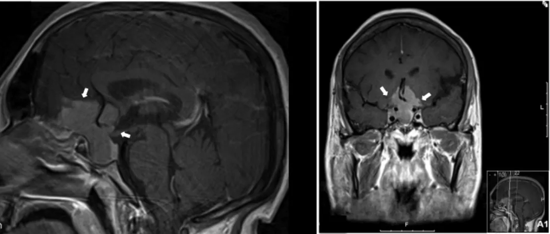
In more than 90% of cases, acromegaly is caused by a monoclonal benign pituitary tumor3 (adenoma – figure 1). GH overproduction will be responsible for the stimulation of hepatic production of insulin-like growth factor 1 (IGF-1) and, elevated levels of both hormones will cause a number of systemic manifestations. Patients usually present with frontal bossing, enlarged nose and lips, prognathism, increased interdental space, oily skin, deep voice and acral enlargement.3 Increased sweating, joint manifestations, carpal tunnel syndrome, visual changes, headache, colon polyps, sleep apnea and reproductive, metabolic and cardiovascular disorders can also be present.4
Diagnosis is based on clinical presentation and levels of fasting or random GH and IGF-1. It is excluded when random GH level is lower than 0.4 µg/L and IGF-1 level is within the age and sex adjusted normal range. If one of these values is altered, an oral 75 g glucose load (OGTT – Oral Glucose Tolerance Test) is taken. To rule out acromegaly, the GH level should fall to 1 µg/L or less.5
The primary treatment of acromegaly is pituitary surgery. Medical therapy can be used after unsuccessful surgery, when the tumor is nonresectable or there are important comorbidities, and while awaiting the effectiveness of radiation therapy. Radiotherapy is left as a last resort, when the tumor continues to grow and is not responsive to medical treatment or when the patient has an aggressive tumor with invasion of local structures.4 Cure is
achieved when IGF-1 levels are in the age and sex adjusted normal range and GH level after OGTT is lower than 1 µg/L.5
! $! Acromegaly is associated with increased mortality when compared with age-matched control population. Cardiovascular, cerebral, cancerous and respiratory disorders are considered to be the main causes of death in acromegaly, being cardiovascular disease the most important one (around 40%). 1, 6 High levels of GH/IGF-1 are associated with increased cardiac mass and diastolic dysfunction, which is followed by systolic dysfunction in long course disease.7, 8 There is also an increase of cardiovascular risk factors in acromegaly. In different studies, the prevalence of hypertension ranges from 18 to 60%.6, 9-12 It is related and involved in worsening of acromegalic
cardiomyopathy13 and an independent mortality predictor in acromegaly.14 The prevalence of impaired glucose tolerance in patients with acromegaly is estimated to be between 16 and 46%.15 Diabetes Mellitus is present in 19
to 56% of acromegalic patients, a higher prevalence than among general population.15, 16 Abnormalities of glucose metabolism are also significantly
correlated with increase in blood pressure in this disease.17
Although there is evidence of an abnormal lipid profile in active acromegalic patients, there is some controversy when trying to determine which values are changed. Total cholesterol (TC) appears unchanged compared with normal population18-20 whereas triglyceride (TG) levels appear
higher18, 21 or unchanged.19 Low21, 22 or unchanged19, 20 levels of high-density
lipoprotein cholesterol (HDL-C) were also found, as well as the presence of small and/or dense low-density lipoprotein cholesterol (LDL-C).19, 22
Acromegalic patients have lower visceral and subcutaneous fat mass and increased intermuscular fat mass, which may be related with insulin resistance in this disease. It is also associated with increased lean body mass.23, 24
Some studies reveal disruption of coagulation and fibrinolytic system. Fibrinogen levels seem to be consistently higher,25-29 whereas plasminogen activator inhibitor-1 (PAI-1) is either elevated27 or unchanged28, 29 in
acromegalic patients. There is some evidence of the presence of higher levels of antithrombin III (ATIII), lower levels of protein S and unchanged levels of protein C and coagulation factor VIII.26, 27 C-reactive protein, an acute phase
! %! Studies report amelioration of cardiac function and some cardiac risk factors with complete or partial biochemical control of acromegaly.28, 31-34 There seems to be a decrease in blood pressure (BP) with control of the disease.11, 28, 31, 33, 34 However, changes in metabolic parameters are not well
established. Some studies show increase in insulin sensitivity and better glucose regulation,28, 32, 34 while others show no modification in majority of
patients.30, 35 As to lipidic profile, some studies can’t find any difference between active and controlled patients,30 while others report increase in HDL-C and decrease in THDL-C, LDL-HDL-C and TG.22, 28 Levels of fibrinogen were found to
be either similar25, 26 or reduced28, 29 and levels of CRP were found to be either similar26 or increased28 after control of acromegaly. There is some
evidence of decrease in PAI-128 and maintenance of Factor VIII levels.26
As discussed above, there are no certainties about the changes in cardiovascular risk factors with control of acromegaly. Also, there are few studies approaching some risk factors, specially coagulation status. The aim of this study is to compare the presence of cardiovascular risk factors between acromegalic patients and a control population (non-functioning pituitary adenomas) and to evaluate the changes in these factors with control of the disease. Body mass index (BMI), blood glucose, BP, TC, HDL-C, LDL-C and TG will be compared among each group. Protein S, Protein LDL-C, Antithrombin III, Factor VIII, Fibrinogen and genetics of PAI-1 will also be evaluated and compared.
Subjects and Methods
This study was approved by the Ethical Committee for Health of Hospital de São João (HSJ). It was a case-control study, performed in patients selected from the Endocrinology department of HSJ database. All patients included signed an informed consent form.
Subjects
Subjects were selected from Endocrinology department database. Patients who had died, whose follow up was lost or had the last surgery more than 10 years ago, were excluded. Patients without data available on computer
! &! internal network and without laboratory analysis made in the previous year were also excluded. Selected subjects were summoned by telephone call and those who refused, couldn’t come to the hospital or didn´t answer were also excluded from the study.
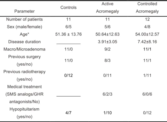
This study included 23 acromegalic patients (table 1), 11 (5 men/6 women) with active disease and 12 (4 men/8 women) with controlled disease. Mean age was 50.64±12.63 in active group and 54.00±12.57 in controlled disease group. In both groups, most patients had macroadenomas at diagnosis. In active disease group, there were 3 newly diagnosed patients, who didn´t make any kind of treatment to the date of study. All other patients in this group underwent previous surgery and had necessity of medical therapy after that. Among controlled patients, only one was treated with radiotherapy and medical treatment. All other patients had previous surgery, 6 of them not having the need for medical therapy.
Eleven controls (6 men/ 5 women) with non-functioning pituitary adenomas were evaluated. They were diagnosed with non-functioning adenoma, which has no hormonal hypersecretion. Elevated urinary free cortisol or cortisol after suppression test with dexamethasone, hyperprolactinemia, elevated thyroxine with high levels of thyroid-stimulating hormone or hypersecretion of GH were not proven. Diagnosis was confirmed with histological analysis after surgical resection. Mean age in this group was 51.36±13.76.
Study Protocol
Disease status was evaluated by IGF-1 levels and GH suppression after OGTT, when appropriate. Active disease was defined as IGF-1 levels over sex and age adjusted normal range or GH after OGTT over 1 µg/L. IGF-1 is expressed as percentage of upper limit of normal range (ULN).
Anthropometric characteristics (height and body mass) were obtained from the last consultation register and body mass index (BMI) was calculated (body mass/height2). Blood pressure was also obtained from the same consultation and was measured by blood pressure monitor.
Last available laboratory analysis of each patient were consulted and blood levels of glucose, total cholesterol (TC), low-density lipoprotein cholesterol
! '! (LDL-C), high-density lipoprotein cholesterol (HLD-C) and triglycerides (TG) were collected.
To evaluate coagulation status of included patients, blood samples were taken and levels of fibrinogen, protein S, protein C, AT III and coagulation factor VIII were measured. An evaluation of plasminogen activator inhibitor 1 (PAI-1) genetic polymorphisms was also made.
For statistical purposes, besides levels measured alone, patients were also categorized as having or not hypertension, hyperglycemia and hyperlipidemia. Hypertension was defined as SBP!140 and/or DBP!90, or being medicated for hypertension. Hyperglycemia was defined as having fasting glucose over 110 mg/dL or being medicated for diabetes mellitus. Hyperlipidemia was defined as having hypercholesterolemia and/or hypertriglyceridemia (TC>200; LDL-C>130; HDL-C<60; TG>150).
Laboratory Analysis
Blood samples from acromegalic patients and controls were taken by venopunctures. Fibrinogen was measured by Clauss Method and AT-III, Protein C and Protein S were measured by functional methods, using reagents obtained from Diagnostica Stago (Asnieres-sur-Seine, France). Factor VIII levels were measured by coagulometric methods (Siemens Healthcare Diagnostics, Newark, Germany).
For 4G/5G PAI-1 gene polymorphisms, DNA was extracted using QIAamp (Hilden, Germany) and real-time polymerase chain reaction (PCR) using LightCycler (Roche Molecular Biochemicals, Mannheim, Germany) was performed. A hybridization probes technique (sequence-specific fluorescent detection with oligonucleotide hybridization probes that are coupled to suitable fluorophores) was applied. A mix with 2 µl genomic DNA (100 ng), 10.8 µl H2O, 1.6 µl MgCl2 (3 mM), 1 µl of each primer (10 µmol), 0.8 µl of each probe (5 µmol) and 2µl DNA-Master hybridization probes was used. Oligonucleotide primers and probes were synthesized by TIB MOLBIOL (Berlin, Germany) and cycling conditions involved initial denaturation (958°C for 600 s), followed by 45 cycles with denaturation at 958°C for 0 s, hybridization at 598°C for 5 s and extension at 728°C for 2s. Melting curves were obtained by slowly heating the sample to 758°C. Melting points of the two alleles were at 598°C
! (!
and 658°C. For better differentiation of heterozygotes, amplified products were run on agarose gel.36
Serum glucose, TC, LDL-C, HLD-C and TG were measured by conventional methods with an Olympus AU5400! automated clinical chemistry
analyzer. (Beckman-Coulter!, Izasa, Porto, Portugal). IGF-1 was measured by
solid phase, enzyme labelled chemiluminescent immunometric assay and GH by solid phase, two-site chemiluminescent immunometric assay (Immulite 2000®).
Statistics
Statistical analysis was made in Statistical Package for Social Sciences (SPSS). Values were tested for normality distribution by Kolmogorov-Smirnov and Shapiro-Wilk tests and log-transformed to achieve normal distribution when necessary. Parametric unpaired t-test was used to compare the groups. Spearman Rank Order Correlation coefficient was used to look for association between levels of IGF-1 or GH and variables in study. Pearson’s correlation was used to search association among the variables studied. In order to find if there was association between being in a group and having hypertension, hyperglycemia or hyperlipidemia, Chi-square test was used. P values under 0.05 were considered significant.
Results
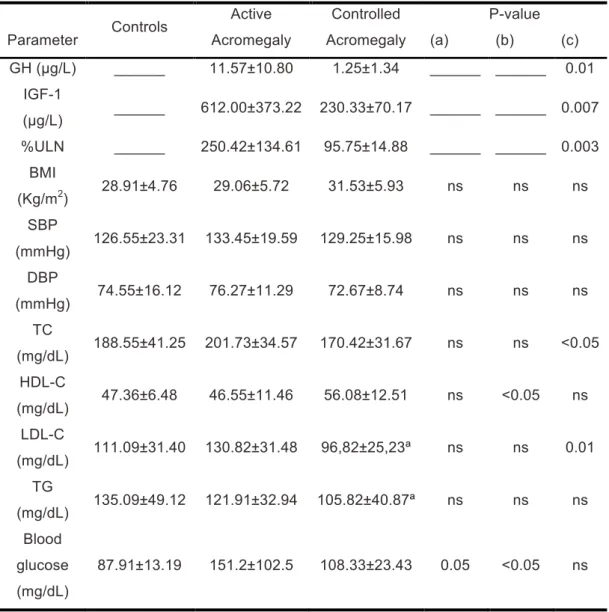
Table 2 summarizes comparison (GH, IGF-1 levels, %ULN, BMI, BP, lipidic profile and glucose status) among the three evaluated groups. Patients with active disease, as expected, had significantly higher levels of GH and IGF-1 than controlled ones.
BMI and levels of SBP and DBP were not significantly different among the three groups, although they were slightly higher in acromegalic patients. Active group had the highest mean blood pressures, and controlled group the highest levels of BMI. However, DBP was lowest in controlled acromegaly group.
TC was slightly higher in active patients than controls, but the result was not statistically significant. When compared with controlled patients, the
! )! difference was significant, with the active group having higher levels. HDL-C was higher in acromegalic patients, being significantly different between controlled patients and controls. In what concerns LDL-C, it was significantly lower in controlled patients when compared with active acromegalic ones. Levels were slightly higher in active patients than controls. Triglycerides were not significantly different among the three groups, being slightly higher in control group.
Blood glucose was significantly higher in controlled acromegaly group and higher with borderline significance in active acromegaly group, when compared separately with control group. No difference was found between active and controlled acromegaly groups (although somewhat higher in active group).
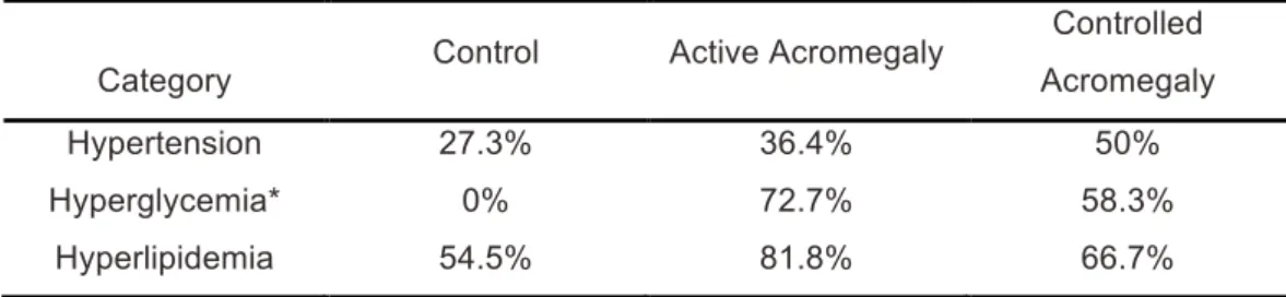
When categorizing patients as having hyperglycemia, hypertension and hyperlipidemia (table 3), higher percentages of all three variables were found in acromegalic patients when compared with controls. Hypertension prevalence was higher in controlled acromegalic group. Hyperglycemia and hyperlipidemia had the higher percentages in active disease group. However, significant correlation between those categories and the three studied groups was found only in hyperglycemia (p=0.002).
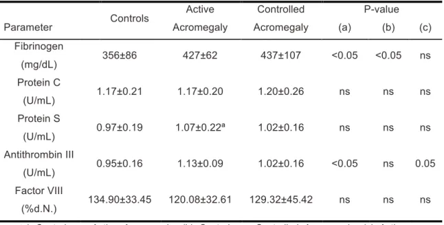
Coagulation status of the three evaluated groups is summarized on table 4. Levels of protein C and S were similar among the groups. As to fibrinogen levels, these were significantly higher in acromegalic groups when compared with control group. There were no significant differences in these values between active and controlled patients (controlled patients having slightly higher levels). ATIII was significantly higher in active acromegaly when compared with control group and when compared with controlled acromegaly. There was no difference between control and controlled acromegaly groups. Although levels of coagulation factor VIII were slightly higher in controls than controlled acromegalic patients and in controlled acromegalic than active acromegalic patients, these values were not significantly different.
When considering all acromegalic patients, significant positive correlation was found between ATIII and IGF-1 levels (r=0.654; P=0.001) and ATIII and GH levels (r=0.498; P=0.013). Dividing acromegalic patients in active and controlled groups, negative correlation was found between TC and GH in the
! *+! active group (r=-0.675, P=0.032) and positive correlation was found between LDL-C and IGF-1 levels in controlled disease group (r=0.630, P=0.038). Within the active acromegaly group, a negative correlation was found between GH and fibrinogen levels (r=-0.610; P= 0.046).
A positive, significant correlation was found between glucose levels and BMI (r=0.478;P=0.005) and glucose and SBP (r=0.428; P=0.013). There was also found positive significant correlation between TC and LDL-C, TC and DBP, HDL-C and TG, LDL-C and DBP, SBP and DBP, BMI and SBP, protein C and DBP, protein C and HDL-C, coagulation factor VIII and fibrinogen.
No correlation was found between hypopituitarism and the variables studied.
Genetic polymorphisms of PAI-1 were evaluated. In control group, 54.5% were heterozygotes, 27.3% homozygotes by deletion and 18.2% homozygotes by insertion. Within active acromegaly group, 36.4% were heterozygotes, 18.2% homozygotes by deletion and 36.4% homozygotes by insertion (genetic polymorphism was not available for one patient). In controlled acromegaly group, 58.3% were heterozygotes, 25.0% homozygotes by deletion and 16.7% homozygotes by insertion. No differences among the groups were found.
Discussion
Since patients with non-functioning pituitary adenoma have no hormones production, these were used as control subjects.
In this study, prevalence of hypertension in both acromegalic groups was within the range found in other studies (18 to 60%).6, 9-12 Although not significant, BP levels and prevalence of hypertension were slightly higher in acromegalic groups than in control group, results previously found in other studies.10 When comparing active and controlled acromegalic patients, BP levels seem to be slightly lower in controlled group, but hypertension prevails over active group. This happens probably because a higher percentage of patients in controlled group were taking anti-hypertensive medication. The tendency to slightly lower levels of BP in controlled acromegalic patients is in concordance with other studies, that show higher levels in active disease than
! **! in controlled disease.28, 31, 33, 34 One study shows lower frequency of
hypertension in active than in controlled acromegalic patients.11 As in other studies, no correlation was found between IGF-1 and GH levels and BP.10, 17, 33 Fedrizzi et al. found no correlation among these values when including all
acromegalic patients (active and controlled), but found correlation between SBP and IGF-1 levels in active acromegalic patients.11
BMI was reported as being higher in acromegalic patients than controls in several studies.25, 37, 38 In the present study, no significant difference was found in BMI, with a slight tendency for controlled patients to have higher values, results that are in agreement with other studies.32, 39 However, there is evidence that acromegalic patients have different body composition: lower visceral and subcutaneous fat mass and increased intermuscular fat mass, with an increase in lean body mass and body water.23, 24 These changes probably make BMI less indicated when trying to deduce body composition or obesity presence in these patients, suggesting other measurements should be used.
Prevalence of Diabetes Mellitus has been reported as being higher among acromegalic patients than in general population.15, 16 Higher insulin resistance and higher glucose and insulin levels in acromegalic patients compared with healthy subjects were also reported.26, 38 Findings in this study show
significantly higher levels of fasting glucose and prevalence of hyperglycemia in all acromegalic patients when compared with control subjects. Although there seems to be a tendency for controlled acromegalic patients to have lower levels of fasting glucose, this difference is not statistically significant. Studies show different results after treatment of acromegaly: some report increase in insulin sensitivity and amelioration of glucose regulation;28, 32, 34 others show no modification in glucose, insulin and insulin resistance.30, 35
The results here reported suggest that there is an improvement in fasting glucose in controlled patients, but with glucose status remaining worse than in control subjects, which should be taken in account when evaluating acromegalic patients, even when having disease control. A significant correlation between fasting glucose levels and SBP was found, an association also previously established.15, 17
! *"! Comparing lipidic values between control and active acromegaly groups, there appears to be higher TC and LDL-C, similar HDL-C and lower TG levels and higher hyperlipidemia prevalence. Yet, these values are not statistically significant. Other authors have found TC levels to be similar, HDL-C lower or unchanged and TG higher or unchanged when compared with a control population18-22. Results regarding LDL-C appear to report either similar or
higher levels in acromegalic patients;19, 20, 22 in these studies, higher levels of small and/or dense LDL-C were consistently reported.19, 22 Controlled acromegalic patients appear to have a better lipidic profile with significant lower levels of TC and LDL-C than active patients. When compared with control population, there seems to be a better lipidic profile with significant higher levels of HDL-C. However, there is a higher percentage of hyperlipidemic controlled patients, because more of them were under anti-dyslipidemic medication. This suggests close attention and control of dyslipidemia in acromegalic controlled patients is effective and probably addictive to control of the disease. Studies reported increase in HDL-C and decrease in TC, LDL-C and TG with control of the disease, supporting the present findings.22, 28
Within active acromegaly group, there seems to be a significant negative correlation between GH and TC levels. When considering controlled acromegaly group there is a positive association between IGF-1 and LDL-C levels. Maison et al. suggest different effects of GH and IGF-1 on lipidic profile.40 One study reports that administration of GH antagonist in acromegaly increase TC41 and another that GH is responsible for lowering
LDL-C by accelerating its elimination rate.42 Szkodziñski et al, report that administration of statins and low fat diet in hypercholesterolemic patients reduce IGF-1, TC and LDL-C.43
Different studies find elevated levels of fibrinogen in acromegalic patients, with decrease of those levels after treatment.25-29 Residual risk with higher
levels in treated patients than controls was found in some studies.25, 28, 29 In the present study, higher levels of fibrinogen were also found in both active and controlled acromegaly groups when compared with control group. No differences were found between active and controlled group. Few studies evaluate the role of protein C, protein S and ATIII in acromegaly. Erem et al
! *#! showed higher levels of ATIII, lower levels of protein S and no difference in protein C levels between acromegalic patients and a control population27. In this study, higher levels of ATIII were found in active acromegalic patients when compared with both control population and controlled patients, with no differences found between control and controlled acromegaly groups. Similar levels of proteins C and S were found among the three groups. Levels of coagulation factor VIII were similar in evaluated groups, as in other studies that found no significant difference in factor VIII levels between acromegalic patients and control subjects or between remission and active disease.26, 27 High fibrinogen levels in acromegalic patients suggest the existence of a pro-coagulation status in acromegaly, with residual risk in controlled disease. However, ATIII levels may be elevated in response to high fibrinogen, counterbalancing its effect. Coagulation status in acromegalic patients should be further studied.
PAI-1 polymorphisms were no different among the three groups, which suggest no genetic predisposition for this factor is present in acromegalic patients.
In conclusion, patients with active acromegaly have subtle changes in BP, and relevant changes in lipidic profile, glucose and coagulation status. With control of the disease, there is amelioration of these parameters, but probably with a residual risk superior to control subjects. Although trying to deduce cardiovascular risk, this study is not able to access morbidity and mortality risk in acromegalic patients.
Acknowledgements
We would like to express our gratitude to Prof Fernando Araujo, Drª Manuela Carvalho and Drª Diana Gonçalves of the Imunohemoterapy Department for the collaboration and suggestions regarding the research protocol.
! *$!
References
1. Holdaway IM, Rajasoorya C. 1999;Epidemiology of acromegaly. Pituitary. 2(1):29-41. Epub 2000/11/18.
2. Paiva I Ribeiro C GL, Carvalho D, Sequeira Duarte J, Barreiros E., editor. Acromegaly Portuguese epidemiology data. Annual meeting of the american endocrine society; 2006.
3. Chanson P, Salenave S. 2008;Acromegaly. Orphanet journal of rare diseases. 3:17. Epub 2008/06/27.
4. Scacchi M, Cavagnini F. 2006;Acromegaly. Pituitary. 9(4):297-303. Epub 2006/11/02.
5. Giustina A, Barkan A, Casanueva FF, et al. 2000;Criteria for cure of acromegaly: a consensus statement. The Journal of clinical endocrinology and metabolism. 85(2):526-9. Epub 2000/02/26.
6. Mestron A, Webb SM, Astorga R, et al. 2004;Epidemiology, clinical characteristics, outcome, morbidity and mortality in acromegaly based on the Spanish Acromegaly Registry (Registro Espanol de Acromegalia, REA). European journal of endocrinology / European Federation of Endocrine Societies. 151(4):439-46. Epub 2004/10/13.
7. Sacca L, Napoli R, Cittadini A. 2003;Growth hormone, acromegaly, and heart failure: an intricate triangulation. Clinical endocrinology. 59(6):660-71. Epub 2004/02/21.
8. Colao A, Marzullo P, Di Somma C, Lombardi G. 2001;Growth hormone and the heart. Clinical endocrinology. 54(2):137-54. Epub 2001/02/24.
9. Bondanelli M, Ambrosio MR, degli Uberti EC. 2001;Pathogenesis and prevalence of hypertension in acromegaly. Pituitary. 4(4):239-49. Epub 2002/12/28.
10. Vitale G, Pivonello R, Auriemma RS, et al. 2005;Hypertension in acromegaly and in the normal population: prevalence and determinants. Clinical endocrinology. 63(4):470-6. Epub 2005/09/27.
11. Fedrizzi D, Rodrigues TC, Costenaro F, Scalco R, Czepielewski MA. 2011;Hypertension-related factors in patients with active and inactive acromegaly. Arquivos brasileiros de endocrinologia e metabologia. 55(7):468-74. Epub 2011/12/08.
12. Minniti G, Moroni C, Jaffrain-Rea ML, et al. 1998;Prevalence of hypertension in acromegalic patients: clinical measurement versus 24-hour ambulatory blood pressure monitoring. Clinical endocrinology. 48(2):149-52. Epub 1998/05/14.
13. Lopez-Velasco R, Escobar-Morreale HF, Vega B, et al. 1997;Cardiac involvement in acromegaly: specific myocardiopathy or consequence of systemic hypertension? The Journal of clinical endocrinology and metabolism. 82(4):1047-53. Epub 1997/04/01.
14. Holdaway IM, Rajasoorya RC, Gamble GD. 2004;Factors influencing mortality in acromegaly. The Journal of clinical endocrinology and metabolism. 89(2):667-74. Epub 2004/02/07.
15. Kreze A, Kreze-Spirova E, Mikulecky M. 2001;Risk factors for glucose intolerance in active acromegaly. Brazilian journal of medical and biological research = Revista brasileira de pesquisas medicas e biologicas / Sociedade Brasileira de Biofisica [et al]. 34(11):1429-33. Epub 2001/10/23.
! *%! 16. Shaw JE, Sicree RA, Zimmet PZ. 2010;Global estimates of the prevalence of diabetes for 2010 and 2030. Diabetes research and clinical practice. 87(1):4-14. Epub 2009/11/10.
17. Jaffrain-Rea ML, Moroni C, Baldelli R, et al. 2001;Relationship between blood pressure and glucose tolerance in acromegaly. Clinical endocrinology. 54(2):189-95.
18. Nikkila EA, Pelkonen R. 1975;Serum lipids in acromegaly. Metabolism: clinical and experimental. 24(7):829-38. Epub 1975/07/01.
19. Tan KC, Shiu SW, Janus ED, Lam KS. 1997;LDL subfractions in acromegaly: relation to growth hormone and insulin-like growth factor-I. Atherosclerosis. 129(1):59-65. Epub 1997/02/28.
20. Twickler TB, Dallinga-Thie GM, Zelissen PM, Koppeschaar HP, Erkelens DW. 2001;The atherogenic plasma remnant-like particle cholesterol concentration is increased in the fasting and postprandial state in active acromegalic patients. Clinical endocrinology. 55(1):69-75. Epub 2001/07/17. 21. Beentjes JA, van Tol A, Sluiter WJ, Dullaart RP. 2000;Low plasma lecithin:cholesterol acyltransferase and lipid transfer protein activities in growth hormone deficient and acromegalic men: role in altered high density lipoproteins. Atherosclerosis. 153(2):491-8. Epub 2001/02/13.
22. Arosio M, Sartore G, Rossi CM, et al. 2000;LDL physical properties, lipoprotein and Lp(a) levels in acromegalic patients. Effects of octreotide therapy. Italian Multicenter Octreotide Study Group. Atherosclerosis. 151(2):551-7. Epub 2000/08/05.
23. Freda PU, Shen W, Heymsfield SB, et al. 2008;Lower visceral and subcutaneous but higher intermuscular adipose tissue depots in patients with growth hormone and insulin-like growth factor I excess due to acromegaly. J Clin Endocr Metab. 93(6):2334-43.
24. Katznelson L. 2009;Alterations in body composition in acromegaly. Pituitary. 12(2):136-42. Epub 2008/03/29.
25. Kaluzny M, Bolanowski M, Daroszewski J, Szuba A. 2010;The role of fibrinogen and CRP in cardiovascular risk in patients with acromegaly. Endokrynol Pol. 61(1):83-8.
26. Potter BJ, Beauregard C, Serri O. 2008;Serum markers of cardiovascular risk in patients with acromegaly before and after six months of treatment with octreotide LAR. Pituitary. 11(1):49-53.
27. Erem C, Nuhoglu I, Kocak M, et al. 2008;Blood coagulation and fibrinolysis in patients with acromegaly: increased plasminogen activator inhibitor-1 (PAI-1), decreased tissue factor pathway inhibitor (TFPI), and an inverse correlation between growth hormone and TFPI. Endocrine. 33(3):270-6.
28. Delaroudis SP, Efstathiadou ZA, Koukoulis GN, et al. 2008;Amelioration of cardiovascular risk factors with partial biochemical control of acromegaly. Clinical endocrinology. 69(2):279-84.
29. Landin-Wilhelmsen K, Tengborn L, Wilhelmsen L, Bengtsson BA. 1997;Elevated fibrinogen levels decrease following treatment of acromegaly. Clinical endocrinology. 46(1):69-74. Epub 1997/01/01.
30. Sesmilo G, Fairfield WP, Katznelson L, et al. 2002;Cardiovascular risk factors in acromegaly before and after normalization of serum IGF-I levels with the GH antagonist pegvisomant. J Clin Endocr Metab. 87(4):1692-9.
! *&! 31. Colao A, Auriemma RS, Galdiero M, Lombardi G, Pivonello R. 2009;Effects of initial therapy for five years with somatostatin analogs for acromegaly on growth hormone and insulin-like growth factor-I levels, tumor shrinkage, and cardiovascular disease: a prospective study. The Journal of clinical endocrinology and metabolism. 94(10):3746-56. Epub 2009/07/23. 32. Damjanovic SS, Neskovic AN, Petakov MS, et al. 2005;Clinical indicators of biochemical remission in acromegaly: does incomplete disease control always mean therapeutic failure? Clinical endocrinology. 62(4):410-7. Epub 2005/04/06.
33. Minniti G, Moroni C, Jaffrain-Rea ML, et al. 2001;Marked improvement in cardiovascular function after successful transsphenoidal surgery in acromegalic patients. Clinical endocrinology. 55(3):307-13. Epub 2001/10/09. 34. Jaffrain-Rea ML, Minniti G, Moroni C, et al. 2003;Impact of successful transsphenoidal surgery on cardiovascular risk factors in acromegaly. European journal of endocrinology / European Federation of Endocrine Societies. 148(2):193-201. Epub 2003/02/20.
35. Couture E, Bongard V, Maiza JC, Bennet A, Caron P. 2011;Glucose status in patients with acromegaly receiving primary treatment with the somatostatin analog lanreotide. Pituitary. Epub 2011/11/08.
36. Magro F, Dinis-Ribeiro M, Araujo FM, et al. 2003;High prevalence of combined thrombophilic abnormalities in patients with inflammatory bowel disease. European journal of gastroenterology & hepatology. 15(11):1157-63. Epub 2003/10/16.
37. Dimopoulou C, Sievers C, Wittchen HU, et al. 2010;Adverse anthropometric risk profile in biochemically controlled acromegalic patients: comparison with an age- and gender-matched primary care population. Pituitary. 13(3):207-14. Epub 2010/02/05.
38. Boero L, Manavela M, Gomez Rosso L, et al. 2009;Alterations in biomarkers of cardiovascular disease (CVD) in active acromegaly. Clinical endocrinology. 70(1):88-95. Epub 2009/01/09.
39. Maldonado Castro GF, Escobar-Morreale HF, Ortega H, et al. 2000;Effects of normalization of GH hypersecretion on lipoprotein(a) and other lipoprotein serum levels in acromegaly. Clinical endocrinology. 53(3):313-9. Epub 2000/09/06.
40. Maison P, Balkau B, Souberbielle JC, et al. 2007;Evidence for distinct effects of GH and IGF-I in the metabolic syndrome. Diabetic medicine : a journal of the British Diabetic Association. 24(9):1012-8. Epub 2007/05/31. 41. Parkinson C, Drake WM, Wieringa G, et al. 2002;Serum lipoprotein changes following IGF-I normalization using a growth hormone receptor antagonist in acromegaly. Clinical endocrinology. 56(3):303-11. Epub 2002/04/10.
42. Rudling M, Norstedt G, Olivecrona H, et al. 1992;Importance of growth hormone for the induction of hepatic low density lipoprotein receptors. Proceedings of the National Academy of Sciences of the United States of America. 89(15):6983-7. Epub 1992/08/01.
43. Szkodzinski J, Romanowski W, Hudzik B, et al. 2009;Effect of HMG-CoA (3-hydroxy-3-methylglutaryl-CoA) reductase inhibitors on the concentration of insulin-like growth factor-1 (IGF-1) in hypercholesterolemic patients. Pharmacological reports : PR. 61(4):654-64. Epub 2009/10/10.
! *'! Figure 1. Magnetic Resonance of a pituitary macroadenoma with suprasellar
! *(!
Table 1. Clinical Characteristics of control subjects, patients with active acromegaly
and controlled acromegaly.
Parameter Controls Active Acromegaly Controlled Acromegaly Number of patients 11 11 12 Sex (male/female) 6/5 5/6 4/8 Age* 51.36 ± 13.76 50.64±12.63 54.00±12.57 Disease duration __________ 3.91±3.05 7.42±8.16 Macro/Microadenoma 11/0 9/2 11/1 Previous surgery (yes/no) 11/0 8/3 11/1 Previous radiotherapy (yes/no) 0/12 0/11 1/11 Medical treatment (SMS analogs/GHR antagonists/No) __________ 6/2/3 6/0/6 Hypopituitarism (yes/no) 4/7 1/10 0/12
Values are expressed as means ± Std. Deviation
SMS analogs: somatostatin analogs; GHR antagonists: Growth Hormone Receptor antagonists
! *)!
Table 2. GH and IGF-1 levels and %ULN in patients with acromegaly. BMI, BP,
lipidic profile and glucose status in control, active acromegaly and controlled acromegaly groups. Parameter Controls Active Acromegaly Controlled Acromegaly P-value (a) (b) (c) GH (µg/L) ______ 11.57±10.80 1.25±1.34 ______ ______ 0.01 IGF-1 (µg/L) ______ 612.00±373.22 230.33±70.17 ______ ______ 0.007 %ULN ______ 250.42±134.61 95.75±14.88 ______ ______ 0.003 BMI (Kg/m2) 28.91±4.76 29.06±5.72 31.53±5.93 ns ns ns SBP (mmHg) 126.55±23.31 133.45±19.59 129.25±15.98 ns ns ns DBP (mmHg) 74.55±16.12 76.27±11.29 72.67±8.74 ns ns ns TC (mg/dL) 188.55±41.25 201.73±34.57 170.42±31.67 ns ns <0.05 HDL-C (mg/dL) 47.36±6.48 46.55±11.46 56.08±12.51 ns <0.05 ns LDL-C (mg/dL) 111.09±31.40 130.82±31.48 96,82±25,23ª ns ns 0.01 TG (mg/dL) 135.09±49.12 121.91±32.94 105.82±40.87ª ns ns ns Blood glucose (mg/dL) 87.91±13.19 151.2±102.5 108.33±23.43 0.05 <0.05 ns
Values are expressed as means ± Std. Deviation; ª No data available for 1 subject
GH – Growth Horomone; IGF-1 - Insulin-like Growth Factor 1; %ULN - percentage of Upper Limit of Normal Range; BMI – Body Mass Index; SBP – Systolic Blood Pressure; DBP – Diastolic Blood Pressure; TC – Total Cholesterol; HDL-C – High Density Lipoprotein Cholesterol; LDL-C – Low Density Lipoprotein Cholesterol; TG – Triglycerides.
(a) Controls vs Active Acromegaly; (b) Controls vs Controlled Acromegaly; (c) Active Acromegaly vs Controlled Acromegaly; ns: non significant.
! "+!
Table 3. Hypertension, hyperglycemia and hyperlipidemia in studied groups.
Category Control Active Acromegaly
Controlled Acromegaly Hypertension 27.3% 36.4% 50% Hyperglycemia* 0% 72.7% 58.3% Hyperlipidemia 54.5% 81.8% 66.7% *P=0.001
! "*!
Table 4. Coagulation status in control, active acromegaly and controlled acromegaly
groups. Parameter Controls Active Acromegaly Controlled Acromegaly P-value (a) (b) (c) Fibrinogen (mg/dL) 356±86 427±62 437±107 <0.05 <0.05 ns Protein C (U/mL) 1.17±0.21 1.17±0.20 1.20±0.26 ns ns ns Protein S (U/mL) 0.97±0.19 1.07±0.22ª 1.02±0.16 ns ns ns Antithrombin III (U/mL) 0.95±0.16 1.13±0.09 1.02±0.16 <0.05 ns 0.05 Factor VIII (%d.N.) 134.90±33.45 120.08±32.61 129.32±45.42 ns ns ns
(a) Controls vs Active Acromegaly; (b) Controls vs Controlled Acromegaly; (c) Active Acromegaly vs Controlled Acromegaly; ns: non significant.
"#$%&!'!
!
Anexo II
INSTRUCTIONS FOR PREPARATION OF
MANUSCRIPT
HORMONES publishes articles related to:
• Original research findings on humans or experimental animals • Complete reviews of the Literature
• Case reports
• Puzzling cases: Undiagnosed cases or cases in which the final diagnosis was unexpected
• Editorial Comments • News and Views • Book Reviews • Letters to the Editor
• Short notes on etymology of medical terms • History of Endocrinology
PREPARATION OF MANUSCRIPT
• Must be written in English • Use wide margins
• Number all pages
• Use separate pages for title page, references, footnotes, tables, legends
The title page should include
• Full title
• Abbreviated title of not more than 40 characters • Authors' names and institution affiliations
• Corresponding author's address, telephone number, fax and e-mail • Key words up to 8
• Grants or fellowship supports
ABSTRACT
For research papers the abstracts must be structured: OBJECTIVE, DESIGN, RESULTS, CONCLUSIONS. Not exceeding 200 words (not necessary for review articles)
Abstract is not necessary for review articles. Describe briefly the
background, the aim of the study or the hypothesis tested, the methods used, the results and the conclusions.
MAIN TEXT
experimental animals and methodology, results, discussion Laboratory values should be stated in both the international system (SI units) and in metric units in parenthesis. The abstract should be stated only in SI units. Temperatures should be written in degrees Celsius
FOOTNOTES
The text should be numbered consecutively at the foot of each page using a line for separation from the text
FIGURES
For electronic submission use a separate file for each figure in one of the three acceptable formats TIF, EPS, jPG (at least 300dpi). For mail submission send 3 sets of figures for the reviewers and 2 original sets for press requirements. Mark with a soft pencil only on the back the number of the figure, the name of the first author and the presentation (top) with an arrow. Number in Arabic numerals. TABLES should be presented on separate plain pages as printed text. Tables require a concise heading.
REFERENCES
References should be cited consecutively in numerical
order in the text (as superscript outside the punctuation) and the same numerical order must be followed on a separate sheet at the end of the manuscript. The title of the Journals used follow the abbreviated style used in the index medicus. The author is responsible for the accuracy of references.
Examples of References
• Papers published in Journals:
Mahagan T, Lightman SL, 2000 A simple test for growth hormone deficiency in adults. J Clin Endocrinol Metab 85: 1473-1476
When the number of authors exceeds six, the designation et al must be used after the third author
• Books
Mazzaferri EL 1993 Thyroid carcinoma. Papillary and follicular. In: Mazzaferri EL, Samaan N (eds) Endocrine tumors, Blackwell Scientific Publications Inc, Cambridge; pp, 278-333
SUBMISSION PROCEDURES
• Electronic Through our website www.hormones.gr - Create an account as an author - Log in to our system as an author -
includes FAQs for any further information you may need. Manuscripts may also be sent to: [email protected]
• Mail Submission of an original and four copies, typewritten on one side, double spaced on standard paper. Manuscript accompanied by a disk with the text in electronic form. Please label the outside of the diskette with the first author’s name and the word
processing program and version (e.g., Word-Perfect 5.0). Provision of a statement signed by all authors that the manuscript has not been published or submitted elsewhere. Personal communications should not be included in the
references. They may be cited in the text in parentheses. Papers in press may be mentioned in the references given the Journal. • Personal communications should not be included in the references.
They may be cited in the text in parentheses. Papers in press may be mentioned in the references given in the Journal. Manuscripts will become the permanent property of HORMONES and should not be published elsewhere without written permission from the authors and Journal.
Manuscripts are submitted to HORMONES with the understanding that they have not been simultaneously submitted to other journals
ETHICAL CONSIDERATION
Authors should state these, when applicable. The relevant statement should also be accompanied by a notation of approval by the institutional committee on human research, whenever applicable