Miguel Diogo de Freitas Horta
Design and development of novel magnetic
lipid nanoparticles for anti-cancer therapy
Supervisor: Prof. Dr. Salette Reis Co-supervisor: Dr. Cláudia Nunes Mestrado Integrado em Bioengenharia Faculty of Engineering of University of Porto
blank.-i
Abstract
Cancer is the third leading cause of worldwide morbidity, only surpassed by cardiovascular and infectious diseases. Nonetheless, and despite all the efforts being made, humanity is as far away from real progress in fighting this pathology as it was decades ago. Nanotechnology has appeared as a hopeful partner in this battle, in that it has introduced new ways to improve the current therapies based in antineoplastic drugs. These drugs, which were in desperate need of improvement, tend to kill almost as much as the disease itself, through their aggressive systemic side effects.
Among nanotechnology’s tools for such improvements are nanostructured lipid carriers (NLC) and superparamagnetic iron oxide nanoparticles (SPION). NLCs are biocompatible nanometric sphere-like meshes of lipids which have the ability to encapsulate drugs within its pores, travel through the blood stream undetected (when below 200 nanometers), slowly and controllably release the contained drug and deliver it to, and only to, the target location. A magnitude smaller, SPIONs are an agglomeration of iron oxides, usually in a sphere shape, very prone to modulation. They can perform a plethora of accomplishments, ranging from diagnostics to therapeutics and even both at the same time.
This thesis followed a three-pronged approach, and is thus divided as such. First and foremost, a paclitaxel-loaded NLC formulation was optimized through an iterative process. The final iteration yielded a spherical nanoparticle suspension with 117.83 nm of average diameter, a ζ-potential of -29.2 mV, polydispersity index of 0.113 and encapsulation efficiency higher than 99%, which was then used as a stepping stone for the third section of this work. Secondly, a fast, reliable and reproducible synthesis methodology for SPIONs was designed, developed, optimized and characterized. The developed process was based in a microwave-assisted method and resulted in crystalline iron oxide nanoparticles with a near-null coercivity, in a somewhat broad range of diameters (5-15 nm) with a few multi-domain outliers. The possible existence of a thin maghemite coating was not ruled out, as there was evidence of strong interparticle dipolar interactions. In the last section, a paclitaxel-SPION-loaded NLC formulation was optimized through a Box-Behnken experimental design and characterized. The chosen formulation was synthesized with a ratio of solid to liquid lipid of 7, 10 mg of paclitaxel and a 15 minutes sonication time. It resulted in a 94.9 nm NLC with 0.165 of polydispersity index, encapsulation efficiency (of paclitaxel) of 81.14% and a -29.2 mV ζ-potential. SQUID and XRD measurements confirmed the SPIONs’ presence inside the NLCs. The formulation showed a slow and uncharacteristic release profile, withholding three quarters of the encapsulated paclitaxel for more than 68 hours, while releasing more quantity in a simulated neoplastic environment.
ii
Resumo
O cancro é atualmente a terceira causa de morte em todo mundo, sendo apenas ultrapassado por doenças cardiovasculares e infeciosas. Mesmo apesar de vários esforços estarem a ser feitos, a Humanidade está tão longe de atingir avanços na erradicação da doença como estava há décadas atrás. A nanotecnologia surge como um novo parceiro nesta batalha, no sentido em que possibilita melhorar as atuais terapias baseadas em fármacos antineoplásicos, os quais necessitam desesperadamente de aperfeiçoamento uma vez que tendem a matar quase tanto como a própria doença, através dos seus efeitos secundários agressivos e sistémicos.
Entre as diversas nanopartículas que são hoje possível sintetizar encontram-se os transportadores lipídicos nanoestruturados (TLN) e as nanopartículas superparamagnéticas de óxido de ferro (NSOF). Os TLN são esferas lipídicas biocompatíveis, que têm a capacidade de encapsular fármacos, percorrer a corrente sanguínea sem serem detetados (desde que tenham menos de 200 nm), libertar o fármaco contido de forma lenta e controlada e entregá-lo a apenas um local-alvo. De um tamanho cerca de dez vezes menor, as NSOF são aglomerados de óxidos de ferro, comumente esféricos, facilmente modulados. Podem ter várias aplicações biomédicas desde diagnósticos até terapias, ou mesmo ambos simultaneamente.
Esta tese é o resultado de uma abordagem a três vertentes, e está dividida como tal. Primeiramente, várias formulações de TLNs contendo paclitaxel foram desenvolvidas por forma a se chegar a uma formulação otimizada, através de um processo iterativo. A iteração final resultou numa suspensão de nanopartículas esféricas com 118 nm de diâmetro, um potencial-ζ de -29.2 mV, índice de polidispersão de 0.113 e uma eficiência de encapsulação maior que 99%, a qual foi então utilizada como base para a terceira secção deste trabalho. Em segundo lugar, foi desenhada, desenvolvida, otimizada e caracterizada uma metodologia para a síntese de NSOFs que fosse rápida, reproduzível e de confiança. O processo desenvolvido foi baseado num método assistido por micro-ondas e resultou em nanopartículas de óxido de ferro cristalinas com uma coercividade quase nula à temperatura ambiente, com um intervalo de diâmetros um pouco largo (5-15 nm), o que poderá indicar a existência algumas partículas com mais de um domínio magnético. A possível existência de uma fina camada de maghemite na superfície não foi desconsiderada, na medida em que se identificou uma forte interação entre partículas de natureza dipolar. Na última secção, uma formulação de TLN contendo paclitaxel e NSOF foi produzida e otimizada através de um desenho experimental do tipo Box-Behnken, tendo-se procedido também à sua caracterização. A formulação escolhida foi sintetizada com um rácio de lípido sólido para líquido de 7, 10 mg de paclitaxel e um tempo de sonicação de 15 minutos. O resultado final foi uma formulação com um diâmetro médio de 95 nm com 0.165 de índice de polidispersão, eficiência de encapsulação (de paclitaxel) de 81.1% e -29.2 mV de potencial-ζ. Medidas de SQUID e difração de raios X confirmaram a presença das NSOF dentro das TLNs. A formulação demonstrou possuir um perfil de libertação de fármaco lento e em dois regimes distintos, retendo três quartos do paclitaxel encapsulado durante mais de 68 horas e libertando maior quantidade no ambiente de simulação de cancro.
iii
Acknowledgements
First and foremost, I would like to thank Professor Salette Reis for this wonderful learning opportunity. Thank you for accepting me, teaching me and sometimes guiding me, for all the little chats and funny stories in the balcony and, in general, thank you for the past six months.
To Doctor Cláudia Nunes, I send the most sincere of thank yous. I know sometimes you thought you weren’t helping me enough (I bet these last few days turned that thought around) but you did. Not just a little bit either. You see, the time we spent together was not solely science. Of course, you taught me enough science to write a few of these thesis. But you did so much more. Because of you, your help and your attitude in life and work, I know I became a better person and a better scientist (well, isn’t that a cliché). I arrived loving science, and I’ll leave loving it more. I know I wasn’t the easiest person to pass through your hands (procrastination comes to mind) but I’ll bet I was the one who liked it the most. So thank you, for everything.
Next, a great deal of gratitude to my whole family, especially my mother and father. For all the hard work you did during 22 years. For all the support and for believing, well, most of the times at least, that I would graduate in 5 years. I kid, I know you had no doubts. Look at the fruits of your labor. It’s starting to pay off!
To Inês, for staying by my side for so long and enduring so much. All my heart (well, except the bits that went for the last paragraph) goes to you.
Afterwards, a big hug to my lab mates. Trust me girls, I’m doing this in a random order. You can bet it wouldn’t have been the same without you all. To Catarina Alves, for all the tidbits of advice and companionship, and for keeping me grounded. It seems I can’t make a living out of patents. Shame. To Mara, for teaching me the beauty and usefulness of the ultrasound. This thesis would have taken a couple more days to finish hadn’t been for her. And the crosswords. Good times. To Joaninha, for all the companionship she offered, and for the readiness she accepted me as I entered the workspace. To Virgínia, for alerting me about the number of burger houses there are. There really is a big number of them in this town. To Catarina PL, for the quickness of advice whenever it was needed. To Alexandre (sorry about the girls part at the start) and Luise for being so available. And to Nini, for scheduling my DLS. It is you, who made all my work possible, especially the size measurement parts!
Lastly, to all my friends, both old and new, from kindergarten to college, for simply being there. And to Nhetas! We had some good 5 years, didn’t we?
Alas, a thank you all around. I appreciated getting acquainted to all of you. We’ll keep in touch.
iv
Index
Abstract ... i Resumo... ii Acknowledgements... iii Index... iv Figure list ... viTable list ... viii
Chapter 1 ... 1
Nanotechnology in medicine ... 1
Nanomedicine in Cancer ... 2
Objectives and reasoning ... 4
Chapter 2 ... 5
Lipid nanoparticles ... 5
Introduction ... 5
Paclitaxel ... 7
NLC synthesis ... 9
Encapsulation efficiency assessment ... 12
Characterization techniques ... 13
Results and discussion ... 14
Chapter 3 ... 16
Magnetic nanoparticles ... 16
Introduction ... 16
SPIONs synthesis ... 19
Characterization techniques ... 21
Results and discussion ... 21
Chapter 4 ... 26
SPION-Paclitaxel-loaded Nanostructured Lipid Carriers ... 26
Introduction ... 26
SPION-loaded NLC synthesis ... 27
Box-Behnken experimental design ... 27
v
Results and discussion ... 28
Chapter 5 ... 37
Final remarks ... 37
Future work ... 38
vi
Figure list
Figure 1. Graphical representation of the number of articles published in Pubmed during the last
35 years. Information collected on 2015, July 7th... 1
Figure 2. Lipid matrix representation of solid lipid nanoparticles (A) and nanostructured lipid
carriers (B). ... 5
Figure 3. Paclitaxel structural and chemical configuration. ... 7 Figure 4. Representative diagram of the NLC synthesis process by a standalone ultrasonication
technique. From left to right: addition of water to melted lipid, surfactant and drug, sonication and final product. ... 11
Figure 5. Centrifugation step of the encapsulation efficiency assessment methodology. ... 13 Figure 6. Cryo-SEM images of the placebo (A) and Paclitaxel-loaded (B) nanostructured lipid
carrier formulation... 15
Figure 7. Possible orderings of magnetic moments: (A) paramagnetic; (B) ferromagnetic; (C)
antiferromagnetic; and (D) ferrimagnetic states. ... 17
Figure 8. Illustrative magnetic field dependencies of different materials’ magnetization during
several magnetization cycles. Diamagnetism in green, paramagnetism in orange, superparamagnetism in blue and ferromagnetism, with its characteristic hysteresis loop, in yellow. ... 18
Figure 9. Illustrative representation of the three heat generating mechanisms by magnetic
losses: Néel relaxation (A); Brown relaxation (B); and hysteresis (C). ... 19
Figure 10. SPION solution response to a small permanent magnet. From left to right: the baseline
suspension state prior to magnetic exposure; 1 second after the magnet’s placement; and 5 seconds of magnetic influence. ... 22
Figure 11. TEM image of microwave-assisted synthesized SPIONs with TEM-assisted size
measurements. ... 23
Figure 12. XRD diffraction patterns of pure magnetite (top) and the synthesized SPIONs
(bottom). ... 24
Figure 13. M(H) SQUID measurements of the synthesized SPIONs, at 300K and 10K, and a
vii
Figure 14. Zero-Field-Cooled and Field-Cooled measurements of the synthesized SPIONs. ... 25 Figure 15. Illustrative representation of hyperthermia-aided release in a lipid nanoparticle. ... 26 Figure 16. Representative diagram of the SPION-NLC synthesis process by an ultrasonication
technique. From left to right: addition of water and SPIONS to melted lipid, surfactant and drug, sonication and final product. ... 27
Figure 17. Design geometry for the Box-Behnken 3-factor design. ... 28 Figure 18. Graphical representation of three of the dependencies determined by the
Box-Behnken design. Top left: Encapsulation efficiency according to amount of drug (y axis) and solid to liquid lipid ratio (x axis); Top right: Particle diameter according to time (y axis) and solid to liquid lipid ratio (x axis); Bottom: Particle diameter according to amount of drug (y axis) and solid to liquid lipid ratio (x axis). ... 30
Figure 19. Comparison between the NLC-Paclitaxel-loaded (A) and Paclitaxel-loaded
formulations (B) and the magnetic field influence on the first (C). ... 31
Figure 20. Cryo-SEM images of placebo NLCs (A), Paclitaxel-loaded NLCs (B), SPION-loaded NLCs
(C) and SPION-Paclitaxel-loaded NLCs (D). ... 32
Figure 21. XRD diffraction patterns of pure magnetite (top) and the synthesized
SPION-Paclitaxel-loaded NLCs (bottom). ... 33
Figure 22. M(H) SQUID measurements of the synthesized SPION-Paclitaxel-loaded NLCs at 300
K and 10 K, a centered, zoomed version of each (top). ... 33
Figure 23. Corrected values of M(H) SQUID measurements of SPION-Paclitaxel loaded NLCs at
10 K (left) and 300 K (right). ... 34
Figure 24. Zero-Field-Cooled and Field-Cooled measurements of a synthesized SPION-loaded
NLC placebo. ... 34
viii
Table list
Table 1. Marketed and approved nanoparticles for cancer therapy applications. ... 3 Table 2. Small sample of lipidic nanoparticles’ research in cancer therapy. ... 6 Table 3. Physicochemical parameters of some of the synthesized NLC formulations during the
optimization process. ... 14
Table 4. Synthesis variables for the trifactorial Box-Behnken experimental design. ... 29 Table 5. Physicochemical characteristics of the synthesized NLCs. ... 29
1
Chapter 1
Nanotechnology in medicine
Nanotechnology represents the field of sub-micron manipulation, which allows to assemble and control particles of 1/80000th the size of the average human hair [1]. As the
particle size diminishes, its physicochemical properties and environmental interactions begin to change, largely due to increased surface area to volume ratio. Nanoparticles can be seen as a bridge between the macroscopic world ruled by classical physics and the atomic world, ruled by quantum physics.
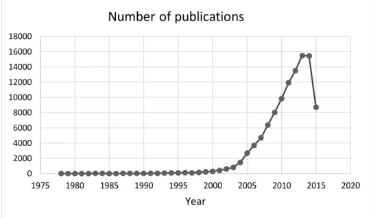
Although it may seem a futuristic science, nanotechnology has emerged over 40 years ago, with Gerd Birrenbach’s attempt at a process he called “micelle polymerization” in 1969 [2]. Since then, research on nanoparticles has increased exponentially (Figure 1).
Figure 1. Graphical representation of the number of articles published in Pubmed during the last 35 years.
Information collected on 2015, July 7th.
One of the fields most responsible for this accelerated growth is nanomedicine, the therapeutic and diagnostic field of nanotechnology.
Among the therapeutic applications, the most frequently employed revolve around utilizing nanoparticles as delivery agents, which act as vehicles for drugs [3-5], heat releasing [6, 7] or light emitting molecules [8] allowing them to reach specific types of cells or extracellular locations. These nanoparticles are engineered in such a way that they are attracted to a specific environment, allowing a directed treatment and, therefore, reducing the therapeutic agent’s
0 2000 4000 6000 8000 10000 12000 14000 16000 18000 1975 1980 1985 1990 1995 2000 2005 2010 2015 2020 Year
Number of publications
2
damage to bystander cells. Other less used nanotherapy techniques have also been reported, such as nanosponges, polymeric nanoparticles coated with a red blood cell membrane which act as decoys and intercept toxins in the blood stream [9] or a lens coated with carbon nanotubes which can convert light to tightly focused sound waves [10], among a multitude of others.
Due to the high diversity among different nanoparticle types, they can be applied to any of the existing diagnostic techniques, frequently increasing their efficiency, both in output velocity and quality. The most commonly known application lies in Magnetic Resonance Imaging (MRI), where superparamagnetic iron oxides coated with a recognition molecule, such as a tumor-binding molecule, provide a localized contrast [11]. Another use of nanoparticles in diagnostic involves an encapsulation or functionalization with a fluorescent molecule, allowing an optical detection [12]. There are also reports of multimodal nanoparticles which comprise both of the previous functions [13]. In a simpler approach, semiconductor nanoparticles known as quantum dots have been extensively studied due to their unique physiochemical properties, conferred by a quantum confinement effect [14], allowing for varied applications such as molecule tracking [15] and fluorescence imaging [16].
Aside from imaging techniques, most applications can be classified as nanosensors. A sensor, by definition, recognizes an agent and consequently produces a signal. In nanodiagnostics, the detection is based on the recognition of a specific molecule or a change in the environment and the signal can range from an absorbance shift due to agglomeration [17], to a release of light [18], or a magnetic resonance change [19].
In the present decade, a more condensed and multifunctional strategy has surfaced, called theranostics, combining both diagnostic and therapy in a single agent. The purpose is to diagnose and treat the disease at its earliest stage, improving the likelihood of a cure. Nanoparticles are excellent candidates for this approach due to their high variability and modification possibilities [20]. All types of nanoparticles can be engineered to achieve such a goal, being them organic, inorganic or even a combination of both [21-24].
A somewhat recent review (2013) on the current state of nanomedicine showed that most approved and investigated products on this field are categorized by the U.S. Food and Drug Administration as drug nanocarriers, more than 80% of which are intended for cancer treatment [25]. It is therefore obvious the existence of a keen oncological interest by today’s population of “nanoresearchers”.
Nanomedicine in Cancer
The World Health Organization attributed 8.2 million worldwide deaths to cancer in the year of 2012, approximately 13% of all reported deaths, with a 70% estimated increase of cancer cases in the next two decades [26]. One of the main reasons for such high numbers is the lack of efficient treatment. Current antineoplastic agents have poor selectivity, which, in practice, corresponds to a dose-limiting toxicity and systemic action, i.e. either the drug is in a too low concentration, resulting in death by cancer progression, or too high, resulting in the death of healthy cells by drug toxicity. In some cases, the drug concentration could actually be the same in both situations. Alas, the need for a targeted delivery of said drugs is of paramount importance to overcome current oncological treatments’ limitations and allow the patient to
3
outlast its illness, to which nanotechnology responded. Nanoparticles have the potential to increase a drug’s in vivo stability, extend its blood circulation time, control its release and alter its biodistribution, through either passive or active targeting, essentially allowing for a controlled modulation of the drug’s pharmacokinetic and pharmacodynamics profiles.
In Table 1 are summarized the approved nanoparticles for cancer therapy, evidencing the focus on liposome-type formulations.
Table 1. Marketed and approved nanoparticles for cancer therapy applications.
Product
(Company) Vehicle Drug Indication Approval Ref.
DaunoXome® (Galen) Liposome Daunorubici n Kaposi’s sarcoma 1996 [27] DepoCyt®
(Pacira) Liposome Cytarabine
Neoplastic meningitis 1999 [28] Doxil® Caelyx® (Johnson & Johnson) Liposome Doxorubicin Kaposi’s sarcoma Ovarian and breast cancer Multiple myeloma 1995, 1999, 2003 and 2007 [29] Genexol-PM® (Samyang Biopharm) PEG-PLA polymeric micelle Paclitaxel Breast, lung and ovarian cancer 2007 [30] Lipo-Dox®
(Taiwan Liposome) Liposome Doxorubicin
Kaposi’s sarcoma Breast and ovarian cancer 1998 [31] Marqibo®
(Talon) Liposome Vincristine
Acute lymphoid leukemia
2012 [32]
Mepact®
(Takeda) Liposome Mifarmutide Osteosarcoma 2009 [33]
Myocet®
(Cephalon) Liposome Doxorubicin
Breast cancer (in conjunction with cyclophospha mide) 2000 [34] NanoTherm® (Magforce Nanotechnologies) Iron oxide nanoparticle None 1 Glioblastoma 2010 [35]
1Intended purpose is thermal ablation instead of drug vehicularization.
The United States clinical trials registry contains, as of September of 2015, 1620 registries under the keyword “liposome” and 206 under “nanoparticle”, which, when compared to the previously listed nine formulations, indicate a growing trend in nanotechnological approaches to cancer therapies.
4
Objectives and reasoning
The elaborated thesis encompassed several objectives, which can be grouped into three different sections.
Current pharmaceutical treatments for neoplasms are mostly based on extremely hydrophobic drugs. Such compounds have stability issues in the blood stream and require third-party molecules, known as excipients, to function. Additionally, they have lack of selectivity, affecting both healthy and diseased tissue alike. In this sense, the first objective was to design and develop a Paclitaxel-loaded nanostructured lipid carrier (NLC) formulation. Paclitaxel was chosen due to its degree of hydrophilicity, which is one of the highest known to drugs, representing the ideal experimental subject. The lipid matrix of NLCs confers the perfect environment for Paclitaxel’s presence, while their surfactant-stabilized surface would hypothetically permit a very high and long-term stability in aqueous media. Additionally, and perhaps most importantly, they may confer occlusion and bar the Paclitaxel’s destructive potential until it reaches its target destination – tumoral tissue.
Magnetic nanoparticles have been establishing an ever-increasing ground in nanomedicine. Albeit more present in diagnostics, they have a wide range of therapeutic capabilities. However, their current commercialization is deficient, largely due to morose and inefficient largescale synthesis methodologies. Such leads to the second objective of this thesis, the development, optimization and characterization of superparamagnetic iron oxide nanoparticles (SPIONS). SPIONs were chosen as focus due to a plethora of reasons. First and foremost, they are comprised of solely iron, a material widely known as biocompatible (in sensible quantities). Secondly, SPIONs are abundant in the scientific literature, with well-described and extensive characterizations, and are already clinically approved and marketed (such as Feridex [36], Feraheme [37] and MION-46L [38]). Thirdly, the reagents needed for their synthesis are cheap and easily acquired and a functioning protocol for their synthesis would be a useful asset to the laboratory. Furthermore, their superparamagnetic behavior confers hyperthermic capabilities - an increase of energy, dissipated as heat, caused by an applied alternated magnetic field.
The thesis’s third and ultimate objective consisted on the innovative combination of both previous sections with the intention of creating a synergistic behavior. The synthesis and characterization of this novel formulation, the Paclitaxel-SPION-loaded NLCs, could lead into insights about unexplored reaches of nanotherapeutics and, ideally, result in a massive advance of the drug’s effectiveness.
5
Chapter 2
Lipid nanoparticles
Introduction
Lipid nanoparticles are defined as sub-micron sphere-like arrangements of lipid-based molecules held together by a surfactant. Due to its unique composition, these particles usually attain GRAS (generally recognized as safe) status, since its components are biomolecules commonly found in the human body or known to be biocompatible [39].
Currently, this classification encompasses many different types of nanoparticles depending on composition and synthesis method, albeit only two are mainly developed and studied, solid lipid nanoparticles (SLNs) and nanostructured lipid carriers (NLCs).
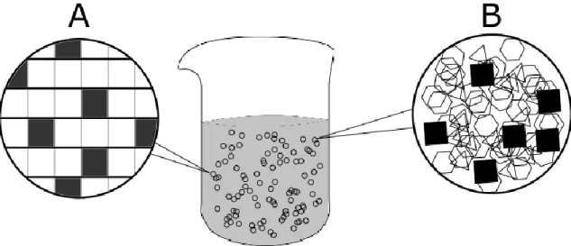
First developed during the early 1990s [40], SLNs are composed of 0.1-30% (w/w) solid lipid dispersed in an aqueous solution of 0.5-5% (w/w) surfactant [41]. These nanoparticles tend to exhibit a good physicochemical stability, protection and controlled release of the encapsulated drug, low cytotoxicity, organic solvent free synthesis and an ease of scale-up [39]. However, due to their high crystallinity and ordered matrix (Figure 2.A), they can suffer from low encapsulation efficiency and a possible drug expulsion during storage [42], depending on the structure of the drug.
To circumvent such limitations, a second generation of lipid nanoparticles was developed. The addition of a liquid lipid to the formulation allows a less ordered lipid matrix and, consequently, more room for the active compound (Figure 2.B) [43].
6
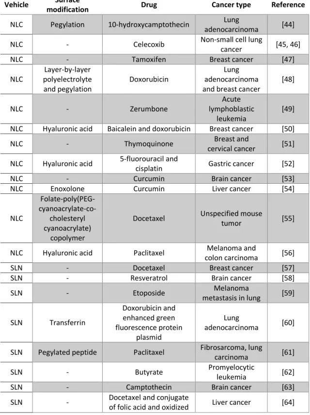
These types of nanoparticles have shown great potential in cancer treatment research. Albeit none is already approved or in ongoing clinical trials, numerous in vitro and animal models assays have been conducted successfully and reported in the literature. A very succinct fraction of which is shown in Table 2.
Table 2. Small sample of lipidic nanoparticles’ research in cancer therapy.
Vehicle Surface
modification Drug Cancer type Reference
NLC Pegylation 10-hydroxycamptothecin Lung
adenocarcinoma [44]
NLC - Celecoxib Non-small cell lung
cancer [45, 46]
NLC - Tamoxifen Breast cancer [47]
NLC Layer-by-layer polyelectrolyte and pegylation Doxorubicin Lung adenocarcinoma and breast cancer
[48] NLC - Zerumbone Acute lymphoblastic leukemia [49]
NLC Hyaluronic acid Baicalein and doxorubicin Breast cancer [50]
NLC - Thymoquinone Breast and
cervical cancer [51] NLC Hyaluronic acid 5-fluorouracil and
cisplatin Gastric cancer [52]
NLC - Curcumin Brain cancer [53]
NLC Enoxolone Curcumin Liver cancer [54]
NLC Folate-poly(PEG- cyanoacrylate-co-cholesteryl cyanoacrylate) copolymer
Docetaxel Unspecified mouse
tumor [55]
NLC Hyaluronic acid Paclitaxel Melanoma and
colon carcinoma [56]
SLN - Docetaxel Breast cancer [57]
SLN - Resveratrol Brain cancer [58]
SLN - Etoposide Melanoma metastasis in lung [59] SLN Transferrin Doxorubicin and enhanced green fluorescence protein plasmid Lung adenocarcinoma [60]
SLN Pegylated peptide Paclitaxel Fibrosarcoma, lung
carcinoma [61]
SLN - Butyrate Promyelocytic
leukemia [62]
SLN - Camptothecin Brain cancer [63]
SLN - Docetaxel and conjugate
7
single-walled carbon nanotubes
SLN Hyaluronic acid Vorinostat
Tongue squamous cell carcinoma and
lung adenocarcinoma
[65]
SLN Folic acid Docetaxel and
ketoconazole
Brain
endothelioma [66]
Paclitaxel
Paclitaxel, as well as the other taxanes, acts by decreasing the critical concentration of tubulin needed for microtubule assembly, promoting its formation and hindering its detachment [67]. This destabilization of functional microtubule dynamics leads to the impossibility of some essential cell processes, such as movement, chromosome segregation and cell division, ultimately resulting in the cell’s unrest and self-induced apoptosis [68].
Along with his structural cousin docetaxel, paclitaxel (Figure 3) is the highlight of the taxane family. His tetracyclic heptadecane skeleton confers a staggeringly low hydrophilicity, which was reported to be as low as 0.357 µg/mL [69]. In fact, its behavior in aqueous media lead to the addition of third party molecules, as excipient or binder, as the only method to stabilize this taxoid, with the examples of the two most important marketed forms of paclitaxel, Taxol®, where it is dissolved in a 50/50 (w/w) solution of polyoxyethylated castor oil (Cremophor EL - CrEl) and dehydrated ethanol [70], and Abraxane®, in which the taxoid molecule is conjugated to albumin (nab-paclitaxel) [71].
Figure 3. Paclitaxel structural and chemical configuration.
The first of the previously enunciated formulations is largely accompanied by undesired pharmacokinetic profiles, due to paclitaxel’s extensive binding to serum albumin, and consequent inactivity [72], broad tissue distribution, albeit incapable of passing the blood-brain
8
barrier [73], and a low half-life of 2.9 ±0.3 hours [74]. Additionally, the excipient CrEl only worsens such effects [75].
The second formulation, on the other hand, demonstrates an increased treatment response and slower disease progression with a simultaneous significantly decreased systemic toxicity [76]. It requires, however, recombinant albumin for its synthesis, relating into a much higher cost and, consequently, turning the better antitumor efficacy relatively marginal.
Nano-sized delivery systems have therefore attracted a heightened attention in the current millennium as a strategy to overcome the limitations of simpler paclitaxel therapy approaches.
Firstly, a nanoparticle system would provide physical and chemical protection for the very water insoluble and metabolic degradation-prone molecule through its entrapment, allowing a higher injectable concentration and completely removing the excipient’s presence and toxicity. Secondly, nano-based formulations could improve the paclitaxel’s pharmacokinetics through drug occlusion (lack of epitopes recognizable by the immune system), a controlled release profile and possible surface modification (such as pegylation). Thirdly, a nanoparticle can take advantage of the Enhanced Permeation and Retention effect (EPR), a passive targeting approach where small particle (less than circa 400 nanometers) accumulate in cancer tissue through defective neo-angiogenic blood vessels [77], or it can be modulated into partaking an active targeting, with molecules such as folic acid, transferrin and hyaluronic acid. Both the previous targeting methods result in an improved biodistribution of the anticancer agent. Finally, the nano-sized systems have a limitless potential in versatility and functionality modulation. They allow for co-delivery of multiple agents, being it synergistic drugs, a theranostics combination or anything else. They can also be positively, neutral or negatively charged, depending on which suits the synthesizer needs. Environment responses are also possible, such as a pH-, heat-, light- or magnetically-responsive structural change.
A myriad lipid-based nanoparticles have already been reported to effectively deliver paclitaxel.
Liposomal formulations encapsulating paclitaxel have showed a steep decrease in side effects while maintaining a similar to slightly higher antitumor activity when compared to Taxol® [78-80] and even managed to show a significant tumor inhibition in a Taxol®-resistant murine model [81]. Liposomes, however, contain an aqueous core, leading to disappointing encapsulation efficiencies of hydrophobic drugs such as paclitaxel.
On the other hand, SLNs are composed of a solely lipid matrix, more easily accommodating such drugs, leading to higher encapsulation efficiencies and slower release profiles, as has been reported by Cavalli et al (0.1% release in PBS during 2 hours [82]), Yegin et al (12.5-16.5% within 14 days [83]) and Lee at al (10% in 24 hours [84]). The in vitro cellular uptake and cytotoxicity has also been shown to be influenced by the lipid matrix composition and surfactant [85-87], which is most likely explained by specific affinities of cell membranes to different lipids. Dong et al presented a clever use of surfactant (Brij 78) which provoked a temporary reduction in intracellular ATP levels partially inhibiting the energy-dependent P-gp efflux (a drug efflux mechanism that some tumors employ) in a P-gp overexpressing human ovarian carcinoma cell line, effectively bypassing its drug resistance. An unexpected find revealed the propensity of SLNs to deliver paclitaxel to the brain, passing through the blood-brain barrier without an active mediator [88].
9
As stated in the previous chapter, the SLNs’ matrix confers potential disadvantage issues, which NLCs were developed to resolve. However, being the most recent addition to the lipid-based nanoparticle family, the reports of successful paclitaxel-loaded NLC formulations are staggeringly scarce. As of the time of writing, the author could only locate five divulged papers on the subject [56, 89-92], three written by the same authors. Yang et al developed started with the development of hyaluronic acid-coated NLC formulation and reported its higher effectiveness in murine melanoma, mouse colon and human colon cancer cell lines, and in vivo in Kumming mice when compared to Taxol® [56]. They later went on to disregard the hyaluronic acid and include a surface modification of a photo-responsive cell-penetrating peptide and again reported a higher antitumor efficacy against non-functionalized NLCs and Taxol®, this time in human fibrosarcoma cells [89]. More recently, in 2015, he included another molecule to the formulation’s surface functionalization, a NGR peptide (Asn-Gly-Arg), reporting an even greater antitumoral efficacy in comparison to all earlier formulations, in both the previously studied cell line and a new human breast adenocarcinoma one [90]. The other two reports were developed for pulmonary delivery. Kaur et al underwent a Box-Behnken design optimization of all conceivable parameters and evaluated the optimal formulation against a plain drug solution, reporting a circa 3-fold increase in paclitaxel lung concentration [91]. Lastly, Taratula et al developed a NLC co-delivery system containing paclitaxel (or doxorubicin) and siRNA for the suppression of proteins responsible for drug resistance in cancers, with the addition of a synthetic hormone analog as a targeting moiety specific to lung cancer cells. His data evidenced a much higher effectiveness with the targeted paclitaxel-NLC than with placebo and free paclitaxel with an almost complete cancer regression when siRNA is co-encapsulated [92].
Hence, it is obvious the utmost importance to explore deeper into the vastly untapped potential of paclitaxel-loaded nanostructured lipid carriers.
NLC synthesis
To date, various techniques have been reported for the synthesis of lipid nanoparticles. One of such is high pressure homogenization, which can be further divided into hot [93] and cold [94] variants. In both, the active compound is dissolved in the previously melted lipid blend. The variants then differ in the addition of a hot or cold aqueous surfactant solution, respectively, and the temperature at which the microemulsifying step is conducted. These techniques offer some advantages, such as a narrow particle size distribution with a low content of microparticles, avoidance of organic solvents and a great ease of scale-up accompanied by a frequent availability of the equipment required in industrial laboratories [95].
Another methodology is o/w microemulsion, which is based on the dispersion of warm microemulsions in cold water under stirring, leading to a nanoprecipitate [96]. Unfortunately, in this method a high concentration of surfactants and co-surfactants are used and results in very small yields. Variations of the previous method, also based on the breaking of an o/w microemulsion into a nanoemulsion, rely on solvent evaporation, diffusion or injection molding. In the emulsification-evaporation process, the lipid is first dissolved in an organic solvent and a microemulsion is created through the addition of an aqueous phase containing a surfactant. Subsequently, the solvent is left to evaporate under normal or reduced pressure while at room temperature, resulting in a nanoprecipitate in the aqueous medium, with the benefit of avoiding possible heating-related issues [97]. The emulsification-diffusion methodology, on the other hand, was developed for lipids which could not be dissolved in the organic solvent under
10
ambient conditions. Consequently, both the lipids and the solvent are mixed while in a heated water bath for the dissolution to occur. An addition of water to the organic solution results in coacervation and formation of lipid nanoparticles, which are then collected by ultracentrifugation, solvent evaporation or cross-flow filtration [98]. The third method, injection molding, represents a faster, mechanically-aided solvent-diffusion technique, as lipids dissolved in a water miscible organic solvent are rapidly injected into an aqueous phase, resulting in coacervation and a nanoparticle dispersion. This process has the specific advantage of a simultaneous efficiency, tight conditional control (by parameter variation) and simple mechanical implementation (the most technically complex apparatus is a syringe) [99]. However, all 3 of these methods sin on the utilization of organic solvents and their accompanying toxicological and environmental hazards. Additionally, the first two present a fairly dilute nanoparticle suspension, largely due to the limited solubility of lipids in the solvents. A clever way to ensure a higher hydrophilic drug encapsulation efficiency resulted from the double emulsion technique. This method stands on the drug’s dissolution and entrapment in the internal aqueous phase of a w/o/w emulsion, along with a stabilizer, in order to prevent its partitioning to the outer phases during solvent evaporation. It is initiated as a warm w/o microemulsion, resulting from the addition of drug, water, stabilizer and melted lipids, which is then dispersed into another stabilizer-containing water solution forming a w/o/w [100]. The nanoparticles are ultimately collected through diafiltration. This technique yields good results with sensitive drugs, such as peptides and nucleic acids, if an organic solvent is utilized for the lipid’s dissolution instead of heat, although with the advent of common organic solvent disadvantages [101]. Another disadvantage is the relatively small aqueous inner compartment compared to the whole lipid matrix, resulting in, likewise, comparatively inferior encapsulation efficiencies.
NLCs can also be synthesized by ultrasonication in conjunction with [102], or absence of [103, 104], high shear homogenization. In this technique, lipids, surfactant and drug are all mixed together in a water bath at a high enough temperature to melt the lipids. Water, at the same temperature, is then added to the previous mixture, resulting in an emulsion, which is, optionally, subjected to high shear forces through high speed stirring, such as with a rotor-stator homogenizer, to form a microemulsion. Lastly, the particles undergo high strain during ultrasonication, due to cavitations caused by sonic waves, extremely elevated and localized temperature and pressure occurrences by collapsing bubbles, resulting in a suspension of nanoparticles. The greatest advantages of this methodology are the complete absence of organic solvents, ease and velocity of process and commonly available equipment. However, there is a possible metal contamination from particles released by the sonicator’s tip, commonly associated with its usage, being it recently acquired or age-old. The other, much more frequent disadvantage is the broad particle size distribution, largely resolved by a higher surfactant concentration (which could, in turn, lead to toxicity issues) or the absence of high speed stirring. Additionally, it is possible to employ this technique in a standalone high sheer homogenization version [105], although unadvisable due to the stated broad distribution problem.
The standalone ultrasonication method, was the chosen technique for this work, due to the extensive experience in its employment, good advantage to disadvantage ratio and the presence of a sonicator in the laboratory.
11
Reagents and equipment
The chosen lipid blend comprise Gelucire® 43/01, Compritol® 888 ATO and Miglyol® 812 while the chosen surfactant Tween® 80.
Gelucires are blends of mono-, di- and triglycerides with polyethylene glycol esters of fatty acids marketed by Gattefossé. In particular, Gelucire® 43/01 has a melting point of 43oC
and HLB of 1. Compritol® 888 ATO, also marketed by Gattefossé, is a blend of different esters of behenic acid with glycerol. It has a melting point of 70°C and HLB of 1. On the other hand, Miglyol® 812 is marketed by Acofarma Distribuición and comprised of a mixture of caprylic and capric triglycerides. It has a melting point lower than 0°C and HLB of 15.36. Tween® 80, the surfactant, is the trademark name for Polyethylene glycol sorbitan monooleate marketed under Sigma-Aldrich, a biocompatible and amphoteric molecule.
The described lipid combination was chosen taking into account the need of having a formulation which melted when the temperature arose higher than 40-45oC. Additionally,
paclitaxel’s solubility was taken into account by conducting lipophilicity tests. Compritol® 888 ATO was also added to counterbalance the melting point of Miglyol® 812 and to introduce an even higher degree of disorder to the nanoparticle’s matrix, thereby increasing the drug load.
Paclitaxel was bought from LC Laboratories and utilized without further treatment. The probe-sonicator utilized was a VCX130 from Sonics & Materials, Inc. (130 W of maximum potency) and the double-deionized water was filtered through a nanopore membrane (conductivity lesser than 0.1 μS.cm-1).
Ultrasonication technique
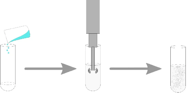
The methodology herein described was an adaptation of the protocols followed by the research group and is summarized in Figure 4.
Figure 4. Representative diagram of the NLC synthesis process by a standalone ultrasonication technique. From left
12
The optimized formulation was synthesized as followed. 70% (w/w) of Gelucire® 43/01, 6% (w/w) of Compritol® 888 ATO, 10% (w/w) of Miglyol® 812, 12% (w/w) of Tween® 80 and 2% (w/w) of Paclitaxel were weighted and stored in a glass tube. The tube was then placed in a water bath at 75oC until all lipids had melted and magnetically stirred to suspend the drug.
Afterwards, 16 mL of double distilled water, pre-heated at 75oC were added, obtaining an
opaque, white suspension, which was placed under the probe-sonicator at 104 W of potency during 10 minutes. The resulting white nanoemulsion was stored in a sealed glass vial and left to cool at room temperature.
Encapsulation efficiency assessment
Encapsulation, or entrapment, efficiency is defined as the percentage of encapsulated drug relative to the total added amount [106]. Hence, in this work, the percentage was determined by measuring the unentrapped paclitaxel’s amount and calculating its inverse relativized to the absolute quantity initially added, as per the equation:
%𝐸𝐸 =𝑇𝑜𝑡𝑎𝑙 𝑎𝑚𝑜𝑢𝑛𝑡 𝑜𝑓 𝑝𝑎𝑐𝑙𝑖𝑡𝑎𝑥𝑒𝑙−𝑀𝑒𝑎𝑠𝑢𝑟𝑒𝑑 𝑓𝑟𝑒𝑒 𝑝𝑎𝑐𝑙𝑖𝑡𝑎𝑥𝑒𝑙
𝑇𝑜𝑡𝑎𝑙 𝑎𝑚𝑜𝑢𝑛𝑡 𝑜𝑓 𝑝𝑎𝑐𝑙𝑖𝑡𝑎𝑥𝑒𝑙 ∗ 100 (2.1)
The study of the in vitro behavior of Paclitaxel in an aqueous medium is a gargantuan task, due to the difficulty of maintaining a good sink condition derived from Paclitaxel’s infinitesimal water solubility. In order to overcome such difficulty, a hydrotropic agent was used, sodium salicylate, which has already been reported to increase Paclitaxel’s solubility by 100 times at 1 M concentration [107].
The sodium salicylate was purchased from Merck and utilized without further modification.
Initially, the assessment of particle stability in sodium salicylate had to be made. Therefore, size and ζ-potential measures and aggregates determination were conducted under different concentrations of the hydrotrope. The Paclitaxel-loaded NLCs presented as stable at concentrations as high as 0.5 M, upon which they started to deform and aggregate.
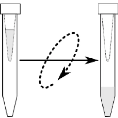
For the unentrapped paclitaxel assessment, a formulation sample was diluted in double deionized water with sodium salicylate at 0.5 M concentration, at a 1:100 ratio, and placed inside an Amicon® Ultra-4 Centrifugal Filter Device (Millipore). Centrifugation was then performed using a Jouan BR4i multifunction centrifuge (Thermo Electron) with a fixed 23o-angle
rotor at 3500 rotations per minute for 30 minutes (Figure 5). Afterwards, the supernatant, which contained the unentrapped paclitaxel, was collected and the concentration was quantified through a V-660 spectrophotometer (Jasco) at 200-400 nm.
13
Figure 5. Centrifugation step of the encapsulation efficiency assessment methodology.
Characterization techniques
Cryo-scanning electron microscopy (Cryo-SEM)
The SEM technique relies on the emission of an electron beam from a tungsten filament, which, when it contacts with a sample’s surface, provokes the ejection of low-energy secondary electrons by inelastic scattering, high-energy backscattered electrons by elastic scattering and x-ray radiation. These signals can be measured by different detectors to gather information about the topology and chemical composition of the sample [108].
SEMs operate under high vacuum, which tends to cause the nanoparticle’s deformation, or even rupture, since NLCs are known to be very labile. Additionally, the ones synthesized during this work have a melting point of little higher than 40oC, increasing their fluidity to even
higher proportions. To circumvent such limitations, a cryogenic variant of sample preparation was utilized, where the sample is previously fixed with liquid nitrogen.
The images obtained for this work were measured by a FEI Quanta 400FEG SEM system and conducted in Centro de Materiais da Universidade do Porto (CEMUP).
Dynamic and Electrophoretic Light Scattering (DLS and ELS)
When a light source emits radiation upon small particles, the light scatters, a phenomena known as Rayleigh scattering. If the source is a laser (monochromatic and coherent) the scattering suffers an intensity fluctuation due to the particles’ Brownian motion. The information acquired, post autocorrelation, from this fluctuation permits the calculation of a particle’s translational diffusion coefficient, which can be equated into the particle’s hydrodynamic radius by the Stokes-Einstein-Sutherland equation:
𝑟ℎ =6𝜋𝜂𝐷𝑘𝐵𝑇 (2.2)
where rh is the hydrodynamic radius, kb the Boltzmann’s constant, T the absolute temperature,
η the solution’s viscosity and D the diffusion coefficient [109]. The hydrodynamic radius of a particle represents the radius of a hard sphere that diffuses at the same rate as the particle, which can be considered the same when applied to nanostructured lipid carriers since they are quasi-spheres.
14
Electrophoretic light scattering is a variant of the dynamic technique, where, instead of Brownian motion, an oscillating electric field is responsible for the particles’ mobility. Through the data obtained, the particles’ electrophoretic mobility is determined, which can then be related to their ζ-potential by a variety of models, the most common being Smoluchowski’s:
𝜈𝐸 = 4𝜋𝜀0𝜀𝑟6𝜋𝜂𝜁 (1 + 𝑘𝑟) (2.3)
where νE represents the electrophoretic mobility, εr and ε0the relative dielectric constant and
electrical permittivity of vacuum respectively, r the particles’ radius and k the Debye-Hückel parameter [110]. The ζ-potential represents the nanoparticle’s electric potential at the slipping plane, the boundary beyond which ions in solution do not suffer any interaction by the particle’s mobility. This potential is closely related with the particle’s stability in suspension.
For the purpose of this work, both size and ζ-potential measures were performed with a NanoBrook 90Plus PALS Particle Size Analyzer from Brookhaven Instruments Corporation, with the addition of a BI-ZEL electrode for ELS.
Results and discussion
The synthesis methodology described above corresponds to the optimized NLC formulation, in respect to size, ζ-potential, polydispersity index and encapsulation efficiency. This optimization was conducted following an iterative approach, which is based on a
create-assess-learn-recreate routine, applying changes in lipid ratios, water and surfactant content and
sonication time and potency as needed.
In Table 3 are depicted the physicochemical characteristics of a small fraction of the synthesized formulations. This table was kept summarized due to the sheer amount of NLCs synthesized.
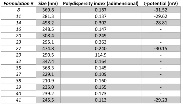
Table 3. Physicochemical parameters of some of the synthesized NLC formulations during the optimization process.
Formulation # Size (nm) Polydispersity index (adimensional) ζ-potential (mV)
8 369.8 0.187 -31.52 11 281.3 0.137 -29.62 14 498.2 0.302 -28.81 16 248.5 0.147 - 20 308.4 0.249 - 23 295.1 0.263 - 27 474.8 0.240 -30.15 29 290.5 114.9 - 32 347.4 0.164 - 35 368.3 0.145 - 37 229.1 0.109 - 38 210.9 0.160 - 39 235.0 0.155 - 40 239.2 0.173 - 41 245.5 0.113 -29.23
15
The ζ-potential started as being measured at every new formulation. However, when noticing it would only change within the electrode’s sensibility range, it started to be measured at more spacious intervals. In a similar note, the relationship between encapsulation efficiency and the synthesis’s parameters was not thoroughly examined, as this section was only meant to be used as a starting point in size, polydispersity and ζ-potential for the later experimental work and it was expected for it to drastically diverge when the magnetic nanoparticles were introduced, and therefore only on the last formulation was it assessed.
The optimized formulation presented a mean hydrodynamic diameter of 245.5 ±0.1 nm, with 99.96% of all nanoparticles below 117.83 nm, and polydispersity index of 0.113 ±0.008, both measured by DLS. Besides being greatly monodisperse, these results are in agreement with the initial objective of having a nanoparticle smaller than 200 nm to avoid capture by the Mononuclear Phagocyte System [111] and to take advantage of the Enhanced Permeation and Retention effect (EPR) [77]. The ζ-potential of -29.2 ±0.5 mV, measured by ELS, is considered optimal, since it leads to a lack of aggregation and low cytotoxicity [112]. The entrapment efficiency of the optimized formulation was found to be above 99%. It was not possible to ascertain a specific percentage due to the supernatant’s absorbance being lower than the spectrophotometer’s sensitivity. This result is slightly higher than the efficiencies reported by other authors, which ranged from 90% to 95% [56, 89-91].
Cryo-SEM imaging was utilized instead of the conventional TEM and SEM. Since the nanoparticles were engineered with the purpose of melting at around 40oC, they have a high
fluidity at room temperature. Due to this plasticity, the vacuum required for TEM/SEM imaging distorted the nanoparticles, leading to the necessity of freezing them prior to observations. The images collected (Figure 6) were in agreement with the results obtained by the light scattering techniques. Additionally, no noticeable change in diameter resulted from Paclitaxel’s encapsulation.
16
Chapter 3
Magnetic nanoparticles
Introduction
Magnetism occurs from the motion of electrically charged particles, such as protons, electrons and neutrons (although neutrons are neutral, they consist of smaller electrically charged particles) [113]. Hence, since every material in nature is composed by them, all have an intrinsic property called magnetization, M, which is regarded as the vector sum of all its individual constituents’ magnetic moments. When a material is placed in contact with an external magnetic field, H, its magnetization creates a larger induced magnetic field, B, in a phenomena called magnetic induction, where µ0 represents the permeability of free space [114,
115]:
𝑩 = µ𝟎(𝑴 + 𝑯) (3.1)
Since the magnetic properties of a material should be measured as a direct magnetization response dependent on an applied magnetic field, the magnetic materials categorization relies on the ratio between those two physical entities, which is called the magnetic susceptibility, χ [114, 115]:
𝛘 = 𝐌/𝐇, −1 < χ < +∞ (3.2)
There are two categories under which magnetic materials can be classified depending on their magnetic susceptibility: paramagnetism, when it is positive, and diamagnetism, when it is negative. Diamagnetism refers to the case where the magnetization in a material physically opposes the applied field H, consequently reducing it. Lenz’s law states that “If an induced current flows, its direction is always such that it will oppose the change which produced it”. Such statement means that, under an applied magnetic field, the electrons in atomic orbitals will slightly adjust their orbits with the intent of creating current loops that oppose said field [116]. The result is that every material has an innate diamagnetic property, albeit very small in most cases [117]. Nonetheless, even though all materials present diamagnetic properties, in some it is negligible compared to a positive magnetic susceptibility created by the magnetic moments of unpaired electrons when they align with an applied field, a property known as paramagnetism [118].
A less broad classification method relies on the relative ordering of the magnetic moments contained in a material (Figure 7). The absence of diamagnetic properties is due to the inexistence of magnetic moments in such substances. In paramagnetic materials, each magnetic moment is randomly oriented due to thermal energy, as shown in Figure 7.A. However, if the temperature decreases, the magnetic interaction between the magnetic moments predominates over the thermal energy’s influence, causing a characteristic ordered state. In ferromagnetic materials (Figure 7.B), this ordering is parallel and happens below the Curie-Weiss temperature, while for antiferromagnetic materials (Figure 7.C) the ordering is antiparallel and occurs below the Néel temperature. These critical transition temperatures are
17
properties individual to each material’s chemistry and magnetic characteristics. Lastly, there is a variant of antiferromagnetism where each interacting pair of magnetic moments have different magnitudes (Figure 7.D) [114, 115, 117, 118].
There is one other magnetic state which these classifications fail to encompass, since it cannot be described by an ordering of domains. When a magnetic material is very small, ranging from atomic scale to a couple dozens of nanometers depending on the material (as example, circa 20 nanometers for magnetite [119]), it starts behaving as a single giant magnetic domain. In this state, materials behave similarly to paramagnets, by avoiding any reminiscent magnetization after the magnetic field removal, but have a much higher susceptibility [120]. Like the previously described magnetic states, superparamagnetic materials also have a critical transition temperature specific to each material’s composition. However, since they are a single magnetic domain particle, below this temperature they are in a blocked state, ignoring any external field, hence it is known as blocking temperature [121].
The easiest way to distinguish between the different magnetic states is to analyze a material’s M-H curve (Figure 8). If we apply an increasing magnetic field on a ferromagnet and subsequently decrease it, the magnetization does not follow the initial magnetization curve. This irreversibility is called hysteresis, and it occurs because the magnetic susceptibility is not a scalar, but a vector. At high applied fields, the magnetization approaches saturation magnetization, (Msat), where every magnetic domain is aligned, and, when such field is removed, remanent
magnetization (Mr), where some of the domains do not return to the original state. The sole
manner to return these domains to their basal state is to apply a negative field, having a specific magnitude denominated coercive field (Hc) where the magnetization returns to zero. The
hysteresis curve follows a symmetric pattern if the applied field varies from positive to negative values. Materials with a coercive field greater than 1000 A.m-1 (circa 12 Oe) are called hard
magnets, whereas soft magnetic materials are those with below that value [122]. In biological Figure 7. Possible orderings of magnetic moments: (A) paramagnetic; (B) ferromagnetic; (C) antiferromagnetic; and
18
applications, namely hyperthermia, high values of Ms and, more importantly, low values of Hc
are required, as will be the case of superparamagnetism, where the coercivity is null.
Hyperthermia is a type of cancer treatment in which localized body tissue is exposed to high temperatures. At temperatures higher than 40oC the natural enzymatic processes that keep
cells alive start to wane, rendering them more susceptible to the effect of radiation or chemotherapy while inducing apoptosis. At even higher temperatures (circa 45oC) human cells
stop working all-together and die (necrosis) in a process known as thermoablation [123]. Magnetic nanoparticles have the ability to dissipate heat when subjected to an alternating magnetic field via magnetic losses, which can be distinguished into three different mechanisms – hysteresis, Néel and Brown relaxations. There are also other mechanisms: the magnetic loss by friction in viscous suspension and Foucault currents in metallic materials. However, both shall be omitted since they mainly affect larger particles.
Hysteresis losses (Figure 9.C) are a result of the uneven magnetization of the material during sequential magnetization cycles and may be derived from the integration of the hysteresis loops, representing the energy dissipated per cycle. They are the predominant loss mechanism in ferrimagnetic nanoparticles [124] and are inexistent in superparamagnetic materials, due to the obvious absence of hysteresis loops.
Figure 8. Illustrative magnetic field dependencies of different materials’ magnetization during several magnetization
cycles. Diamagnetism in green, paramagnetism in orange, superparamagnetism in blue and ferromagnetism, with its characteristic hysteresis loop, in yellow.
In smaller nanoparticles, the heat dissipation is mainly caused by a delay in the relaxation of the magnetic moment. This moment behaves according to a uniaxial anisotropy, which means it only has two stable antiparallel orientations, both separated by an energy barrier. Therefore, two mechanisms are possible: either the magnetic moment manages to surpass the anisotropy barrier and shift while the particle remains fixed (Figure 9.A) or it fails and the particle rotates to compensate (Figure 9.B), causing energy dissipation by friction. Evidence shows that the first, Néel relaxation, holds a higher influence on superparamagnetic
19
nanoparticles [125, 126], while the second, Brown relaxation, is heavily influenced by the surrounding fluid’s viscosity [124]. Additionally, the heating rate is very dependent on particle size, while being maximized by a low polydispersity [126, 127].
Besides having a higher heating efficiency in comparison to larger multi-domain magnetic materials [128], superparamagnetic nanoparticles have many characteristics that make them ideal for a therapeutic approach. With the lack of a hysteresis loop, superparamagnetic materials do not retain magnetization once the external magnetic field is removed, thus avoiding particle agglomeration and complications thereof [129]. Despite extensive cell viability assays performed in regards to the cytotoxicity of superparamagnetic iron oxide nanoparticles (SPIONs), no considerable effect has been found at concentrations where other materials do exhibit it [130, 131].
SPIONs synthesis
In past years, multiple synthetic approaches have been taken in order to obtain the perfect iron oxide nanoparticle. One of the earliest, and still much in use, was reported by Massart in 1981 [132], which he called co-precipitation. It consists in the addition of a base to an aqueous solution of ferrous (Fe2+) and ferric (Fe3+) ions in a 1:2 stoichiometric ratio under an
oxygen free environment, preventing further oxidation, resulting in a black precipitate of less than 20 nanometers magnetite particles. Albeit this method offers an ease of controlling the nanoparticle’s size by adjusting the pH, reaction temperature, stoichiometric ratio and iron precursors, such ease is severely limited [133]. Additionally, without further surface modification, the nanoparticles are prone to be oxidized into γ-Fe3O4 [134]. To circumvent these
limitations, other methods were elaborated, such as microemulsion (water in oil) [135, 136], limiting the crystal growth to the inside of micelles, hydrothermal [137, 138] and solvothermal decomposition [139, 140], disassembling large iron-containing molecules in aqueous and organic solvents, respectively, and the sonochemical method [141, 142], utilizing acoustic cavitation to create extreme heat and pressure localized conditions. However, each of these methodologies is accompanied by their own disadvantages, such as a difficult scale-up or the use of highly toxic solutions. As if to keep up with technology advancements, some variants of Figure 9. Illustrative representation of the three heat generating mechanisms by magnetic losses: Néel relaxation
20
the previous methods were adapted to take advantage of microwaves [143-145]. This type of radiation allows for a much faster and uniform heating, which consequently leads to quick, reproducible and efficient reactions [146].
During this work, due to a lack of experimentation in the field, multiple methods and optimizations in regards to SPION synthesis had to be employed. Choices were focused on co-precipitation techniques, since these required no organic solvents and, theoretically, resulted in a large yield of smaller than 20 nanometers magnetic iron oxide nanoparticles.
The iron-based powders were attained through Sigma-Aldrich and the ammonium hydroxide 25% solution was purchased from Merck with no extra purification step conducted. All water utilized was double-deionized with a conductivity inferior to 0.1 μS.cm-1.
Conventional co-precipitation method
The synthetic process utilized was based on the one described by Mahdavi et al, with some slight modifications [147].
In a typical procedure, ferric chloride hexahydrate (FeCl3 · 6H2O) and ferrous chloride
tetrahydrate (FeCl2 · 4H2O), in a 2:1 molar ratio, were dissolved in 150 mL of double distilled
water inside a three-necked closed flask, under a constant flow of nitrogen and vigorous magnetic stirring. After heating to 100oC, 20 mL of ammonium hydroxide (25%) was quickly
added, turning the solution black. 30 minutes later, 3 mL of oleic acid was injected dropwise. The solution was then left stirring for one hour. Finally, the black precipitate was collected by magnetic separation with a strong permanent magnet and washed several times with double distilled water.
Microwave-assisted co-precipitation method
The microwave synthesis was carried out in a Discover® SP microwave system, automated by Explorer-12 software, using 25 mL glass vessels with Teflon caps, all purchased from CEM Corporation. The reaction temperature was controlled by built-in sensors, allowing the wattage to increase or decrease as needed, with a 300 W maximum. Pressure was also left unregulated, allowing the vessels to self-vent as needed, with a 200 psi maximum.
The optimized procedure was conducted as followed. FeCl2 · 4H2O and FeCl3 · 6H2O were
dissolved in 20 mL of double distilled water in a vessel under vigorous magnetic stirring. The stoichiometric ratio used was 1:1.8 to account for the probable oxidation of Fe2+ to Fe3+ and the
total iron concentration was chosen as 0.07 molar, due to it being inside the optimum range. 3 mL of 25% ammonium hydroxide was then rapidly added and the vessel removed from the stirring. After the precipitate settled, the vessel was placed inside the microwave system and the temperature set to 100oC, under maximum stirring, during 10 minutes. After the elapsed
time, pressured nitrogen gas was injected into the system to quickly lower the temperature until 70oC to stop the reaction. Lastly, the black precipitate was collected with a strong permanent
magnet, washed 2 times and suspended in 25 mL of double distilled water. The nanoparticles were then stored in a 50 mL Falcon® at room temperature.
21
Characterization techniques
Transmission Electron Microscopy – TEM
TEM systems utilize a higher energy electron beam than SEM techniques, which, consequently, manages to interact with a sample at higher depths and achieve better resolution [148].
The SPION images were obtained from a JEM-1400Plus Transmission Electron Microscope in Instituto de Biologia Molecular e Celular (IBMC). Uranyl acetate (1%) was used as contrast agent.
X-ray Powder Diffraction – XRD
When x-ray waves are emitted into a crystalline solid they are absorbed by the atoms in the lattice, which then re-emit electromagnetic waves of the same frequency. The angle upon which the x-ray waves approach the atom plane will cause the emitted waves to undergo either a constructive or destructive interference, originating a maximum intensity when the wavelength is an integer multiple of the distance between two atoms.
A diffraction pattern can therefore be assembled by registering the emitted wave’s intensity as a function of the incident angle. In it, the peaks’ positions depend on the periodicity of the structure (i.e. the dimensions of the unit cell), whereas the peaks’ relative intensities relate to the distribution of scattering matter (i.e. the atoms or molecules) within the unit cell, which, in the case of XRD, is the electron density. Such relations equate into a unique diffraction pattern for every material, which allows their identification by a comparison with known patterns stored in databases [149, 150].
The powder diffractometer utilized during this work was a Rikagu Smartlab (9KW) from the Institute of Material Physics of University of Porto.
Superconducting Quantum Interference Device – SQUID
SQUIDs are a type of extremely sensitive magnetometer which can measure magnetic fields as low as 10-14 T, due to a component called Josephson junction, an electronic circuit very
sensitive to magnetic fluctuations. Since these circuits need to operate at near zero K temperatures, SQUIDs are equipped with a liquid helium refrigeration system [151].
For the purpose of this work, the magnetometer was utilized to measure the material’s magnetic response as a function of the applied magnetic field to determine the magnetic saturation, coercivity and residual magnetization and as a function of temperature in both zero field cooled and field cooled techniques to ascertain the blocking temperature and degree of superparamagnetism.
All measures were taken in a Quantum Design superconducting quantum interface device (SQUID) from the Institute of Material Physics of University of Porto.
Results and discussion
The initial particles, synthesized by the conventional co-precipitation method, were analyzed by DLS, which determined their size to be circa 1 micrometer. Since the objective of this work requires magnetic particles of smaller diameters, an exhaustive optimization process