Anti-in
fl
ammatory and Antinociceptive Activity of Epiisopiloturine,
an Imidazole Alkaloid Isolated from
Pilocarpus microphyllus
Valdela
nia G. Silva,
̂
†Renan O. Silva,
†Samara R. B. Damasceno,
†Nathalia S. Carvalho,
†Rafael S. Prude
ncio,
̂
†Karoline S. Araga
o,
̃
‡Maria A. Guimara
es,
̃
†Stefano A. Campos,
†Leiz M. C. Ve
ras,
́
†Markus Godejohann,
§Jose
́
Roberto S. A. Leite,
†Andre
́
L. R. Barbosa,
†and Jand-Venes R. Medeiros
*
,††
Biotechnology and Biodiversity Center Research (BIOTEC), Federal University of Piauí, Campus of Parnaíba, Avenida Sa
o
̃
Sebastia
̃
o, 64202-020, Parnaíba-PI, Brazil
‡Department of Physiology and Pharmacology, Federal University of Ceara
, R. Cel. Nunes of Melo, 60430-270, Fortaleza-CE, Brazil
́
§
Bruker BioSpin GmbH, Rheinstetten 76287, Germany
ABSTRACT:
The aim of this study was to investigate the antinociceptive and
anti-in
fl
ammatory activities of epiisopiloturine (
1
), an imidazole alkaloid found
in the leaves of
Pilocarpus microphyllus
. The anti-in
fl
ammatory activity of
1
was
evaluated using several agents that induce paw edema and peritonitis in Swiss
mice. Paw tissue and peritoneal
fl
uid samples were obtained to determine
myeloperoxidase (MPO) activity or tumor necrosis factor (TNF)-
α
and
interleukin (IL)-1
β
levels. The antinociceptive activity was evaluated by acetic
acid-induced writhing, the hot plate test, and pain induction using formalin.
Compared to vehicle treatment, pretreatment with
1
(0.1, 0.3, and 1 mg/kg,
ip) of mice signi
fi
cantly reduced carrageenan-induced paw edema (
p
< 0.05).
Furthermore, compound
1
at a dose of 1 mg/kg e
ff
ectively inhibited edema
induced by dextran sulfate, serotonin, and bradykinin, but had no e
ff
ect on
histamine-induced edema. The administration of
1
(1 mg/kg) following
carrageenan-induced peritonitis reduced total and di
ff
erential peritoneal leukocyte counts and also carrageenan-induced paw
MPO activity and TNF-
α
and IL-1
β
levels in the peritoneal cavity. Pretreatment with
1
also reduced acetic acid-induced writhing
and inhibited the
fi
rst and second phases of the formalin test, but did not alter response latency in the hot plate test. Pretreatment
with naloxone reversed the antinociceptive e
ff
ect of
1
.
T
he genus
Pilocarpus
, popularly known as
“
jaborandi
”
, is one
of the most important among the Brazilian
fl
ora, with native
species in the north and northeast of the country.
1Pilocarpus
microphyllus
Stapf (Rutaceae) is the most well-known
representative of the jaborandi group economically, because it
contains high concentrations of the imidazole alkaloid
pilocarpine, which is used to treat glaucoma, to stimulate the
lachrymal and sweat glands, and to control xerostomia.
2In
addition to pilocarpine, other imidazole alkaloids such as
isopilosine, epiisopilosine, and epiisopiloturine (
1
) have been
isolated from
P. microphyllus
.
3However, the biological properties
of alkaloids such as
1
are still largely unknown.
The in
fl
ammatory process involves a complex cascade of
biochemical and cellular events that occur in response to cellular
injury.
4This process triggers formation of in
fl
ammatory
neuromediators that activate nociceptors when released,
facilitating pain transmission and peripheral in
fl
ammatory
responses.
5Although a large number of analgesics and
anti-in
fl
ammatory agents are currently in clinical use, it would be
useful to identify novel drugs having a wider speci
fi
city and lower
toxicity than those currently available.
4The aim of the present study was to investigate the role of
epiisopiloturine (
1
) in acute peripheral in
fl
ammation and in the
analgesic processes induced by intraplantar and intraperitoneal
injection of di
ff
erent phlogistic agents.
■
RESULTS AND DISCUSSION
The anti-in
fl
ammatory and antinociceptive potential of
1
was
evaluated using a range of classical pharmacological models. The
present results revealed that
1
possesses anti-in
fl
ammatory and
antinociceptive activity in models of in
fl
ammation (paw edema
Received: February 4, 2013 Published: June 4, 2013
pubs.acs.org/jnp
and neutrophil migration) and pain (acetic acid-induced writhing
and the hot plate and the formalin tests).
In
fl
ammation is a primary response of an organism to injurious
stimuli, and some of these responses are characterized by pain,
heat, redness, edema, and loss of function.
6Among these e
ff
ects,
edema and pain are fundamental and essential outcomes to be
considered when evaluating potential anti-in
fl
ammatory and
antinociceptive compounds.
7The generation of carrageenan-induced mice paw edema is a
widely used test to determine the anti-in
fl
ammatory activity of
test compounds.
8Carrageenan injection into the mouse paw
induces a biphasic edema: the
fi
rst phase is characterized by an
edema of little intensity and di
ff
use cellular in
fi
ltrate with a
predominance of neutrophils that are capable of amplifying the
in
fl
ammatory response via production of reactive oxygen species
and release of in
fl
ammatory mediators; the second phase
develops after 24 h, displaying a more pronounced edema with
a maximum e
ff
ect between 48 and 72 h with an intense accumulation
of macrophages, eosinophils, and lymphocytes.
9In the present experiments, the anti-in
fl
ammatory e
ff
ect of
1
was investigated in the
fi
rst phase of carrageenan-induced paw
edema. It was shown that carrageenan induced severe paw edema
occurring 1 h after injection, which was maintained for 4 h
thereafter. There was a gradual increase in paw volume resulting
from edema in the carrageenan-treated group, with a maximum
value observed at 3 h. Indomethacin (10 mg/kg, ip) suppressed
paw edema signi
fi
cantly between 1 and 3 h after administration,
with a maximal inhibition of 55.0% (
fi
rst hour). Pretreatment
with
1
induced signi
fi
cant inhibition of paw edema throughout
the experimental period. At 3 h, compared to the
carrageenan-treated mice, the animals precarrageenan-treated with
1
at 0.1, 0.3, and 1 mg/kg
showed maximal reductions in edema of 55.0, 70.0, and 85.0%,
respectively (Table 1). Since
1
at a dose of 1 mg/kg a
ff
orded the
maximum protection against carrageenan-induced paw edema,
this dose was selected for subsequent studies.
The development of phase 1 edema induced by carrageenan
also involves the rapid production of various in
fl
ammatory
mediators such as histamine, serotonin, and bradykinin.
8To
investigate the potential of
1
to reduce edema, its e
ff
ects on paw
edema induced by di
ff
erent in
fl
ammatory mediators were
evaluated. The administration of dextran sulfate (0.065
±
0.010 mL; Figure 1A), serotonin (0.225
±
0.008 mL; Figure 1B),
Table 1. E
ff
ect of Epiisopiloturine (1) on Carrageenan-Induced Paw Edema in Mice
apaw edema (mL)b
treatment dose (mg/kg) 1 h 2 h 3 h 4 h
DMSOc 0.006±0.004 0.004±0.004 0.006±0.004 0.004±0.004
control 0.050±0.005# 0.048±0.006# 0.066±0.006# 0.055±0.005# indomethacind 10 0.022±0.004*(55.0) 0.024±0.002*(50.9) 0.037±0.008*(43.7) 0.035±0.002 (37.0)
1 0.1 0.032±0.007*(36.0) 0.026±0.006*(46.8) 0.030±0.007*(55.0) 0.028±0.008*(49.7) 0.3 0.026±0.006*(48.0) 0.014±0.005*(71.3) 0.020±0.007*(70.0) 0.018±0.005*(67.6) 1.0 0.017±0.008*(65.0) 0.010±0.007*(79.5) 0.010±0.010*(85.0) 0.015±0.015*(73.0)
aEdema was measured 1, 2, 3, and 4 h after carrageenin administration.bValues of paw edema expressed as mean±SEM (n= 5). Percent inhibition
of paw edema is indicated in parentheses. Control = carrageenan.#
p< 0.05 vs DMSO group.*p< 0.05 vs control.cNegative control for edema. dPositive control for edema.
Figure 1.Effects of epiisopiloturine (1) on paw inflammation induced by different inflammatory agents. Edema was induced by (A) dextran sulfate
(Dex); (B) serotonin (5-HT); (C) bradykinin (BK); and (D) histamine (Hist). Animals were pretreated with1(1 mg/kg ip), 2% dimethyl sulfoxide (DMSO; control, ip), or indomethacin (10 mg/kg, ip). Each point represents the mean±SEM offive to six animals (#p< 0.05 compared to the DMSO
bradykinin (0.052
±
0.004 mL; Figure 1C), or histamine (0.074
±
0.009 mL; Figure 1D) produced edema over time. In contrast,
paw volumes in the control DMSO-injected group were 0.008
±
0.007 mL, 0.005
±
0.002 mL, 0.010
±
0.004 mL, and 0.008
±
0.007 mL, respectively. Pretreatment with
1
(1 mg/kg)
e
ff
ectively inhibited paw edema induced by dextran sulfate
(54.08% inhibition; Figure 1A), serotonin (46.66% inhibition;
Figure 1B), and bradykinin (76.92% inhibition; Figure 1C) (
p
<
0.05). However,
1
did not inhibit paw edema induced by histamine
(Figure 1D). The reference drug, indomethacin (10 mg/kg, ip),
inhibited paw edema induced by all these in
fl
ammatory mediators
signi
fi
cantly (Figure 1).
Dextran sulfate is known to promote in
fl
ammation by
increasing vascular permeability, because of mast cell
degranu-lation and the subsequent release of histamine and serotonin.
10In the present study, it was observed that
1
inhibited paw edema
induced by dextran sulfate, serotonin, and bradykinin, but not by
serotonin. These results suggested that the antiedematogenic
e
ff
ects of
1
are related to in
fl
ammatory events involving
neutrophil migration, as well as inhibition of the release or
activity of in
fl
ammatory mediators.
One in
fl
ammatory event of great importance in
carrageenan-induced paw edema is the migration of leukocytes, primarily
neutrophils.
11This response can be measured using the
neutrophil-speci
fi
c enzyme myeloperoxidase (MPO), which is
an indicator of neutrophil accumulation.
12MPO can be released
on the outside of the cell, inducing damage to adjacent tissue and
thus contributing to the pathogenesis of in
fl
ammation.
13The
results obtained showed that carrageenan produced a marked
increase in MPO activity (10.9
±
1.2 U/mg of tissue). This increase
was reduced by treatment with indomethacin (3.1
±
0.3 U/mg of
tissue), the positive control. The administration of
1
(3.8
±
0.9 U/mg
of tissue) also reduced MPO activity, suggesting that its
anti-in
fl
ammatory action may involve inhibition of leukocyte migration
and neutrophil in
fi
ltration (Figure 2). A direct relationship between
tissue neutrophil concentration and MPO activity has been
reported,
14and the anti-in
fl
ammatory activities of certain compounds
have been attributed, in part, to the inhibition of MPO activity.
15Leukocytes play an important role in acute in
fl
ammatory
processes, and tissue damage is a deleterious consequence of
intense neutrophil migration, as observed in in
fl
ammatory
diseases.
16Compound
1
was evaluated to determine if it can
inhibit cell recruitment into the peritoneal cavity in
carrageenan-induced peritonitis. This experimental model provides a
well-characterized pharmacological tool to examine acute peritoneal
in
fl
ammation, which allows quanti
fi
cation of cell migration,
resident macrophage activation, and levels of several in
fl
amma-tory mediators.
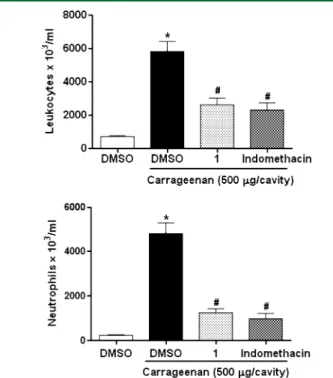
17In the present study, administration of carrageenan promoted the
formation of an intense cellular in
fi
ltrate (Figure 3). Carrageenan
(500
μ
g/cavity) induced cell migration into the peritoneal cavity 4 h
after administration, with a total leukocyte count of (5.81
±
0.61
×
10
6cells/mL) (Figure 3A). However, the administration of
1
(1 mg/kg, ip) 30 min before carrageenan reduced this peritoneal
leukocyte count to (2.60
±
0.61
×
10
6cells/mL), a 55.2% inhibition.
The same dose of
1
also reduced neutrophil migration into the
peritoneal cavity (1.26
±
0.17
×
10
6cells/mL), 73.8% lower than the
control group (5.82
±
0.47
×
10
6cells/mL) (Figure 3B). Similarly,
when compared to carrageenan treatment, pretreatment with
indomethacin produced a 60.2% reduction in total leukocyte count
and a 79.6% reduction in neutrophil migration to the peritoneal
cavity.
Previous studies have demonstrated that carrageenan injection
into the peritoneal cavity induces the release of certain
pro-in
fl
ammatory cytokines, such as TNF
α
and IL-1
β
.
15As shown in
Figure 4, the ip administration of carrageenan induced a marked
increase in TNF-
α
and IL1-
β
concentrations in the peritoneal
fl
uid. Pretreatment of animals with
1
(1 mg/kg, ip) reduced
TNF-
α
(Figure 4A) and IL1-
β
(Figure 4B) levels signi
fi
cantly.
Cytokines have been shown to have many biological activities
in the in
fl
ammatory response, including accumulation of
Figure 2.Effect of epiisopiloturine (1) on carrageenan-induced pawtissue myeloperoxidase (MPO) activity. Mice were injected with 2% dimethyl sulfoxide (DMSO) ip or carrageenan. Thirty minutes earlier, 1(1 mg/kg, ip) or indomethacin (10 mg/kg, ip) was administered to the animals. The control group (DMSO) was treated only with DMSO. MPO activity in the paw tissue was determined after 4 h. Results are expressed as the mean±SEM for at leastfive or six animals per group
(*p< 0.05 compared to the DMSO plus carrageenan group).
Figure 3.Anti-inflammatory effect of epiisopiloturine (1) on neutrophil
migration in mice. Mice were injected ip with dimethyl sulfoxide (DMSO),1 (1 mg/kg), or indomethacin (10 mg/kg, reference control) and were injected with 250μL of carrageenan (500μg/cavity, ip) 30 min later. Neutrophil migration was evaluated after 4 h. The white bars represent the peritoneal neutrophils in animals injected with DMSO (untreated group). (A) Total counts and (B) differential counts. The values are means±SEM offive or six
neutrophils in local tissues and induction of acute-phase protein
synthesis.
18Thus, on the basis of literature precedent and current
data, it can be inferred that the anti-in
fl
ammatory e
ff
ect of
1
is
dependent, at least in part, on inhibition of neutrophil in
fi
ltration and
the release of the pro-in
fl
ammatory cytokines TNF-
α
and IL-1
β
.
Considering the relationship between in
fl
ammation and the
development of a painful sensation, it was decided to investigate
the antinociceptive e
ff
ects of
1
, using three pain models (acetic
acid-induced writhing, the hot plate test, and the
formalin-induced nociception test for peripheral and central activity).
Acetic acid-induced writhing is a visceral pain model widely used
to screen antinociceptive potential.
19The ip administration of this
agent irritates the serous membranes and provokes a stereotypical
behavior in experimental animals, characterized by abdominal
contractions, movement of the body as a whole, and twisting of the
dorsal abdominal muscles.
20This model involves a range of
nociceptive mechanisms, such as release of biogenic amines (e.g.,
bradykinin and serotonin), cyclooxygenases, and their metabolites
(e.g., PGE2 and PGF2
α
),
21activation of peritoneal receptors and
in
fl
ammatory pain by induction of capillary permeability,
22reduction of nociception threshold, and stimulation of nociceptive
nerve terminals.
23Recent evidence has suggested that spinal MAP
kinases and other signaling pathways in the spinal cord mediate the
acetic acid-induced writhing response in mice.
24In the present study, pretreatment with
1
(1 mg/kg, ip),
30 min prior to acetic acid administration, produced inhibition of
the abdominal writhing response by 66.4% (
p
< 0.05). Compared
to the group administered with acetic acid only, the morphine
(reference drug)-administered group showed a reduction in
writhing movements by 98.3% (Figure 5). These results suggested
that
1
may reduce in
fl
ammatory mediator release or block
in
fl
ammation-promoting receptors.
In order to con
fi
rm the antinociceptive activity and investigate the
involvement of central mechanisms on the e
ff
ects of
1
, the hot plate
test was performed according to a protocol described previously.
25This test is a well-known model for acute thermal nociception, is
used to evaluate speci
fi
cally central nociception,
26and measures
complex responses to in
fl
ammation and nociception,
27in which
opioid agents exert their analgesic e
ff
ects via supraspinal and
spinal receptors.
28The present results showed that treatment with
1
(1 mg/kg, ip) failed to modify the hot plate response, indicating that
it does not act centrally. On the other hand, treatment with
morphine (5 mg/kg, sc), an opioid receptor agonist, induced a
signi
fi
cant increase in latency time in the hot plate test, as expected,
which persisted for at least 120 min (Figure 6).
The rota-rod apparatus was used to assess whether treatment
with
1
could in
fl
uence the motor activity of the animals and
consequently impair the assessment of the nociceptive behavior
in experimental models.
29Using the rota-rod test, mice treated
with
1
did not demonstrate any signi
fi
cant motor performance
changes, while diazepam signi
fi
cantly increased the number of
falls (data not shown).
To distinguish between the peripheral and central antinociceptive
actions of
1
, the formalin test was performed. In this test, a
peripheral noxious stimulus causes a distinct biphasic nociceptive
response involving two mechanisms. The
fi
rst phase (neurogenic
Figure 4.Effect of epiisopiloturine (1) on carrageenan-induced cytokineproduction in peritonitis. Levels of tumor necrosis factor (TNF)-α(A), interleukin (IL)-1β(B), in the peritoneal cavity were measured 4 h after carrageenan injection. Mice were administered 1(1 mg/kg, ip) and 250μL of carrageenan (500μg/cavity, ip) 1 h later. Each point repre-sents the mean±SEM offive or six animals for each group (*p< 0.05 compared to dimethyl sulfoxide (DMSO)-treated animals;#
p< 0.05 compared to the carrageenan group).
Figure 5.Effect of epiisopiloturine (1) on the writhing response induced
by acetic acid in mice. Mice received dimethyl sulfoxide (DMSO),1 (1 mg/kg, ip), or morphine (5 mg/kg, ip; reference control), 30 min prior to 0.6% acetic acid (250μL/cavity; ip). Data are expressed as the means±SEM offive or six animals for each group (*p< 0.05 indicates a significant difference from the acetic acid group).
Figure 6.Effect of epiisopiloturine (1) on reaction times to thermal stimuli
(hot plate). Mice received dimethyl sulfoxide (DMSO),1(1 mg/kg, ip), or morphine (5 mg/kg, sc). Data are expressed as the means±SEM offive or
pain) seems to be caused by the direct e
ff
ect of formalin on sensory
C-
fi
bers that re
fl
ect centrally mediated pain, with the release of
substance P.
30The direct activation of primary sensory
fi
bers by
formalin also involves release of mediators such as histamine and
serotonin.
31The second phase (in
fl
ammatory pain) involves a
combination of in
fl
ammatory and nociceptive mediators released
from injured tissues, such as prostaglandins, serotonin, histamine,
and bradykinin, responsible for sensitization of primary and spinal
sensory neurons and subsequent activation of the nociceptors.
30The results obtained demonstrated that mice treated with
2.5% formalin had mean licking times of 65.55
±
20.23 s in the
fi
rst phase (neurogenic; 0
−
5 min; Figure 7A) and 35.88
±
12.39 s in
the second phase (in
fl
ammatory; 20
−
25 min; Figure 7B).
Pretreatment with
1
(1 mg/kg, ip) reduced paw licking induced by
intraplantar administration of formalin signi
fi
cantly in both test
phases: the neurogenic (phase 1, 58.18% reduction) and the
in
fl
ammatory (phase 2, 89.21% reduction) phases. Similarly,
morphine (5 mg/kg, sc) inhibited both phases of the formalin
test (94.0% and 97.21% reduction in phase 1 and 2, respectively)
(Figure 7). These results corroborated those obtained in the paw
edema model. In this test, pretreatment with
1
inhibited paw edema
induced by serotonin and bradykinin, which are important
mediators involved in both phases of formalin-induced nociception.
In the formalin test, a decrease in licking time in both phases is
characteristic of drugs that act centrally and indicates a possible
interaction with opioid receptors, while peripherally acting drugs,
such as nonsteroidal anti-in
fl
ammatory drugs and
cortico-steroids, inhibit only the second phase.
32The e
ff
ect of
1
on
both phases of the formalin test suggested that it acts peripherally
and centrally to reduce in
fl
ammatory pain.
To better understand the antinociceptive e
ff
ect of
epiisopilo-turine (
1
), the formalin
fi
rst phase was evaluated in animals
pretreated with or without naloxone. It was interesting to note
that pretreatment with naloxone, an opioid-receptor
antago-nist,
33reversed the antinociceptive e
ff
ect of
1
(Figure 8). Thus,
1
seems to possess antinociceptive mechanisms that are partially
mediated by the activation of the opioid system.
■
EXPERIMENTAL SECTION
General Experimental Procedures.A 1H NMR spectrum was
acquired on an AVANCE III NMR spectrometer (600 MHz), equipped with a 5 mm TXI probe head (Bruker Biospin, Rheinstetten, Germany). An AmaZon SL mass spectrometer was used (Bruker Daltonics,
Bremen, Germany). A Shimadzu HPLC instrument was employed for compound purification, with a LiChrospher 60 RP Select B column.
Plant Material.A specimen ofPilocarpus microphylluswas collected in October 2008 near Matias Olimpio city (Piauí , Brazil) and waś identified by Dr. Ivanilza Moreira de Andrade, Department of Biology,
Federal University of Piaui. A voucher specimen (TEPB 27.152) waś deposited at the Graziella Barroso Herbarium (Teresina, Piaui, Brazil).́
Extraction and Isolation.Epiisopiloturine (1) was obtained from waste produced by pilocarpine extraction fromP. microphyllus leaves according to Veras et al.34The organic phase was submitted to liquid− liquid extraction, alkalinized with ammonium hydroxide solution to precipitate the1in the neutral form, and then the solution wasfiltered
under reduced pressure.35After further workup,1was shown to be pure
by HPLC (>95% w/w) and exhibited 1H NMR and MS/MS data
consistent with literature values.36,37
Drugs and Reagents. λ-Carrageenan, indomethacin, bradykinin, serotonin, dextran sulfate, histamine, captopril, acetic acid, form-aldehyde, and dimethyl sulfoxide (DMSO) were purchased from Sigma Chemical (Saint Louis, MO, USA). Heparin and morphine were provided by Merck, São Paulo, Brazil. All drugs were dissolved in sterile 0.9% (w/v) NaCl (saline). The epiisopiloturine (1) was dissolved in 2% DMSO. All other chemicals were of analytical grade and obtained from standard commercial suppliers.
Animals.Male Swiss mice (25−30 g) were housed at a temperature of 25±2°C under a 12/12 h light/dark cycle with food and water ad libitum. Animals were fasted for 18−24 h before the experiments. All experiments were performed in accordance with theGuide for Care and Use of Laboratory Animals(National Institute of Health, Bethesda, MD, USA) and were approved by the Ethics Committee in Research of the Federal University of Piauí(protocol no. 0066/10).
Carrageenan-Induced Paw Edema.The animals were randomly divided into six groups (n= 5), and edema was induced by injection of 50μL of a suspension of carrageenan (500μg/paw) in 0.9% sterile saline into the right hind paw (group I). Mice were pretreated intraperitoneally (ip) with either 2% DMSO (group II untreated control), indomethacin 10 mg/kg (group III reference control), or epiisopiloturine (1) 0.1, 0.3, or 1 mg/kg (groups IV, V, and VI, respectively). Paw volume was measured immediately before (Vo) and at 1, 2, 3, and 4 h after carrageenan treatment (Vt) using a plethysmometer (Panlab, Barcelona, Spain), as previously described.38 The effect of pretreatment was
calculated as percent inhibition of edema relative to the paw volume of the DMSO-treated controls by using the following formula:39
=
− − −
−
×
V V V V
V V
%inibition of edema ( ) ( )
( ) 100
t o control t o treated
t o control
Paw Edema Induced by Different Inflammatory Agents.To induce edema, the animals were administered 50 μL injections of Figure 7.Effect of epiisopiloturine (1) on the formalin test in mice. The
time spent licking was determined during thefirst 0
−5 min (phase 1; panel A) and during 20−25 min (phase 2; panel B) after injection with 2.5% formalin. Dimethyl sulfoxide (DMSO), 1 (1 mg/kg, ip), or morphine (5 mg/kg, ip; positive control) were administered 30 min before intradermal administration of formalin. Data are expressed as the mean±SEM offive or six animals for each group (*p< 0.05 indicates significant difference from the formalin group).
Figure 8.Effect of pretreatment with naloxone (3 mg/kg, sc) on the
analgesic effect of epiisopiloturine (1) on the formalin test. Mice were
pretreated with 2% DMSO or naloxone (3 mg/kg, sc, opioid antagonist). After 30 min, the animals were treated with1(1 mg/kg, ip) or morphine (5 mg/kg, sc). After 60 min, the time spent licking was determined during the
first 0−5 min (phase 1) after injection with 2.5% formalin. Data are
expressed as the means±SEM of six animals for each group (*p< 0.05 indicates significant difference from the formalin group;#
dextran sulfate (DEX; 500μg/paw), bradykinin (BK; 6.0 nmol/paw), serotonin (5-HT; 1% w/v), or histamine (HIST; 100μg/paw) into the right hind paw, as adapted from the reports of Barbosa et al.40 and Vasconcelos et al.40The contralateral paw received 50μL of 2% DMSO and served as an untreated control. In the experiment with bradykinin, the animals were pretreated with captopril (5 mg/kg, ip) 1 h prior to bradykinin induction to prevent bradykinin degradation.42
Epiisopilo-turine (1) (1 mg/kg) or indomethacin (10 mg/kg, reference control) was injected ip 30 min before these intraplantar injections of phlogistic agents.
Myeloperoxidase Activity.MPO is an enzyme found primarily in neutrophil azurophilic granules that has been used extensively as a biochemical marker for granulocyte infiltration into tissues, including
paw tissue.43 MPO activity was determined to evaluate neutrophil accumulation in the mouse paw. Briefly, 50−100 mg of paw tissue was
homogenized in potassium phosphate buffer containing 0.5%
hexadecyltrimethylammomium bromide. The homogenate was centri-fuged at 40000gfor 7 min at 4°C. The pellet was resuspended, and MPO activity was assayed by measuring the change in absorbance at 450 nm using
o-dianisidine dihydrochloride and 1% hydrogen peroxide. MPO activity is reported as units/mg of tissue. A unit of MPO activity was defined as that
converting 1μmol of hydrogen peroxide to water in 1 min at 22°C.
Evaluation of Neutrophil Migration.For the determination of neutrophil migration into the peritoneal cavity, mice were injected intraperitoneally with 2.0% DMSO, indomethacin 10 mg/kg, or epiisopiloturine (1) 1 mg/kg. Thirty minutes later, the animals were injected with carrageenan (250 μL; 500 μg/cavity). Mice were euthanized 4 h later, and the peritoneal cavity was washed with 1.5 mL of heparinized phosphate-buffered saline (PBS) to harvest peritoneal
cells. The volumes recovered were similar in all experimental groups and were equivalent to∼95% of the injected volume. Total cell counts were performed in a Neubauer chamber, and differential cell counts (100 cells
total) were carried out on cytocentrifuge slides stained with hematoxylin and eosin. The results are presented as the number of neutrophils per milliliter of peritoneal exudate. Aliquots of the peritoneal exudates were stored at−70°C for later analysis of cytokine content.
Cytokine Measurements.The levels of TNF-αand IL-1βwere evaluated using sandwich ELISA. Briefly, microliter plates were coated
overnight at 4°C with antibody against mice TNF-αor IL-1β(2μg/ mL). Blocking of nonspecific binding sites was accomplished by
incubating plates with PBS containing 2% BSA for 90 min at 37°C. After blocking the plates, the test samples and each standard at various dilutions were added in duplicate and incubated at 4°C for 24 h. The plates were washed three times with buffer. After washing the plates,
50μL of biotinylated sheep polyclonal anti-TNF-α, anti-IL-1β, (diluted 1:1000 with assay buffer 1% BSA) was added to the wells. After further
incubation at room temperature for 1 h, the plates were washed, and 50μL of streptavidin-HRP diluted 1:5000 was added to all wells. The reagento-phenylenediamine dihydrochloride (50μL) was added 15 min later, and the plates were incubated in the dark at 37°C for 15−20 min. After color development, the reaction was stopped with the addition of sulfuric acid (1 M), and absorbance was measured at 490 nm. The results are expressed as pg/mg protein and reported as mean±SD.
Writhing Test.Acetic acid administration causes irritation, resulting in painful contortions, followed by hind limb extension. Each experimental group was pretreated with DMSO, epiisopiloturine (1) (1 mg/kg, ip), or morphine (5 mg/kg, sc, reference control). After 30 min, 0.6% acetic acid (10 mL/kg body weight, ip) was administered. After waiting for 10 min, the number of constrictions, including abdominal muscle contractions and hind paw extension, was recorded over 20 min, as described by Koster et al.44
Hot Plate Test.The mice were treated according to the method described by Eddy and Leinback.26Each mouse was dropped twice on a
heated plate (55±1°C), separated by a 30 min interval. Thefirst trial
familiarized the animal with the test procedure, and the second served as the control reaction time (licking of a paw or jumping), recorded as the response latency on a hot plate (Insight, Ribeirão Preto, Sao Paulo,̃ Brazil; model EFF-361). Animals with baseline latencies of more than 20 s were excluded from the study. The mice were treated with epiisopiloturine (1) (1 mg/kg, ip) or morphine (5 mg/kg, sc; reference
drug) 30 min before the test, and the control group received the same volume of 2% DMSO. Measurements were performed before (zero time) and 30, 60, 90, and 120 min after treatment, with a cut-offtime of
45 s to prevent development of paw lesion.
Formalin Test.The mice were pretreated with either 2% DMSO, epiisopiloturine (1) (1 mg/kg, ip), or morphine (5 mg/kg, sc; reference control). Thirty minutes after administration, 2.5% formalin (20μL) was administered intradermally (id) into the right hind paw. Licking time was recorded from 0 to 5 min (phase 1, corresponding to a direct chemical stimulation of nociceptors) and 20−25 min after formalin injection (phase 2, involving release of inflammatory mediators).45
Evaluation of Opioid Pathway Involvement in the Anti-nociceptive Effect of Epiisopiloturine (1). To examine the involvement of opioid receptors in the antinociceptive activity of1for formalin-induced pain, mice (n= 6) were pretreated with 2% DMSO or naloxone (3 mg/kg, sc; opioid antagonist). After 30 min, the animals were treated with1(1 mg/kg, ip) or morphine (5 mg/kg, sc; opioid agonist). Sixty minutes after administration, 2.5% formalin (20μL) was administered intradermally into the right hind paw. Licking time was recorded from 0 to 5 min (phase 1, corresponding to a direct chemical stimulation of nociceptors).46
Evaluation of Motor Activity. The rota-rod test permits the detection of muscle-relaxing agents or drugs that produce motor incoordination. Earlier, the animals were evaluated to select those that showed ability in walking on the revolving bar under the same conditions used in the test, and these were divided into groups (n= 6). On the day of the test, animals were treated by gavage with 0.5 mL/25 g vehicle (2% DMSO, control),1(1 or 10 mg/kg), or diazepam (5 mg/kg, positive control for motor impairment). After 60 min, the animals were placed on the horizontal rotating bar (12 rpm), which was a nonslip plastic rod located 28 cm over the base, for 1 min. The number of falls was counted, with a maximum of three replacements on the bar.47
Statistical Analysis.Results are expressed as means±SEM of at leastfive animals per group, and statistical analysis was performed using
one-way analysis of variance (ANOVA) followed by the Newman−Keuls post hoc test, when appropriate. Statistical significance was set atp< 0.05.
■
AUTHOR INFORMATION
Corresponding Author
*
Tel: 99862374/33234750. Fax:
+55-86-33235406. E-mail: [email protected].
Notes
The authors declare no competing
fi
nancial interest.
■
ACKNOWLEDGMENTS
The authors gratefully acknowledge
fi
nancial support from the
National Counsel of Technological and Scienti
fi
c Development,
CNPq (Brazil); the Research Foundation for the State of Piaui
,
́
FAPEPI; Nanobiomed Network Capes/Brazil; ANIDRO do Brasil
S.A.; and Phytobios. This work forms part of the requirements to
obtain a Master of Science degree in Biotechnology, Federal
University of Piaui
, for V.G.S.
́
■
REFERENCES
(1) Santos, A. P.; Moreno, P. R. H.Braz. J. Pharm. Sci.2004,40, 115− 137.
(2) Pinheiro, C. U.Econ. Bot.1997,51, 49−58.
(3) Andrade-Neto, M.; Mendes, P. H.; Silveira, E. R.Phytochemistry 1996,42, 885−887.
(4) Carvalho, W. A.; Lemônica, L.Rev. Bras. Anestesiol.1998,48, 137− 158.
(5) Wright, A.Manual Ther.1999,4, 196−202. (6) Nathan, C.Nature2002,420, 846−852.
(7) Morris, C. J.Methods Mol. Biol.2003,225, 115−121.
(9) Henriques, M. G.; Silva, P. M.; Martins, M. A.; Flores, C. A.; Cunha, F. Q.; Assreuy-Filho, J.; Cordeiro, R. S.Braz. J. Med. Biol. Res.1987,20, 243−249.
(10) Rowley, D. A.; Benditt, E. P.J. Exp. Med.1956,103, 399−415. (11) Carvalho, J. C.; Teixeira, J. R.; Souza, P. J.; Bastos, J. K.; dos Santos Filho, D.; Sarti, S. J.J. Ethnopharmacol.1996,53, 175−178.
(12) Ajuebor, M. N.; Singh, A.; Wallace, J. L.Am. J. Physiol. Gastrointest. Liver Physiol.2000,279, 238−244.
(13) Klebanoff, S. J.Proc. Assoc. Am. Physicians1999,111, 383−389. (14) Van der Veen, B. S.; Winther, M. P.; Heeringa, P.; Augusto, O.; Chen, J. W.; Davies, M.; Ma, X. L.; Malle, E.; Pignatelli, P.; Rudolph, T.
Antioxid. Redox Signaling2009,11, 2899−2937.
(15) Chaves, L. S.; Nicolau, L. A. D.; Silva, R. O.; Barros, F. C.; Freitas, A. L.; Aragão, K. S.; Ribeiro, R. A.; Souza, M. H. L. P.; Barbosa, A. L. R.; Medeiros, J. V. R.Immunopharmacol. Immunotoxicol.2013,35, 93−100. (16) Smiderle, F. R.; Olsen, L. M.; Carbonero, E. R.; Baggio, C. H.; Freitas, C. S.; Marcon, R.; Santos, A. R. S.; Gorin, P. A. J.; Iacomini, M.
Eur. J. Pharmacol.2008,597, 86−91.
(17) Montanher, A. B.; Zucolotto, S. M.; Schenkel, E. P.; Fröde, T. S.J. Ethnopharmacol.2007,109, 281−288.
(18) Pinheiro, M. M.; Fernandes, S. B.; Fingolo, C. E.; Boylan, F.; Fernandes, P. D.J. Ethnopharmacol.2013,7, 324−330.
(19) Utsunomiya, I.; Ito, M.; Oh-ishi, S.Cytokine1998,10, 956−963. (20) Zeashan, H.; Amresh, G.; Rao, C. V.; Singh, S.J. Ethnopharmacol. 2009,122, 492−496.
(21) Ribeiro, R. A.; Vale, M. L.; Thomazzi, S. M.; Paschoalato, A. B.; Poole, S.; Ferreira, S. H.; Cunha, F. Q.Eur. J. Pharmacol.2000,387, 111−118.
(22) Medzhitov, R.Nature2008,454, 428−435.
(23) Duarte, I. D. G.; Nakamura, M.; Ferreira, S. H.Braz. J. Med. Biol. Res.1988,21, 341−343.
(24) Martinez, V.; Thakur, S.; Mogil, J. S.; Taché, Y.; Mayer, E. A.Pain 1999,81, 163−185.
(25) Pavao-de-Souza, G. F.; Zarpelon, A, C.; Tedeschi, G. C.; Mizokami, S. S.; Sanson, J. S.; Cunha, T. M.; Ferreira, S. H.; Cunha, F. Q.; Casagrande, R.; Verri, W. A., Jr.Pharmacol., Biochem. Behav.2012,
101, 320−328.
(26) Eddy, N. B.; Leimbach, D.J. Pharmacol. Exp. Ther.1953,107, 385−393.
(27) Bhandare, A. M.; Kshirsagar, A. D.; Vyawahare, N. S.; Hadambar, A. A.; Thorve, V. S.Food. Chem. Toxicol.2010,48, 3412−3417.
(28) Nemirovsky, A.; Chen, L.; Zelma, V.; Jurna, I.Anesth. Analg.2001,
93, 197−203.
(29) Quintans-Junior, L. J.; Melo, M. S.; De Sousa, D. P.; Araú ́jo, A. A. S.J. Orofac. Pain2010,24, 305−312.
(30) Amaral, J. F.; Silva, M. I. G.; Neto, M. R. A.; Neto, P. F. T.; Moura, B. A.; Melo, C. T. V.; Araujo, F. L. O.; Sousa, D. P.; Vasconcelos, P. F.; Vasconcelos, S. M.; Sousa, F. C. F.Biol. Pharm. Bull.2007,30, 1217− 1220.
(31) McNamara, C. R.; Mandel-Brehm, J.; Bautista, D. M.; Siemens, J.; Deranian, K. L.; Zhao, M.; Hayward, N. J.; Chong, J. A.; Julius, D.; Moran, M. M.; Fanger, C. M.Proc. Natl. Acad. Sci. U.S.A.2007,104, 13525−13530.
(32) Parada, C. A.; Tambeli, C. H.; Cunha, F. Q.; Ferreira, S. H.
Neuroscience2001,102, 937−944.
(33) Miranda, F. G. G.; Vilar, J. C.; Alves, I. A. N.; Cavalcanti, S. C. H.; Antoniolli, A. R.BMC Pharmacol.2001,1, 1−16.
(34) Veras, L. M.; Guimarães, M. A.; Campelo, Y. D.; Vieira, M. M.; Nascimento, C.; Lima, D. F.; Vasconcelos, L.; Nakano, E.; Kuckelhaus, S. S.; Batista, M. C.; Leite, J. R. S. A.; Moraes, J.Curr. Med. Chem.2012,
19, 2051−2058.
(35) Leite, J. R. S. A.; Miura, L. M. C. V.; Lima, D. F.; Carneiro, S. M. P.; Carvalho, F. A. A.; Moraes, J.; Batista, M. C. S. Br. Patent 2,108, 2011. (36) Casabianca, L. B.; De Dios, A. C.J. Chem. Phys.2008,128, 52201− 52210.
(37) Voigtländer, H.-W.; Balsam, G.; Engelhardt, M.; Pohl, L.Arch. Pharm.1978,311, 927−935.
(38) El Habazi, K.; Aboufatima, R.; Benharref, A.; Zyad, A.; Chait, A.; Dalal, A.J. Ethnopharmacol.2006,107, 406−411.
(39) Winter, C. A.; Risley, E. A.; Nuss, G. W.Proc. Soc. Exp. Biol. Med. 1962,111, 544−547.
(40) Barbosa, A. L.; Pinheiro, C. A.; Oliveira, G. J.; Moraes, M. O.; Ribeiro, R. A.; Vale, M. L.; Souza, M. H.Inflammat. Res.2009,58, 235− 240.
(41) Vasconcelos, D. I. B.; Leite, J. A.; Carneiro, L. T.; Piuvezam, M. R.; Lima, M. R. V.; Morais, L. C. R.; Rumjanek, V. M.; Mascarenhas, S. R.
Mediators Inflammat.2011,11, 1−12.
(42) Correa, C. R.; Calixto, J. B.Br. J. Pharmacol.1993,110, 193−198. (43) Bradley, P. P.; Christensen, R. D.; Rothstein, G.Blood1982,60, 618−622.
(44) Koster, R.; Anderson, M.; Debber, E. J.Fed. Proc.1959,18, 412− 414.
(45) Fasmer, O. B.; Berge, O. G.; Hole, K.Neuropharmacology1985,
24, 729−734.
(46) Hunskaar, S.; Fasmer, O. B.; Hole, K.J. Neurosci. Methods1985,
14, 69−76.