(Annals of the Brazilian Academy of Sciences)
Printed version ISSN 0001-3765 / Online version ISSN 1678-2690 http://dx.doi.org/10.1590/0001-3765201620150246
www.scielo.br/aabc
Bioactivities of the ethanol extract from
Ageratum fastigiatum
branches: antioxidant, antinociceptive and anti-inflammatory
Glauciemar Del-Vechio-Vieira1
, Bruna c.S. SantoS1
, maria SilVana alVeS1, aílSon l.a. araújo1, célia h. Yamamoto1, míriam a.o. Pinto1,
maria auxiliaDora c. KaPlan2
and orlanDo V. SouSa1
1
Departamento de Ciências Farmacêuticas, Faculdade de Farmácia, Universidade Federal de Juiz de Fora, Rua José Lourenço Kelmer, s/n, São Pedro, Campus Universitário, 36036-330 Juiz de Fora, MG, Brasil 2
Instituto de Pesquisas de Produtos Naturais, Centro de Ciências da Saúde, Universidade Federal do Rio de Janeiro, Avenida Carlos Chagas Filho, 373, Bloco H, Ilha do Fundão, 21941-902 Rio de Janeiro, RJ, Brasil
Manuscript received on April 2, 2015; accepted for publication on March 1, 2016
aBStract
The present study was designed to investigate the antioxidant, antinociceptive and anti-inflammatory activities of the ethanol extract from Ageratum fastigiatum branches. Phytochemical screening and total phenol and flavonoid contents were determined. The antioxidant activity was assessed by 2,2-diphenyl-1-pycrilhydrazin (DPPH) and iron reducing power methods. The antinociceptive effect was evaluated using the acetic acid-induced writhing, formalin, hot plate and tail immersion assays; while the carrageenan-induced paw edema and pleurisy tests were performed to examine the anti-inflammatory activity against acute inflammation. The extract revealed the presence of flavonoids, tannins, coumarins, terpenes, sterols and saponins. Expressive levels of total phenols and flavonoids and a promising antioxidant effect were quantified. At the doses of 50, 100 and 200 mg/kg, the extract inhibited the writhing, reduced both phases of paw licking time and increased the reaction time on the hot plate. In the tail immersion test, the extract (50, 100 and 200 mg/kg) caused a significant inhibition of pain. In these doses, the paw edema, exudate volume and leucocyte mobilization were significantly reduced. These results suggest that A. fastigiatum can be an active source of substances with antioxidant, antinociceptive and anti-inflammatory activities, adding scientific support to the appropriate use in the Brazilian folk medicine.
Key words: Ageratum fastigiatum, analgesic, anti-inflammatory, antioxidant, flavonoids, total phenols.
Correspondence to: Orlando Vieira de Sousa E-mail: orlando.sousa@ufjf.edu.br
introDuction
In recent decades, a large number of studies have
shown the significance of the free radicals and
oxidants associated with pathological processes,
particularly pain and inflammation (Harijith et
al. 2014). The painful stimulation, for example,
increases the production of free radicals and it
complement system, and lymphokines (Ahmed 2011). In addition, pathologic disorders, as cardiovascular and metabolic complications, peptic ulcer, cancer and others, are related to the oxidative processes, pain and inflammation (Alie et al. 2014, Klöting and Blüher 2014, Ganty and Chawla 2014, Sen et al. 2010). However, the nonsteroidal anti-inflammatory drugs, for example, promote adverse effects, such as irritation of gastric mucosa and ulcer, water retention and nephrotoxicity, compromising the use of these therapeutic agents (Teslim et al. 2014, Slater et al. 2010). In this sense, an alternative option of treatment is the use of medicinal plants with antioxidant, analgesic and anti-inflammatory actions, a common worldwide practice (Kumar et al. 2013, Sen et al. 2010). For this purpose, the evaluation of the pharmacological effects of the extracts can be used as a strategy to find new drugs with scientific sustainability, with less adverse effects for the patients and low manufacturing cost to the pharmaceutical industries and consequent better prices to the population.
Asteraceae is a large and wide spread family of Angiosperms with about 23000 species belonging to 1620 genera (Amim et al. 2013). Asteraceae species have been reported as medicinal and most of them have been found as antioxidant, analgesic and anti-inflammatory agents, including the genus Ageratum (Kumar et al. 2013, Shah et al. 2011, Okunade 2002). This genus consists of approximately 30 species and their pharmacological properties and chemical constituents have been investigated (Okunade 2002).
Ageratum fastigiatum (Gardn.) R. M. King et H. Rob. (Asteraceae), popularly known as “mata -pasto”, is a well distributed plant in Minas Gerais State, Southeast region of Brazil (Almeida et al. 2004). The branches of this species are indicated in traditional medicine as cicatrizing, anti-inflamma -tory, analgesic and antimicrobial (Del-Vechio-Vie-ira et al. 2008). Phytochemical studies have iden-tified diterpenes, triterpenes, steroids, coumarins
and derivatives (Gonçalves et al. 2011, Del-Vechio et al. 2007, Bohlmann et al. 1981, 1983). The main compounds found in the essential oil were α-pinene, limonene, germacrene D, α-humulene and β-cedrene (Gonçalves et al. 2011, Del-Vechio-Vieira et al. 2009a, b). In addition, the essential oil and the ethanol extract from A. fastigiatum leaves revealed antinociceptive, anti-inflammatory and antimicrobial activities (Del-Vechio et al. 2007, Del-Vechio-Vieira et al. 2009a, b).
In this context, although antinociceptive and anti-inflammatory activities of A. fastigiatum had been previously described by our research group, this is the first report to establish a full scientific understanding for medicinal use of the branches, an important part of this plant widely used as extract by population after maceration (Gonçalves et al. 2011). Therefore, in the present study, the antioxidant, antinociceptive and anti-inflammatory properties of the ethanol extract from A. fastigiatum branches using appropriate experimental animal models were investigated.
materialS anD methoDS
Plant Material
The plant material was collected in March 2010, in the city of São João del-Rei, Minas Gerais State, Southeast region of Brazil. The species was identified by Dr. Roberto Lourenço Esteves and a voucher specimen (HB number 10329) was deposited in the Herbarium of the Universidade do Estado do Rio de Janeiro (Rio de Janeiro, Brazil).
Plant extraction
to remove the humidity [moisture content (0.1%); total ash (0.01%)], which allowed a yield of 62.40 g. The dried extract was dissolved using 1% DMSO in normal saline for pharmacological studies.
cheMicals
Drugs and reagents employed (and their sources) were as follows: 99.7% acetic acid (Vetec Química Farm. Ltda, Rio de Janeiro, RJ, Brazil); 37% formaldehyde and 99.0% acetylsalicylic acid (Reagen Quimibrás Ind. Química S. A., Rio de Janeiro, RJ, Brazil); 99.0% aluminum chloride, potassium ferrocyanide P.A., 99.8% metanol, 99.5% etanol, 99.0% pyridine and 99.0% sodium carbonate (Labsynth, Diadema, SP, Brazil); Folin-Ciocalteu reagent P.A., 99.0% trichloroacetic acid and 99.0% ascorbic acid (Cromoline Química Fina, Diadema, SP, Brazil); morphine hydrochloride 99.9% (Merck Inc., Whitehouse Station, NJ, USA); 1,1-Diphenyl-2-picrylhydrazyl (DPPH), 98.0% gallic acid, 94.0% rutin, 99.0% naloxone hydrochloride, 99.0% indomethacin and λ-carrageenan commercial grade Type II (Sigma Chemical Co, St Louis, MO, USA) and 5.0% ketamine chloride and 2.0% xylazine chloride (Syntec, Hortolândia, SP, Brazil). All chemicals used in the experiments presented purity certified by the suppliers.
aniMals
Male Wistar rats (90-110 days) weighing 180-220 g and male Swiss albino mice (50-70 days) weighing 25-30 g were provided by the Central Animal facility of the Universidade Federal de Juiz de Fora (UFJF). The animals were divided into groups and kept in plastic cages (47 x 34 x 18 cm) under a 12 h light/12 h dark cycle at room temperature (22 ± 2 °C), with free access to Purina® rations and water. The experimental procedures were performed at the Laboratory of Pharmacology of Natural Products, Faculty of Pharmacy, UFJF. Animal care and the experimental protocol followed the principles and guidelines suggested by the Brazilian College of
Animal Experimentation (COBEA) and were approved by the local Ethical Committee (protocol number 037/2010).
PreliMinary cheMical tests
The ethanol extract was subjected to preliminary screening for various active phytochemical con-stituents such as alkaloids, tannins, flavonoids, coumarins, saponins, steroids and terpenes (Tiwari et al. 2011).
total Phenolic DeterMination
The total phenolic content was determined by Fo-lin-Ciocalteu method using gallic acid as reference standard (Sousa et al. 2007). The sample was oxi-dized with Folin-Ciocalteu reagent and the reaction was neutralized with sodium carbonate. The absor-bance of the resulting blue color was measured at 765 nm after 60 min. The analysis was performed in triplicate and results were expressed as gram of gallic acid equivalent.
total FlavonoiDs DeterMination
Aluminum chloride colorimetric method was used for total flavonoid determination using rutin as standard (Sobrinho et al. 2008). The extract (0.4 mL) was separately mixed with 0.12 mL of acetic acid, 2 mL of pyridine:ethanol (2:8), 0.5 mL of 8% aluminum chloride, and 1.98 mL of distilled water and after that remained at room temperature for 30 min. The absorbance of the reaction mixture was measured at 420 nm with a double beam UV/ Visible spectrophotometer. The calibration curve was prepared with rutin solutions in ethanol (2 to 30 μg/mL) and result was expressed as gram of rutin equivalent.
DPPh raDical scavenging activity
volume, to methanol solution of DPPH (0.03 mM). After 60 min at room temperature, the absorbance was recorded at 518 nm. The experiment was performed in triplicate. Rutin was used as standard control. EC50 values denote the concentration (μg/ mL) of sample, which is required to scavenge 50% of DPPH free radicals.
testoF iron reDucing Power
The reducing power of iron was determined using a serial dilution of the extract (53.48 to 6.68 µg/ mL) with 2.5 mL of 0.2 mM phosphate buffer pH 6.6, and 2.5 mL of 1% potassium ferrocyanide [K3Fe(CN)6] (Oyaizu 1986). The mixture was incubated at 50ºC for 20 min. Five milliliters of this mixture received 2.5 mL of 10% trichloroacetic acid followed by centrifugation at 3000 g for 10 minutes. The supernatant was separated and mixed with 2.5 mL distilled water containing 0.5 mL 1% ferric chloride. The absorbance of this mixture was measured at 700 nm in triplicate. Ascorbic acid (6.68 to 1.67 µg/mL) was used as reference material. The measurement was considered the possible antioxidant activity.
acute toxicity
Groups of ten mice received orally doses of 0.5, 1, 1.5, 2 and 3 g/kg of the ethanol extract from A. fastigiatum branches, while the control group was administered with the vehicle (saline). The groups were observed for 48 h and at the end of this period the mortality was recorded for each group (Lorke 1983). The 50% lethal dose (LD50) was determined by probit test using a percentage of death versus doses’ log (Litchfield and Wilcoxon 1949.) The determination of LD50 was applied to define the doses used in the experiments of antinociceptive and anti-inflammatory activities.
acetic aciD-inDuceD writhing resPonsein Mice
The acetic-acid writhing test is used for the evaluation of analgesic activity (Collier et al.
1968). Mice (n = 8 per group) were injected (i.p.) with 0.6% acetic acid (10 mL/kg body weight), and the intensity of nociception was quantified by counting of the total number of writhes that occurred between 10 and 30 min after injection. The experimental groups were pre-treated 60 minutes previously to the beginning of experiments with ethanol extract (50, 100 and 200 mg/kg, p.o.), 1% DMSO in sterile saline (NaCl 0.9%) as a control group and acetylsalicylic acid (200 mg/kg, p.o.) as reference drug.
ForMalin-inDuceD nocicePtionin Mice
Groups of mice treated as above were injected with 20 µL of 2.5% formalin (in 0.9% saline, subplantar) and the duration of paw licking was determined 0-5 min (first phase) and 15-30 min (second phase) after formalin injection (Hunskaar and Hole 1987). The animals (n = 8) were pre-treated with the extract (50, 100 and 200 mg/kg, p.o.; 0.1 mL per 10 g body weight) or morphine (5 mg/kg, s.c.) 1 hour before formalin administration. Control animals were treated with similar volume of 1% DMSO in sterile saline (NaCl 0.9%). Morphine (5 mg/kg, s.c.) was used as reference drug.
hot Plate latency assayin Mice
tail iMMersion testin Mice
The mice were divided into six groups of eight animals and the reaction time was recorded by observing tail flick response when tail is immersed in water maintained at constant temperature (55±1 °
C) (Ramabadran et al. 1989). A cut off period of 10 s is observed to avoid tissue damage. 1% DMSO in sterile saline (NaCl 0.9%) as a control group, 50, 100 and 200 of the ethanol extract (p.o.) and 1 mg/kg morphine (positive control, s.c.) were administered. The reaction time of animals was recorded at zero, 30, 60, 90 and 120 min after the drug administration.
carrageenan-inDuceD eDeMain rats
Anti-inflammatory activity was assessed on the basis of paw edema inhibition induced by the injection of 0.1 mL 2% carrageenan (an edematogenic agent) into the subplantar region of the right hind paw of the rat (Winter et al. 1962). Male Wistar rats were divided into groups of six animals which received oral doses of the extract (50, 100 and 200 mg/ kg), 1% DMSO in sterile saline (NaCl 0.9%) as a control group or indomethacin (10mg/kg)
and were pre-treated 60 minutes previously to the beginning of experiment. In the left paw, used as a control, 0.1 mL of sterile saline was injected. 1, 2, 3 and 4 h after the carrageenan injection, the measure of edema was made by the difference between the volume displaced by the right and the left paw using a plethysmometer (model LE 7500, Letica Scientific Instruments, Barcelona, Spain).
carrageenan-inDuceD Pleurisyin rats
Pleurisy was induced in male Wistar rats by intra-pleural administration of 0.5 mL 1% carrageenan suspension in sterile saline between the third and fifth ribs on the right side of the mediastinum (Vin -egar et al. 1973). Rats were orally pre-treated 60 minutes previously to the beginning of experi-ment with the extract (50, 100 and 200 mg/kg), 1%
DMSO in sterile saline (NaCl 0.9%) as a control group or indomethacin (10 mg/kg). After that, the animals were killed 4 h after carrageenan injection, and the skin and pectoral muscles were retracted. A longitudinal incision was made between the third and fifth ribs on each side of the mediastinum. The exudate was collected and transferred to a 15 mL conical centrifuge tube and the total volume was determined. A 20 µL aliquot of the exudate was used to determine the total leucocyte using Neu-bauer chamber under microscopy analysis.
statistical analysis
Data are expressed as mean ± S.E.M. Statistical significance was analyzed by the one-way analysis of variance (ANOVA) followed by the Student Newman-Keuls test. P values below 0.05 were considered significant. The percentage of inhibition was calculated by using
100 − T x 100/C(%) or T x 100/C − 100(%)
where C and T indicate non-treated (vehicle) and drug-treated, respectively.
reSultS
PreliMinary cheMical analysis
The phytochemical screening revealed that the eth-anol extract from A. fastigiatum branches contains flavonoids, tannins, coumarins, terpenes, sterols and saponins.
total PhenolicanD FlavonoiDs contentsanD
antioxiDant activity
acute toxicity
The ethanol extract from A. fastigiatum branches was not toxic for mice since produced LD50 greater than 3g/kg. After the oral administration of the extract, no immediate behavioural changes were noted. The animals moved and fed normally and did not vomit, neither was there ptosis.
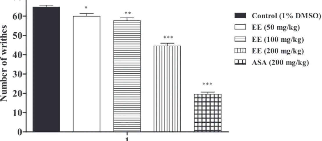
eFFectsonthe writhing resPonse inDuceDby
acetic aciD
The extract from A. fastigiatum branches at the dos-es of 50 mg/kg, 100 mg/kg and 200 mg/kg caused 3.09 (59.88±1.35; p < 0.05), 10.83 (57.62±1.40; p < 0.01) and 31.13% (44.50±1.46; p < 0.001) in-hibition of acetic-acid-induced abdominal writh-ing, respectively, when compared to control group (64.62±1.07) (Figure 1). There was a significant
difference between the doses of 50 and 200 mg/kg (p < 0.001), and 100 and 200 mg/kg (p < 0.001). The abdominal contortions were reduced 69.82% (19.50±1.12) by acetylsalicylic acid (ASA).
eFFectsonthe nocicePtion inDuceDby ForMalin
The intraplantar injection of formalin promoted a biphasic characteristic response (Figure 2). The time spent licking in the first phase (0–5 min) was 86.25±1.98 s and in the second phase (15–30 min) was 91.62±1.89 s for the control group. After 60 min of treatment, a significant reduction in the licking time (p < 0.01 or p < 0.001) was observed during the first phase (neurogenic) by 10.00 (77.62±1.43; p < 0.01), 26.67 (63.25±1.83; p < 0.001) and 41.60% (50.37±1.89; p < 0.001) with 50, 100 and 200 mg/ kg of extract, respectively (Figure 2). In the second taBle i
Total phenolic and flavonoids contents and antioxidant activity of the ethanol extract
obtained from Ageratum fastigiatum branches.
Methods Content (g/100g) EC50 (μg/mL)
Total phenolic 9.15±0.06
-Total flavonoids 2.58±0.03
-DPPH - 064.54±0.25
Fe+3 reducing power - 105.05±0.35
phase, these doses also significantly inhibited at 9.55 (82.87±1.77; p < 0.01), 23.47 (70.12±2.03; p < 0.001) and 35.20% (59.37±2.00; p < 0.001), respectively, when compared to the control. After the statistical analysis, the antinociceptive effect was different between the doses of the ethanol extract (p < 0.001) in both phases of the formalin test. As expected, morphine (5 mg/kg, s.c.) significantly reduced the formalin response in both phases.
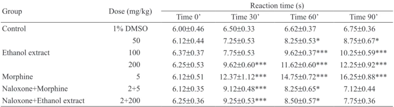
eFFectson hot-Plate latency assay
Based on the analgesic effect detected in the first phase of formalin test, the ethanol extract was
evaluated using hot plate method, a crucial model of central antinociceptive activity investigation. The effect of the extract from A. fastigiatum in the hot plate assay varied according to the doses and the time of observation (Table II). At time zero, no significant antinociceptive effect was observed, while at time 30 min, the dose of 200 mg/kg significantly increased the latency time in 48.00%. After 60 and 90 min of treatment, at the doses 50 (24.62 and 29.63 %; p < 0.05, respectively), 100 (42.32 and 51.85 %; p < 0.001, respectively) and 200 mg/kg (75.53 and 81.48 %; p < 0.001, respectively), were observed a significantly increase of the reaction time. Considering the treatment of 90 min, Figure 2 - Effects of the ethanol extract from A. fastigiatum branches on formalin-induced nociception
in mice. First phase = 0-5 min after formalin injection; second phase = 15-30 min. The experimental groups were pre-treated 60 minutes previously to the beginning of experiment. EE, ethanol extract. Data are mean±S.E.M. of 8 mice. **p < 0.01; ***p < 0.001 vs control group.
taBle ii
Effects of the ethanol extract from A. fastigiatum branches on the latency time of mice exposed to the hot plate test.
Group Dose (mg/kg) Reaction time (s)
Time 0’ Time 30’ Time 60’ Time 90’
Control 1% DMSO 6.00±0.46 6.50±0.33 6.62±0.37 6.75±0.36
50 6.12±0.44 7.25±0.53 8.25±0.53* 8.75±0.67*
Ethanol extract 100 6.37±0.37 7.75±0.53 09.62±0.37*** 10.25±0.59***
200 6.25±0.53 9.62±0.60*** 11.62±0.60*** 12.25±0.92***
Morphine 5 6.12±0.51 12.37±1.12*** 14.75±0.72*** 16.25±0.88***
Naloxone+Morphine 2+5 6.12±0.35 9.12±0.48*** 8.25±0.65* 7.12±0.44
Naloxone+Ethanol extract 2+200 6.25±0.36 9.25±0.53*** 8.50±0.57* 7.75±0.36
the doses of the ethanol extract (50, 100 and 200 mg/kg) were different (p < 0.001) after statistical comparison. The procedure was also performed in the presence of naloxone, an opioid antagonist. Naloxone inhibited the previously results observed with the extract and morphine (Table II).
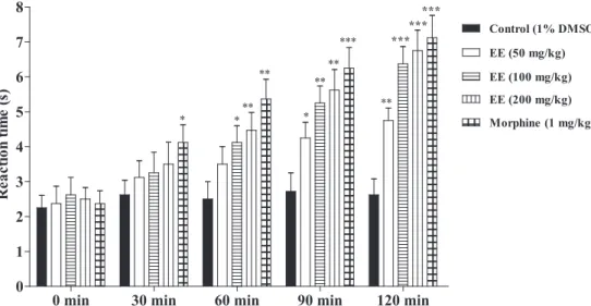
eFFectson tail iMMersion test
Figure 3 shows the effect of the ethanol extract on the latency of tail withdrawal from hot water. The
doses of 100 (4.12±0.48; p < 0.05) and 200 mg/ kg (4.87±0.51; p < 0.01) significantly increased the latency time on the tail-immersion in hot water after 60 minutes of treatment. The maximum effect was observed after 120 min of treatment at the doses of 50 (4.75±0.36; p < 0.01), 100 (6.37±0.50; p < 0.001) and 200 mg (6.75±0.59; p < 0.001) when compared to the control group (2.62±0.46). In this time, the dose of 50 mg/kg was different in relation to the doses of 100 and 200 mg/kg (p < 0.001).
Figure 3 - Effects of the ethanol extract from A. fastigiatum branches on tail-immersion test in mice. EE, ethanol extract. Data are mean±S.E.M. of 8 mice. *p < 0.05; **p < 0.01; ***p < 0.001 vs control group.
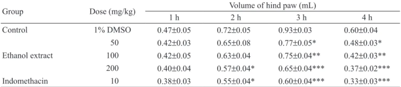
eFFectson eDeMa inDuceDby carrageenan
The anti-inflammatory effect of the ethanol extract from A. fastigiatum evaluated by the paw edema method induced by carrageenan is shown in Table III. After 2 h of carrageenan application, the paw edema was reduced in 20.83% at the dose of 200 mg/kg. The inhibition of edema was observed 3 and 4 h after injection of carrageenan at the doses of 50 (17.20 and 20.00%; p < 0.05, respectively), 100 (19.35 and 30.00%; p < 0.01, respectively) and 200 mg/kg (30.11 and 38.33%; p < 0.001, respectively) when compared with control group. In these times, indomethacin (reference drug) also inhibited the paw edema (35.48 and 45.00%; p <
0.001, respectively). After statistical comparison, the doses of 50 and 200 mg/kg were different (p < 0.01) in the treatment of 4 hours.
eFFectson carrageenan-inDuceD Pleurisy
of 100 (p < 0.01), 200 (p < 0.001) and 400 mg/kg (p < 0.001) (Table IV). Considering the exudates volume, the dose of 50 mg/kg was different of 100 (p < 0.01) and 200 mg/kg (p < 0.001). The doses of the ethanol extract (50, 100 and 200 mg/ kg) produced different values in the number of leukocytes (p < 0.01 or p < 0.001). As expected, indomethacin reduced the exudate volume and the leucocyte migration.
DiScuSSion
The phytochemical analysis of the ethanol extract from A. fastigiatum branches revealed the presence of flavonoids, tannins, coumarins, terpenes, sterols and saponins and showed expressive total phenolic and flavonoid contents (Table I). Considering these data, it is important to mention that a variety of in vitro and in vivo experiments have reported the actions of flavonoids, tannins, triterpenoids and
other secondary plant metabolites against oxidative, nociceptive and inflammatory processes (Soares-Bezerra et al. 2013, Souza et al. 2014, Serafini et al. 2010). In addition, among the active compounds identified in A. fastigiatum, the essential oils have been associated with important biological properties (Del-Vechio-Vieira et al. 2009a, b).
The ethanol extract showed a promising anti-oxidant effect since was able to inhibit the stable radical DPPH and chelate iron (Table I). Among others, this effect is related to the presence of phe-nolic constituents (phytochemical screening and total phenol and flavonoids) in A. fastigiatum that exhibit mechanism against these radicals (Rice-Evans et al. 1997, Van Acker et al. 1996). Further -more, this action can also justify the inhibition of signaling pathways that modulate pain and inflam -mation (Geronikaki and Gavalas 2006).
On the other hand, considering that the use of opioids and non-steroidal anti-inflammatory drugs
taBle iV
Effects of the ethanol extract from A. fastigiatum branches on pleural exudation and number of leucocytes in carrageenan-induced pleurisy in rats.
Group Dose (mg/kg) Exudate volume (mL) Inhibition (%) Nº Leucocytes (x 103
cells/mm3
) Inhibition (%)
Control 1% DMSO 1.68±0.08*** - 14.18±0.22***
-50 1.30±0.07*** 22.62 12.93±0.24*** 08.81
Ethanol extract 100 0.97±0.07*** 42.26 10.80±0.39*** 23.84
200 0.88±0.06*** 47.62 09.51±0.24*** 32.93
Indomethacin 10 0.72±0.06*** 57.14 08.70±0.23*** 38.64
Data are mean±S.E.M. of six rats. **p < 0.01, ***p < 0.001 vs control group. The experimental groups were pre-treated 60 minutes previously to the beginning of experiment.
taBle iii
Effects of the ethanol extract from A. fastigiatum branches on the rat paw edema induced by carrageenan.
Group Dose (mg/kg) Volume of hind paw (mL)
1 h 2 h 3 h 4 h
Control 1% DMSO 0.47±0.05 0.72±0.05* 0.93±0.03*** 0.60±0.04***
050 0.42±0.03 0.65±0.08* 0.77±0.05*** 0.48±0.03***
Ethanol extract 100 0.42±0.05 0.63±0.04* 0.75±0.04*** 0.42±0.03***
200 0.40±0.04 0.57±0.04* 0.65±0.04*** 0.37±0.02***
Indomethacin 010 0.38±0.03 0.55±0.04* 0.60±0.04*** 0.33±0.03***
exert a wide range of side effects (Slater et al. 2010), there is currently a strong interest in developing new therapeutic agents from natural products (Oh et al. 2015, Rohini and Mahesh 2015, Lehra et al. 2014). These products can inhibit different mediators that are involved in the evolution of inflammatory processes, including the pain (Lehra et al. 2014, Paduch et al. 2007). In this context, studies have been carried out with natural products in models of pain and inflammation in order to assess their pharmacological potential, as well as developing new therapeutic options (Kumar and Pandey 2013, Cragg and Newman 2013).
The acute toxicity test showed that the ethanol extract from A. fastigiatum branches was not toxic to mice. In addition, it is important to observe that the largest dose administered (200 mg/kg) was less than the lowest dose applied for determination of the LD50 (0.5 g/kg or 500 mg/kg). Previous study performed by our research group demonstrated that the essential oil from A. fastigiatum leaves showed LD50 of 2.50 g/kg (Del-Vechio-Vieira et al. 2009a). Although the constituents of the essential oil may contribute to the toxicological effects, the quantity found in the extract can not be sufficient to produce toxicity.
The writhing induced by chemical substances, as acetic acid, is due to sensitization of nociceptors by prostaglandins and this test is a classical experimental model used for the screening of drugs with analgesic activity (Collier et al. 1968). Moreover, the acetic acid causes the release of bradykinin, histamine and serotonin in the peritoneal fluid of mice (Deraedt et al. 1980). The results showed that the antinociceptive activity of the extract is dose-dependent (Figure 1) and probably this action could be due the presence of bioactive substances such as those constituents observed in the essential oil (Del-Vechio-Vieira et al. 2009a). This effect was also demonstrated in the ethanol extract from leaves (Del-Vechio et al. 2007) and it could be mediated by peripheral actions, including the prostaglandin synthesis inhibition.
The ethanol extract also produced significant inhibition in the both phases of formalin-induced pain. The formalin test is considered a model to clinical pain because it causes a local tissue injury to the paw and is also indicative of tonic and localized inflammation pain resulting in two distinct phases (Hunskaar and Hole 1987). The first phase (0-5 min after formalin injection) is due to a direct effect on nociceptors and the second phase (15-30 min after formalin injection) produces an inflammatory response and this involves different chemical mediators, such as excitatory amino acids, neuropeptides, PGE2, nitric oxide, and kinins (Hunskaar and Hole 1987, Shibata et al. 1989). Moreover, this model can be used to clarify the possible mechanism of antinociceptive effect of a proposed analgesic (Tjølsen et al. 1992). Centrally acting drugs such as opioids inhibit both phases equally, but peripherally acting drugs as aspirin, indomethacin and dexamethasone only inhibit the second phase (Rosland et al. 1990). The ethanol extract from A. fastigiatum branches was able to decrease the time that the animal spent licking the injected paw on the first and second phases (Figure 2). Taken together, these results revealed a probably similar action to the opioid and nonsteroidal anti-inflammatory drugs.
response latency time to the heat stimulus (Table II and Figure 3). These tests are also considered to be sensitive to drugs acting at the supraspinal modulation level of the pain response (Yaksh and Rudy 1977), suggesting at least a modulatory effect of the investigated extract. Our results indicate that the analgesia induced by the ethanol extract can be dependent on the opioid system, since previous treatment with naloxone changed the observed data (Table II). As expected, morphine (1 or 5 mg/ kg) significantly increased the latency time to the nociceptive response when compared with the control group.
Carrageenan-induced rat paw edema is a suitable test for evaluating anti-inflammatory drugs which has frequently been used to assess the anti-edematous effect of the natural products (Del-Vechio-Vieira et al. 2009a, Oh et al. 2015, Rohini and Mahesh 2015). This is a model of acute inflammation that involves different phases (Vinegar et al. 1969). The first phase (1-2 h) is related with the release of serotonin and histamine; kinins play a role in the middle phase (Di Rosa and Sorrentino 1968), while prostaglandins appear to be the most important mediators in the second phase (3–5 h) of the postcarrageenan response as a resulted of induction of isoforms of cyclooxygenase (Di Rosa et al. 1971, Di Rosa 1972, Nantel et al. 1999). The result of the present study indicates that the suppression of the first phase may be due to inhibition of the release of early mediators, such as histamine and serotonin, and the action in the second phase may be explained by an inhibition of cyclooxygenase with reduced expression of prostaglandins. In this context, the ethanol extract (50, 100 and 200 mg/kg) from A. fastigiatum branches and indomethacin play a crucial role as protective factors against the carrageenan-induced acute inflammation (Table III).
To better understanding of the anti-inflamma -tory effect demonstrated in the paw edema model, the induction of pleurisy with injection of carra-geenan into the pleural cavity of rats was made.
This test elicits an acute inflammatory response, characterized by the accumulation of fluid contain -ing large number of leucocytes (Ammendola et al. 1975, Almeida et al. 1980, Capasso et al. 1975). It is an interesting method that evaluates the leuco-cyte migration during the inflammatory process. Anti-inflammatory drugs, such as indomethacin and dexamethasone, inhibit the accumulation of exudates and mobilization of leucocytes between 3 and 6 h after application of carrageenan (Vinegar et al. 1973, Almeida et al. 1980, Miyasaka and Mi -kami 1982). Our results showed that the ethanol extract from A. fastigiatum branches inhibited the formation of pleural exudate and the leucocyte mi-gration confirming the anti-inflammatory activity (Table IV).
concluSion
The results obtained through the in vitro and in vivo experiments performed in the present study add more subsidies to the use of the ethanol extract from A. fastigiatum branches by population in the Brazilian folk medicine as antioxidant, analgesic and anti-inflammatory. Based on our data, A. fas-tigiatum can be an active source of bioactive sub-stances, representing promising targets for future medicines to new therapeutic purposes. However, further studies should be conducted to ensure the safety, feasibility and sustainability of usage.
acKnoWleDGmentS
The authors are grateful to Fundação de Amparo à Pesquisa do Estado de Minas Gerais (FAPEMIG) and to Coordenação de Aperfeiçoamento de Pes-soal de Nível Superior (CAPES) for financial sup -port and to Dr. Roberto Lourenço Esteves for plant identification.
reFerenceS
ABBOTT FV AND MELzACK R. 1982. Brainstem lesions dissociate neural mechanisms of morphine analgesia in
AHMED AU. 2011. An overview of inflammation: mechanism and consequences. Front Biol 6: 274-281.
ALIE N, ELDIB M, FAyAD zA AND MANI V. 2014. Inflam-mation, atherosclerosis, and coronary artery disease: PET/ CT for the evaluation of atherosclerosis and inflammation. Clin Med Insights Cardiol 8: 13-21.
ALMEIDA AM, PRADO PI AND LEWINSOHN TM. 2004. Geographical distribution of Eupatorieae (Asteraceae) in South-eastern and South Brazilian Mountain Ranges. Plant Ecol 174: 163-181.
ALMEIDA AP, BAyER BM, HORAKOVA z AND BEAVEN MA. 1980. Influence of indomethacin and other anti-inflammatory drugs on mobilization and production of neutrophils: Studies with carrageenan-induced inflammation in rats. J Pharmacol Exp Ther 214: 74-79. AMIM S, KALOO zA, SINGH S AND ALTAF T. 2013.
Mi-cropropagation of medicinally important plant species of
family Asteraceae – a review. Int J Rec Sci Res 4:
1296-1303.
AMMENDOLA G, DI ROSA M AND SORRENTINO L. 1975. Leucocyte migration and lysosomal enzymes release in rat carrageenin pleurisy. Agents Actions 5: 250-255. BANSODE VJ, VyAWAHARE NS, MUNJAL NB, GORE
PN, AMRUTKAR PS AND SONTAKKE SR. 2014. Ameliorative effect of ethyl pyruvate in neuropathic pain induced by chronic constriction injury of sciatic nerve. Indian J Pain 28: 82-88.
BOHLMANN F, AHMED M, KING RM AND ROBINSON H. 1981. Labdane and eudesmane derivatives from Ageratum fastigiatum. Phytochemistry 20: 1434-1435. BOHLMANN F, LUDWIG GW, JAKUPOVIC J, KING RM
AND ROBINSON H. 1983. Daucanolide further farnesene derivatives from Ageratum fastigiatum. Phytochemistry 22: 983-986.
CAPASSO F, DUNN CJ, yAMAMOTO S, WILLOUGHBy DA AND GIROUD JP. 1975. Further studies on carrageen-an-induced pleurisy in rats. J Pathol 116: 117-124. COLLIER HO, DINNEEN LC, JOHNSON CA AND
SCHNEIDER C. 1968. The abdominal response and its suppression by analgesic drugs in the mouse. Br J Pharmacol Chemother 32: 295-310.
CRAGG GM AND NEWMAN DJ. 2013. Natural products: a continuing source of novel drug leads. Biochim Biophys Acta 1830: 3670-3695.
DEL-VECHIO G, SOUSA OV, yAMAMOTO CH AND KAPLAN MAC. 2007. Antinociceptive and antimicrobial activities of Ageratum fastigiatum (Gardn.) R. M. King et H. Hob. (Asteraceae). Rev Bras Farm 88: 181-184. DEL-VECHIO-VIEIRA G, BARBOSA MVD, LOPES BC,
SOUSA OV, SANTIAGO-FERNANDES LDR, ESTEVES RL AND KAPLAN MAC. 2008. Caracterização morfoanatômica de Ageratum fastigiatum (Asteraceae). Rev Bras Farmacogn 18: 769-776.
DEL-VECHIO-VIEIRA G, SOUSA OV, MIRANDA MA, SENNA-VALLE L AND KAPLAN MAC. 2009a. Analgesic and anti-inflammatory properties of essential oil from Ageratum fastigiatum. Braz Arch Biol Technol 52: 1115-1121.
DEL-VECHIO-VIEIRA G, SOUSA OV, yAMAMOTO CH AND KAPLAN MAC. 2009b. Chemical composition and antimicrobial activity of the essential oils of Ageratum fastigiatum. Rec Nat Prod 3: 52-57.
DERAEDT R, JOUQUEy S, DELEyALLéE F AND FLAHAUT M. 1980. Release of prostaglandins E and F in an algogenic reaction and its inhibition. Eur J Pharmacol 61: 17-24.
DI ROSA M. 1972. Biological properties of carrageenan. J Pharm Pharmacol 24: 89-102.
DI ROSA M, GIROUD JP AND WILLOUGBy DA. 1971. Studies of the mediators of the acute inflammatory response induced in rats in different sites by carrageenan and turpentine. J Pathol 104: 15-29.
DI ROSA M AND SORRENTINO L. 1968. The mechanism of the inflammatory effect of carrageenan. Eur J Pharmacol 4: 340-342.
EDDy NB AND LEIMBACH D. 1953. Synthetic analgesics. II. Dithienylbutenyl-and dithienylbutylamines. J Pharma-col Exp Ther 107: 385-393.
GANTy P AND CHAWLA R. 2014. Complex regional pain syndrome: recent updates. Contin Educ Anaesth Crit Care Pain 14: 79-84.
GERONIKAKI AA AND GAVALAS AM. 2006. Antioxidants and inflammatory disease: synthetic and natural antioxi-dants with anti-inflammatory activity. Comb Chem High Throughput Screen 9: 425-442.
GONçALVES LD, ALMEIDA HR, OLIVEIRA PM, LOPES NP, TURATTI ICC, ARCHANJO FC AND GRAEL CFF. 2011. Contribution for the phytochemical studies of Ageratum fastigiatum. Rev Bras Farmacogn 21: 936-942. HARIJITH A, EBENEzER DL AND NATARAJAN V. 2014.
Reactive oxygen species at the crossroads of inflammasome and inflammation. Front Physiol 5: 1-11.
HUNSKAAR S AND HOLE K. 1987. The formalin test in mice: Dissociation between inflammatory and non-inflammatory pain. Pain 30: 103-114.
KLöTING N AND BLüHER M. 2014. Adipocyte dysfunction, inflammation and metabolic syndrome. Rev Endocr Metab Dosord 15: 277-287.
KUMAR S, BAJWA BS, KULDEEP S AND KALIA AN. 2013. Anti-inflammatory activity of herbal plants: A review. Int J Adv Pharm Biol Chem 2: 272-281.
KUMAR S AND PANDEy AK. 2013. Chemistry and biological activities of flavonoids: An overview. Sci World J 2013: 1-16.
LORKE D. 1983. A new approach to practical acute toxicity testing. Arch Toxicol 54: 275-287.
LITCHFIELD JT AND WILCOxON FA. 1949. A simplified method of evaluating dose-effect experiments. J Pharmacol Exp Ther 96: 99-113.
MENSOR LL, MENEzES FS, LEITãO GG, REIS AS, SANTOS TC, COUBE CS AND LEITãO SG. 2001. Screening of Brazilian plant extracts for antioxidant activity by the use of DPPH free radical method. Phytother Res 15:127-130.
MIyASAKA K AND MIKAMI T. 1982. Comparison of the anti-inflammatory effects of dexamethasone, indomethacin and BW755C on carrageenin-induced pleurisy in rats. Eur J Pharmacol 77: 229-236.
NANTEL F, DENIS D, GORDON R, NORTHEy A, CIRINO M, METTERS KM AND CHAN CC. 1999. Distribution and regulation of cyclooxygenase-2 in carrageenan-induced inflammation. Br J Pharmacol 128: 853-859. NEMIROVSKy A, zELMAN V AND JURNA I. 2011. The
antinociceptive effect of the combination of spinal mor-phine with systemic mormor-phine or buprenormor-phine. Anesth Analg 93: 197-203.
OH yC, JEONG yH, CHO WK, HA JH, GU MJ AND MA Jy. 2015. Anti-inflammatory and analgesic effects of pyeong-wisan on LPS-stimulated murine macrophages and mouse models of acetic acid-induced writhing response and xy-lene-induced ear edema. Int J Mol Sci 16: 1232-1251. OKUNADE AL. 2002. Ageratum conyzoides L. (Asteraceae).
Fitoterapia 73: 1-16.
OyAIzU M. 1986. Studies on product of browning reaction-antioxidative activities of products of browning reaction prepared from glucosamine. Jpn J Nutr 44: 307-315. PADUCH R, KANDEFER-SzERSzéN M, TRyTEK M AND
FIEDUREK J. 2007. Terpenes: substances useful in human healthcare. Arch Immunol Ther Exp 55: 315-327.
RAMABADRAN K, BANSINATH M, TURNDORF H AND PUIG MM. 1989. Tail immersion test for the evaluation of a nociceptive reaction in mice: methodological consider-ations. J Pharmacol Methods 21: 21-31.
RICE-EVANS C, MILLER N AND PAGANGA G. 1997. An-tioxidant properties of phenolic compounds. Trends Plant Sci 2: 152-159.
ROHINI RM AND MAHESH D. 2015. Evaluation of anti-inflammatory and antinociceptive activity and isolation of
two new alkaloids from leaves extract of Tabernaemontana sananho. J Chem Pharm Res 7: 31-36.
ROKyTA R, HOLECEK V, PEKáRKOVá I, KREJCOVá J, RACEK J, TREFIL L AND yAMAMOTOVá A. 2003. Free radicals after painful stimulation are influenced by antioxidants and analgesics. Neuroendocrinol Lett 24: 304-309.
ROSLAND JH, TJøLSEN A, MAEHLE B AND HOLE K. 1990. The formalin test in mice: effect of formalin concen-tration. Pain 42: 235-242.
SCHMAUSS C AND yAKSH TL. 1984. In vivo studies on spinal opiate receptor systems mediating antinociception. II. Pharmacological profiles suggesting a differential
association of mu, delta and kappa receptors with visceral
chemical and cutaneous thermal stimuli in the rat. J Pharmacol Exp Ther 228: 1-12.
SCHMID-SCHöNBEIN GW. 2006. Analysis of inflammation. Annu Rev Biomed Eng 8: 93-151.
SEN S, CHAKRABORTy R, SRIDHAR C, REDDy ySR AND DE B. 2010. Free radicals, antioxidants, diseases and phytomedicines: current status and future prospect. Int J Pharm Sci Rev Res 3: 91-100.
SERAFINI M, PELUSO I AND RAGUzzINI A. 2010. Flavonoids as anti-inflammatory agents. Proc Nutr Soc 69: 273-278.
SHAH BN, SETH AK AND MAHESHWARI KM. 2011. A review on medicinal plants as a source of anti-inflammatory agents. Res J Med Plant 5: 101-105.
SHIBATA M, OHKUBO T, TAKAHASHI H AND INOKI R. 1989. Modified formalin test: characteristic biphasic pain response. Pain 38: 347-352.
SLATER D, KUNNATHIL S, MCBRIDE J AND KOPPALA R. 2010. Pharmacology of nonsteroidal antiinflammatory drugs and opioids. Semin Intervent Radiol 27: 400-411. SOARES-BEzERRA RJ, CALHEIROS AS, FERREIRA
NCS, FRUTUOSO VS AND ALVES LA. 2013. Natural products as a source for new anti-inflammatory and anal-gesic compounds through the inhibition of purinergic P2x receptors. Pharmaceuticals 6: 650-658.
SOBRINHO TJSP, SILVA CHTP, NASCIMENTO JE, MONTEIRO JM, ALBUQUERQUE UP AND AMORIM ELC. 2008. Validação de metodologia espectrofotométrica para quantificação dos flavonóides de Bauhinia cheilantha (Bongard) Steudel. Rev Bras Ciênc Farm 44: 683-689. SOUSA CMM, SILVA HR, VIEIRA-JUNIOR GM, AyRES
CLSC, ARAUJO DS, CAVALCANTE LCD, BARROS EDS, ARAUJO PBM, BRANDAO MS AND CHAVES MH. 2007. Fenóis totais e atividade antioxidante de cinco plantas medicinais. Quím Nova 30: 351-355.
SOUzA MT, ALMEIDA JR, ARAUJO AA, DUARTE MC, GELAIN DP, MOREIRA JC, SANTOS MR AND QUINTANS-JúNIOR LJ. 2014. Structure–activity
relationship of terpenes with anti-inflammatory profile –
a systematic review. Basic Clin Pharmacol Toxicol 115: 244-256.
TESLIM OA, VyVIENNE M’K, OLATOKUNBO OM, OLUWAFISAyO AJ, MLENzANA NB, SHAMILA M, NESTO T AND GRACE M. 2014. Side effects of non-steroidal anti-inflammatory drugs: The experience of
patients with musculoskeletal disorders. Am J Health Res
2: 106-212.
TJøLSEN A, BERGE OG, HUNSKAAR S, ROSLAND JH AND HOLE K. 1992. The formalin test: an evaluation of the method. Pain 51: 5-17.
VAN ACKER SABE, DE GROOT MJ, VAN DEN BERG DJ, TROMP MNJL, DEN KELDER GDO, VAN DER VIJGH WJF AND BAST A. 1996. A quantum chemical explanation of the antioxidant activity of flavonoids. Chem Res Toxicol 9: 1305-1312.
VINEGAR R, SCHREIBER W AND HUGO R. 1969. Biphasic development of carrageenin edema in rats. J Pharmacol Exp Ther 166: 96-103.
VINEGAR R, TRUAx JF AND SELPH JL. 1973. Some quan-titative temporal characteristics of carrageenin-induced pleurisy in the rat. Proc Soc Exp Biol Med 143: 711-714. WINTER CA, RISLEy EA AND NUSS GW. 1962.
Carrageenin-induced edema in hind paw of the rat as an assay for antiinflammatory drugs. Proc Soc Exp Biol Med 111: 544-547.