Article
J. Braz. Chem. Soc., Vol. 25, No. 11, 2108-2120, 2014. Printed in Brazil - ©2014 Sociedade Brasileira de Química 0103 - 5053 $6.00+0.00
A
*e-mail: [email protected]
Plumeran Alkaloids and Glycosides from the Seeds of
Aspidosperma pyrifolium
Mart.
Patrícia C. N. Nogueira,a Renata M. Araújo,b Glauce S. B. Viana,c
Dayane P. de Araújo,c,d Raimundo Braz Filhoe and Edilberto R. Silveira*,a
aDepartamento de Química Orgânica e Inorgânica, Universidade Federal do Ceará, CP 12200, 60021-940 Fortaleza-CE, Brazil
bInstituto de Química, Universidade Federal do Rio Grande do Norte, CP 1524, 59072-970 Natal-RN, Brazil
cLaboratório de Neurofarmacologia, Universidade Federal do Ceará, CP 3157, 60430-270 Fortaleza-CE, Brazil
dLaboratório de Neurologia Experimental, Faculdade de Ciências da Saúde (FACS), Universidade do Estado do Rio Grande do Norte (UERN), 59607-360 Mossoró-RN, Brazil
eLaboratório de Ciências Químicas, Universidade Estadual do Norte Fluminense, Campos dos Goytacazes, 28013-602 Rio de Janeiro-RJ, Brazil
A fração aquosa resultante da partição líquido-líquido do extrato etanólico de sementes de Aspidosperma pyrifolium apresentou atividades antinociceptiva e anti-inflamatória nos testes de formalina, contorção abdominal induzida pelo ácido acético e edema de pata por carragenina. O estudo fitoquímico conduziu ao isolamento de um novo alcaloide com esqueleto plumerano rearranjado, o (–)-(3S,7S,21R)-rel-(3αH)-15(14→ 3)-abeo-2,16,17,20,6,7-hexahidro-15H,8aH,16a,20a-etano-1H-indolizino[3,1-cd]carbazol, além de seis alcaloides plumeranos conhecidos, um alcaloide tetra-hidro-β-carbolínico, e os heterosídeos de dois iridoides e do ácido salicílico, que estão sendo descritos pela primeira vez para a espécie. Dos alcaloides já descritos na literatura, um está sendo relatado pela primeira vez como produto natural, de origem vegetal. As estruturas dos compostos foram determinadas através de análises por ressonância magnética nuclear (NMR), uni e bidimensionais, infravermelho por transformada de Fourier (FT-IR) e espectroscopia de massas de alta resolução com ionização por electrospray (HRESIMS), além da comparação com dados da literatura. A revisão dos dados de NMR de alguns alcaloides, e as correspondentes correlações estruturais, também são sugeridas.
The residual aqueous fraction of the liquid-liquid partition of the ethanol extract from seeds of
Aspidosperma pyrifolium showed antinociceptive and anti-inflammatory activities in the formalin test, abdominal writhing induced by acetic acid, and paw edema induced by carrageenan. Its phytochemical analysis led to the isolation of a new alkaloid with a rearranged plumeran skeleton, the (–)-(3S,7S,21R)-rel-(3αH)-15(14→3)-abeo-2,16,17,20,6,7-hexahydro-15H,8aH ,16a,20a-ethano-1H-indolizino[3,1-cd]carbazole, in addition to six other known plumeran alkaloids, one tetrahydro-β-carboline alkaloid, and the glycosides of two iridoids and of salycilic acid, that are being reported for the first time for A. pyrifolium. One of the known alkaloids is being reported for the first time from a natural source. The nuclear magnetic resonance (NMR) data, and the correspondent assignments, of some alkaloids already reported in the literature are suggested to be revised. Structures of all compounds were characterized by 1D and 2D NMR and Fourier transform infrared (FT-IR) spectroscopies and high-resolution electrospray ionization mass spectrometry (HRESIMS), and comparison with data from literature.
Keywords: plumeran alkaloids, Aspidosperma pyrifolium, biological activity, 1H and 13C NMR
Introduction
The Aspidosperma genus is reported as a prolific source of indole alkaloids, substances of great interest as a function of their structural diversity. Several biological activities associated with this class of compounds have been reported,1-5 for example, the hypotensive and analgesic activities of A. quebracho-blanco,4 and the antimicrobial and cytotoxic activities of A. marcgranianum.4 In addition, anti-inflammatory, anticancer, antimalarial, antiulcer, antileishmanial and healing activities are attributed to alkaloids from other Aspidosperma species.1,2,4,5
Aspidosperma pyrifolium Mart., popularly known as “pereiro preto” (Port. lit. = dark pereiro), is a shrub, sometimes a small tree, widely distributed in the northeastern Brazil flora. It is largely used in carpentry due to the excellent quality of its wood.6 Previous works report on the hypotensive effect4,6,7 shown by the alkaloids present in the root bark and leaves of A. pyrifolium, as well the antiplasmodial activity8 revealed by some alkaloids with aspidospermane skeleton, for instance aspidospermine, isolated from the stem bark. No antinociceptive or anti-inflammatory activities have been reported so far. Despite the report of about 27 dihydroindole alkaloids with plumeran skeleton, isolated from leaves, roots and trunk bark of A. pyrifolium,4,6,7 there is no report in the literature for the phytochemical study of its seeds.
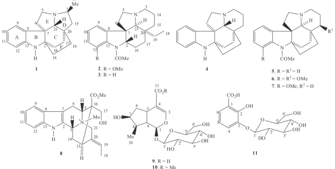
This work reports on the isolation and structural elucidation of an unknown alkaloid with a rearranged plumeran skeleton, the (–)-(3S,7S,21R)-rel-(3αH)-15(14→ 3)-abeo-2,16,17,20,6,7-hexahydro-15H,8aH,16a,20a-ethano-1H
-indolizino[3,1-cd]carbazole (1), as well as of ten known compounds: the plumeran alkaloids aspidospermine (2),6,7 demethoxyaspidospermine (3),9,10 aspidofractinine (4),11,12
N-acetylaspidofractinine (5),13,14 pirifoline (6),6,7 15-demethoxypirifoline (7),6,11 the tetrahydro-β-carboline alkaloid N-methylakuammidine (8),15,16 the iridoid glycoside of loganic acid (9)17-19 and of its methyl derivative loganin (10),20 and the salycilic acid derivative, 2-hydroxy-3-O-β-D-glucopyranosylbenzoic acid (11),19,21 sometimes referred to as pyrocatechuic acid 3-O-β-D-glucoside,22 all obtained after the phytochemical investigation of the ethanol extract from seeds of A. pyrifolium (Figure 1). Although known, compounds 8-11 are being reported for the first time for the species, and compound 5 is reported for the first time from a natural source. The nuclear magnetic resonance (NMR) data assignments of several alkaloids already reported in the literature have been reviewed and are suggested to be revised.
Experimental
General experimental procedures
The 1D and 2D NMR spectra were acquired on a Bruker Avance DRX-500 and/or DPX-300 spectrometers, using CD3OD, C5D5N, (CD3)2SO or CDCl3 as solvents. All standard pulse sequences were provided by the TopSpin software from Bruker. The Fourier transform infrared (FT-IR) spectra were obtained on a Perkin Elmer Spectrum 1000 spectrometer, using an universal attenuated total reflectance accessory (UATR). The high-resolution electrospray ionization mass
spectra (HRESIMS) were acquired using a Shimadzu LCMS-IT-TOF (225-07100-34) spectrometer. Specific rotations were measured on a Jasco polarimeter model P-2000. Column chromatography was performed using silica gel 60 (EMD, 70-230 mesh) and Sephadex LH-20 from Pharmacia Fine Chemicals. Thin layer chromatography (TLC) was performed on precoated silica gel aluminum plates (TLC Silica gel 60 F254, from Merck). The compound spots were visualized by UV light detection and/or sprayed with a solution of vanillin/perchloric acid/EtOH followed by heating, as well as with the Dragendorff reagent. The analyses by high-performance liquid chromatography (HPLC) were performed on a Waters chromatograph, using semi-preparative (250 × 10 mm, 5 µm) and analytical (250
× 4.6 mm, 5 µm) Phenomenex RP-18 columns, with flow rate of 4.72 and 1.0 mL min-1, respectively.
Plant material
The seeds of A. pyrifolium were collected in Cabrobó city neighborhoods (Pernambuco, Brazil), in June 2010. The material was identified by comparison with a specimen collected in December 2004 from “Fazenda não me deixes”, in Quixadá (Ceará, Brazil). A voucher specimen has been deposited at the Herbarium Prisco Bezerra at Universidade Federal do Ceará, under the registration number 35524.
Extraction and isolation
The seeds of A. pyrifolium (835.0 g), dried and crushed, were extracted by maceration with hexane (3 × 2 L) at room temperature, yielding, after solvent evaporation under reduced pressure, 65.2 g of a dark viscous extract. The plant residue was extracted with ethanol (3 × 2 L) and the solution was concentrated in a rotary evaporator, providing 162.5 g of dark solid extract, denominated APSE. An aliquot of 30.0 g of APSE was suspended in 300 mL of water. After resting for a few minutes, there was the formation of an insoluble residue, that was filtered, yielding the fraction denominated APSE/I. The remaining aqueous solution was partitioned successively with hexane (2 × 100 and 1 × 50 mL), CH2Cl2 (2 × 100 and 2 × 50 mL) and EtOAc (2 × 100 and 1 × 50 mL), yielding the correspondent fractions APSE/H, APSE/D and APSE/EA, respectively. The remaining aqueous residue was designated APSE/Aq. This procedure was repeated four times (4 × 30 g) and the fractions were compared by thin layer chromatography and combined accordingly, resulting in 17.2 g of APSE/I (11.47%), 2.5 g of APSE/H (1.67%), 20.8 g of APSE/D (13.87%), 1.0 g of APSE/EA (0.67%) and 91.2 g of APSE/Aq (60.8%). Due to its higher solubility in water, and so, due to the easiness of administering the drug
solutions were extracted with EtOAc and the organic fractions were washed with water, dried with Na2SO4 and concentrated in a rotary evaporator, giving the respective deprotonated compounds.
(–)-(3S,7S,21R)-rel-(3αH)-15(14→ 3)-Abeo-2,16,17,20,6,7-hexahydro-15H,8aH ,16a,20a-ethano-1H-indolizino[3,1-cd]carbazole (1): brown resin, [α]D
20
–21.5 ± 0.4 (c 0.40, MeOH); IR νmax/cm-1 3345 (N–H), 2924 and 2858 (Csp3–H), 1606 and 1459 (skeletal Ø), 1101 (C–N); HRESIMS m/z 281.2016 [M + H]+ (calcd. for C19H25N2
+, 281.2017); 1H and 13C NMR (see Table 1).
(+)-Aspidospermine (2): white needle crystals, m.p. 202.5-204.5 °C, [α]D20+89.0 (c 0.19, CHCl3); IR νmax/cm-1 1606, 1496 and 1452 (skeletal Ø), 1639 (C=O), 1439 (CH2), 1381 (CH3); HRESIMS m/z 355.2369 [M + H]+ (calcd. for C22H31N2O2
+, 355.2385); 13C NMR (see Table 2).
(+)-Demethoxyaspidospermine (3): brown resin, [α]D 20
+25.4 (c 0.11, MeOH); IR νmax/cm-1 1667 (C=O), 1198 and 1130 (C–N), 1599, 1480 and 1405 (skeletal Ø); HRESIMS
m/z 325.2325 [M + H]+ (calcd. for C
21H29N2O+, 325.2279); 13C NMR (see Table 2).
(–)-Aspidofractinine (4): red amorphous solid, [α]D 20
–8.35 (c 0.23, MeOH); IR νmax/cm-1 3341 (N–H), 2925 and 2856 (Csp3–H), 1607 and 1459 (skeletal Ø), 746 (C–Har); HRESIMS m/z 281.2016 [M + H]+ (calcd. for C
19H25N2+, 281.2017); 13C NMR (see Table 2).
(+)-N-Acetylaspidofractinine (5): orange amorphous solid, m. p. 104.9-107.0 °C, [α]D20 +31.5 ± 1 (c 0.15, MeOH); IR νmax/cm
-1 1646 (C=O), 1481 (CH
2), 1379 (CH3), 1177 and 1124 (C–N); HRESIMS m/z 323.2124 [M + H]+ (calcd. for C21H27N2O
+, 323.2123); 1H and 13C NMR (see
Table 3).
(+)-Pirifoline (6): red resin, [α]D20 +56.7 (c 0.05, MeOH); IR νmax/cm
-1 2942 and 2873 (C
sp3–H), 1656 (C=O), 1190 and 1131 (C–N or C–O); HRESIMS m/z 383.2386 [M + H]+ (calcd. for C23H31N2O3
+, 383.2335); 13C NMR (see Table 2).
(+)-Demethoxypirifoline (7): brown resin, [α]D 20 +59.4
(c 0.24, MeOH); IR νmax/cm-1 1659 (C=O), 1194 and 1130 (C–N or C–O), 1459 (CH2) and 1375 (CH3); HRESIMS
m/z 353.2287 [M + H]+ (calcd. for C
22H29N2O2+, 353.2229); 13C NMR (see Table 2).
(–)-N-Methylakuammidine (8): brown amorphous solid, m.p. 221.0-223.5 ºC, [α]D20 –15.7 (c 0.43, MeOH); IR
νmax/cm-1 3343 (O–H and N–H), 2947 (Csp3–H), 1731 (C=O), 1637 (C=C), 1625 and 1454 (skeletal Ø), 744 (C−Har); HRESIMS m/z 367.2072 [M]+ (calcd. for C22H27N2O3
+, 367.2021); 13C NMR (see Table 2).
(–)-Loganic acid (9): yellow amorphous solid, [α]D 20
–58.9 (c 0.12, MeOH); IR νmax/cm-1 3327 (O–H), 2930 and 2880 (Csp3–H), 1674 (C=O), 1633 (C=C), 1065 and 1027 (C–O), 995 (C–H); HRESIMS m/z 375.1360 [M – H]– (calcd. for C16H23O10
–, 375.1291).
(–)-Loganin (10): white crystal solid, m.p. 220-222 ºC, [α]D20 –56.7 (c 0.34, MeOH); IR νmax/cm-1 3348 (O–H), 1680 (C=O), 1632 (C=C), 1066 and 1028 (C–O); HRESIMS m/z
413.1499 [M + Na]+ (calcd. for C
17H26O10Na+, 413.1423).
2-Hydroxy-3-O-β-D-glucopyranosylbenzoic acid (11): gray amorphous solid, m.p. 127.5-129.5 ºC; IR
νmax/cm-1 3239 (O–H), 1707 (C=O), 1586, 1499 and 1468 (skeletal Ø), 1075, 1051 and 1032 (C–O); HRESIMS
m/z 315.0723 [M – H]– (calcd. for C
13H15O9–, 315.0716) and m/z 339.0681 [M + Na]+ (calcd. for C
13H16O9Na +,
339.0692).
Antinociceptive and anti-inflammatory activities
Animals
Male Swiss mice (weighing 25-30 g), 5-8 per group, were used for the tests. The animals were housed in standard environmental conditions (22 ± 1 °C, humidity 60 ± 5%, 12 h light, 12 h dark cycle) with free access to water and food. All experiments were performed in accordance with the NIH Guide for Care and Use of Laboratory Animals.
Experimental protocol
The animals were divided into six groups and received distilled water (control), APSE-Aq (1, 10, 50, 100 and/or 200 mg kg-1) and indomethacin (10 mg kg-1) or morphine (5 mg kg-1) intraperitoneally (ip). After 30 min of the last treatment, the animals were subjected to the following tests: abdominal writhing induced by acetic acid, the formalin test and paw edema induced by carrageenan.
Abdominal writhing induced by acetic acid
Formalin test
Mice were injected with formalin (20 µL 1% formalin) intraperitoneally under the ventral surface of the right hind paw. The amount of time spent licking the injected paw was timed with a chronometer and was considered as indicative of nociception. The initial nociceptive response peaked 5 min after formalin injection (early phase) and 20-25 min after formalin injection (late phase), representing the tonic and inflammatory pain responses, respectively.24
Paw edema induced by carrageenan
The animals received an intraplantar injection of 1% carrageenan (100 µL) to induce the edema in the right hind paw. The volume of the paws was measured before and 1, 2, 3, 4 and 24 h after the administration of carrageenan.25
The volume of the edema, in milliliters, was measured using a pletismometer (Ugo Basile, Italy), where the right hind paw was submerged up to the tibio-tarsal joint, in the measuring chamber of the device. The volume of fluid displaced was recorded and considered the volume of the paw. The results were expressed as the difference between
the volume of the paw at the specified time intervals and the volume before the carrageenan injection (t = 0).
Statistical analyses
The results are presented as the mean ± the standard error of the mean (SEM). The statistical differences between the groups were analyzed by one-way analysis of variance (ANOVA) followed by the Student-Newman-Keuls multiple comparisons test. To analyze the data from the paw edema induced by carrageenan tests, two-way ANOVA followed by a Bonferroni post hoc test was used. GraphPad software (GraphPad Software, San Diego, CA, USA) was used in these analyses. Values of p < 0.05 were considered significant.
Results and Discussion
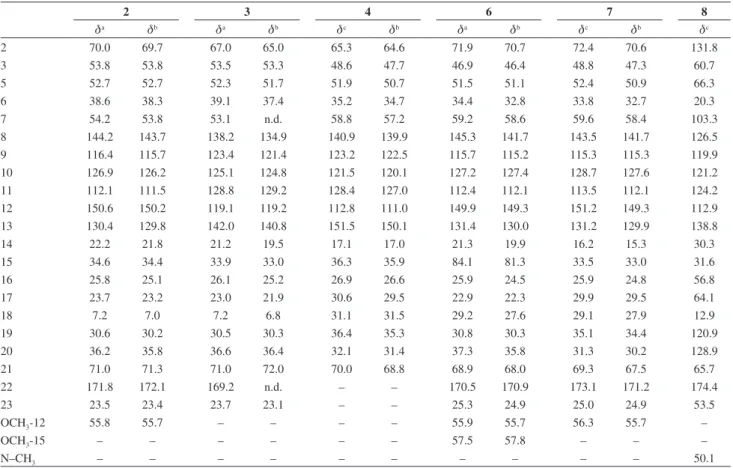
Compound 1 was isolated from APSE/Aq, after purification by HPLC. It was obtained as a brown resin, [α]D20 –21.5 ± 0.4 (c 0.40, MeOH), and gave a positive
Table 1. 1H and 13C NMR (CD
3OD) data assignments for the protonated and deprotonated forms of compound 1
1 (protonated) 1 (deprotonated) HMBC (1 deprotonated)
dCa dHb (m, J / Hz) dCc dHb (m, J / Hz) 2JCH 3JCH
2 65.6 – 65.8 – 2H-16; H-18β H-6α; H-17α
3 71.9 4.12 (sept, 6.0) 69.2 3.52 (m) 3H-14; H-15b 2H-5
5 55.7 3.90 (brq) 56.3 3.53 (m) H-6α H-3; H-21
3.64 (ddd, 12.5, 10.0, 3.0) 3.13 (ddd, 12.0, 10.0, 3.0)
6 35.5 2.99 (dt, 14.0, 10.0) 35.8 2.75 (dt, 14.0, 10.0) – H-21
1.79 (m) 1.51 (m)
7 58.2 – 58.7 – 2H-6; H-21 H-5b; H-9; H-16α; H-18β
8 135.8 – 138.6 – – 2H-5; H-10; H-12; H-21
9 123.4 7.24 (d, 7.5) 123.7 7.28 (d, 7.5) H-10 H-11
10 121.3 6.80 (t, 7.5) 121.2 6.76 (td, 7.5, 1.0) – H-12
11 129.8 7.09 (t, 7.5) 128.9 7.02 (td, 7.5, 1.0) H-10 H-9
12 112.8 6.73 (d, 7.5) 112.6 6.68 (d, 7.5) H-11 H-10
13 151.6 – 151.8 – – H-9; H-11
14 18.1 1.58 (d, 6.0) 21.6 1.34 (d, 6.5) – H-15b
15 44.7 2.09 (dd, 12.5, 6.0) 46.1 1.85 (m) H-3 3H-14; H-17β
1.68 (t, 12.5) 1.41 (m)
16 26.9 2.14 (m) 27.4 2.09 (m) 2H-17 2H-18
1.98 (m) 1.85 (m)
17 29.6 2.13 (m) 30.2 1.94 (m) – H-15b; H-19β; H-21
1.75 (m) 1.55 (m)
18 32.1 1.82 (m) 32.8 1.81 (m) H-19β H-16β
1.53 (m) 1.42 (m)
19 31.4 1.92 (m) 31.9 1.81 (m) – H-15b; H-17α; H-21
1.60 (m) 1.46 (m)
20 42.5 – 42.8 – H-15b; H-17α; H-19β –
21 79.7 4.16 (s) 79.9 3.60 (s) – H-15a; H-17α
test to the Dragendorff reagent after TLC analysis. Its molecular formula, C19H24N2 (9 double bond equivalents), was deduced by HRESIMS, which showed a protonated molecule peak at m/z 281.2016 [M + H]+ (calcd. 281.2017). The FT-IR spectrum exhibited absorptions bands at 3345 cm-1, indicating the presence of N–H bond, 2924 and 2858 cm-1, characteristic of C–H bonds of sp3 carbons, 1101 cm-1, characteristic of C–N bond, and bands at 1606 and 1459 cm-1, typical of skeletal bands of aromatic ring, in addition to the strong band at 744 cm-1, typical of the out-of-plane bending of benzenoid =C–H. The 13C-composite pulse decoupling (CPD) NMR spectrum (Table 1) exhibited 19 lines, compatible with the suggested molecular formula and, by comparison with 13C-distortionless enhancement by polarization transfer
(DEPT) 135 NMR spectrum, allowed the identification of five non-hydrogenated, six mono-hydrogenated (2 methines and 4 sp2), seven methylene and one methyl carbons. Signals of the non-hydrogenated at d 151.6 (C-13) and 135.8 (C-8), and the mono-hydrogenated at d 129.8 (C-11), 123.4 (C-9), 121.3 (C-10) and 112.8 (C-12) sp2 carbons, were compatible with a di-substituted benzene ring. The
deshielded methines at d79.7 (C-21) and 71.9 (C-3), and the methylene at d55.7 (C-5), were assigned to carbons bearing a nitrogen atom. The 1H NMR spectrum (Table 1) showed characteristic signals for four contiguous aromatic protons at d7.24 (d, J 7.5 Hz, H-9), 7.09 (t, J 7.5 Hz, H-11), 6.80 (t, J 7.5 Hz, H-10) and 6.73 (d, J 7.5 Hz, H-12), suggesting a 1,2-di-substituted benzene ring, in agreement with the 13C NMR spectrum. Four signals of protons at d 4.16 (s, H-21), 4.12 (sept, J 6.0 Hz, H-3), 3.90 (brq, H-5β) and 3.64 (ddd, J 12.5, 10.0, 3.0 Hz, H-5α) were assigned to hydrogens attached to nitrogenated carbon atoms, which was in agreement with the 2D heteronuclear single quantum coherence (HSQC) spectrum. Among the other signals, a doublet was observed at d1.58 (J 6.0 Hz, 3H-14), which was assigned by the HSQC spectrum to the methyl carbon at d18.1 (C-14). The HSQC spectrum also permitted the assignment of seven diastereotopic methylenes at d55.7 (C-5) with d 3.90 (brq, H-5β) and d3.64 (ddd, J 12.5, 10.0, 3.0 Hz, H-5α); d44.7 (C-15) with d 2.09 (dd, J 12.5, 6.0 Hz, H-15α) and d 1.68 (t, J 12.5 Hz, H-15β); d35.5 (C-6) with
d 2.99 (dt, J 14.0, 10.0 Hz, H-6α) and d 1.79 (m, H-6β); d
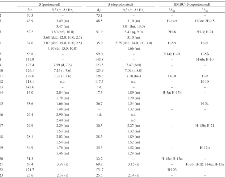
32.1 (C-18) with d 1.82 (m, H-18β) and d 1.53 (m, H-18α); Table 2. 13C NMR data assignments for alkaloids 2-4 and 6-8
2 3 4 6 7 8
da db da db dc db da db dc db dc
2 70.0 69.7 67.0 65.0 65.3 64.6 71.9 70.7 72.4 70.6 131.8
3 53.8 53.8 53.5 53.3 48.6 47.7 46.9 46.4 48.8 47.3 60.7
5 52.7 52.7 52.3 51.7 51.9 50.7 51.5 51.1 52.4 50.9 66.3
6 38.6 38.3 39.1 37.4 35.2 34.7 34.4 32.8 33.8 32.7 20.3
7 54.2 53.8 53.1 n.d. 58.8 57.2 59.2 58.6 59.6 58.4 103.3
8 144.2 143.7 138.2 134.9 140.9 139.9 145.3 141.7 143.5 141.7 126.5
9 116.4 115.7 123.4 121.4 123.2 122.5 115.7 115.2 115.3 115.3 119.9
10 126.9 126.2 125.1 124.8 121.5 120.1 127.2 127.4 128.7 127.6 121.2
11 112.1 111.5 128.8 129.2 128.4 127.0 112.4 112.1 113.5 112.1 124.2
12 150.6 150.2 119.1 119.2 112.8 111.0 149.9 149.3 151.2 149.3 112.9
13 130.4 129.8 142.0 140.8 151.5 150.1 131.4 130.0 131.2 129.9 138.8
14 22.2 21.8 21.2 19.5 17.1 17.0 21.3 19.9 16.2 15.3 30.3
15 34.6 34.4 33.9 33.0 36.3 35.9 84.1 81.3 33.5 33.0 31.6
16 25.8 25.1 26.1 25.2 26.9 26.6 25.9 24.5 25.9 24.8 56.8
17 23.7 23.2 23.0 21.9 30.6 29.5 22.9 22.3 29.9 29.5 64.1
18 7.2 7.0 7.2 6.8 31.1 31.5 29.2 27.6 29.1 27.9 12.9
19 30.6 30.2 30.5 30.3 36.4 35.3 30.8 30.3 35.1 34.4 120.9
20 36.2 35.8 36.6 36.4 32.1 31.4 37.3 35.8 31.3 30.2 128.9
21 71.0 71.3 71.0 72.0 70.0 68.8 68.9 68.0 69.3 67.5 65.7
22 171.8 172.1 169.2 n.d. – – 170.5 170.9 173.1 171.2 174.4
23 23.5 23.4 23.7 23.1 – – 25.3 24.9 25.0 24.9 53.5
OCH3-12 55.8 55.7 – – – – 55.9 55.7 56.3 55.7 –
OCH3-15 – – – – – – 57.5 57.8 – – –
N–CH3 – – – – – – – – – – 50.1
aData in C
5D5N; bdata in CDCl3; cdata in CD3OD. Compound 2 data at 125 MHz; compound 3 data at 125 MHz (in Pyr) and 75 MHz (in CDCl3); compound 4
data at 125 MHz; compound 6 data at 125 MHz (in Pyr) and 75 MHz (in CDCl3); compound 7 data at 75 MHz (in CD3OD) and 125 MHz (in CDCl3);
d 31.4 (C-19) with d 1.92 (m, H-19a) and d 1.60 (m, H-19b); d 29.6 (C-17) with d 2.13 (m, H-17β) and d 1.75 (m, H-17α); and d 26.9 (C-16) with d2.14 (m, H-16a) and
d 1.98 (m, H-16b) (see Table 1). Indole alkaloids, including those with a plumeran skeleton, are well reported in the literature as originated from Aspidosperma in general, and also from A. pyrifolium.4 Aspidospermine (2) and aspidofractinine (4) are examples of pentacyclic and hexacyclic plumeran alkaloids of Aspidosperma. Because of the absence of any sp2 carbon besides the benzene ring, that counts as 4 double bond equivalents (DBEs), compound 1 seemed to belong to the plumeran alkaloids like aspidofractinine (4), which possesses five saturated rings. However, the presence of one methyl and just seven methylene carbons (aspidofractinine has 9 methylenes and no methyl) indicated the constriction of the D-ring of the plumeran skeleton, from 6 to 5 members. An evidence for the presence of this moiety was the scalar couplings
observed in the correlation spectroscopy (COSY) spectrum between the hydrogen H-3 (d4.12) with the diastereotopic protons H-15α (d2.09) and H-15β (d 1.68), as well as with the protons 3H-14 (d1.58). Other evidences of this moiety were the long-range correlations shown in the heteronuclear multiple-bond correlation spectroscopy (HMBC) spectrum for protons H-3 (d 4.12) with the carbons C-5 (d55.7), C-15 (d44.7) and C-14 (d18.1); 3H-14 (d1.58) with C-3 (d71.9) and C-15; H-21 (d4.16) with C-8 (d135.8), C-7 (d 58.2), C-5, C-20 (d 42.5), C-6 (d35.5), C-19 (d31.4) and C-17 (d29.6); H-15α (d 2.09)with C-21 (d 79.7), C-20 and C-17, and, finally, H-15β (d 1.68) with C-3 (d71.9), C-20 (d42.5), C-19 (d31.4), C-17 (d 29.6) and C-14 (d18.1). Other key HMBC correlations are shown in Figure 2. One of the most deshielded methine hydrogens, H-3 (d 4.12), showed a septet (J 6.0 Hz) like splitting pattern on the 1H NMR spectrum (see Supplementary Information), leading to the conclusion that an extra proton Table 3. 1H and 13C NMR (CD
3OD) data assignments for the protonated and deprotonated formsof compound 5
5 (protonated) 5 (deprotonated) HMBC (5 deprotonated)
dCa dHb (m, J / Hz) dCc dHb (m, J / Hz) 2JCH 3JCH
2 70.3 – 73.1 – – –
3 48.9 3.49 (m) 48.5 3.10 (m) H-14α H-3α; 2H-15
3.47 (m) 3.01 (brt, 13.0)
5 52.2 3.80 (brq, 10.0) 51.9 3.41 (q, 9.0) 2H-6 2H-3; H-21
3.68 (ddd, 12.0, 10.0, 2.5) 3.10 (m)
6 34.6 3.07 (ddd, 15.0, 10.0, 2.5) 35.9 2.75 (ddd, 14.0, 9.0, 3.0) H-5α H-21
1.90 (dt, 15.0, 10.0) 1.66 (m)
7 58.8 – 59.0 – 2H-6; H-21 H-5β
8 139.9 – 143.8 – – H-6b; H-10
9 123.4 7.59 (d, 7.6) 123.5 7.47 (brd) – –
10 126.1 7.15 (t, 7.6) 125.9 7.09 (t, 8.0) – –
11 129.6 7.28 (t, 7.6) 128.3 7.18 (brs) H-10 H-9
12 118.1 n.d. 117.5 n.d. – H-10
13 142.6 – n.d. – – –
14 16.0 2.04 (m) 17.3 1.89 (m) H-3a; H-15b –
1.78 (m) 1.29 (m)
15 33.6 1.66 (m) 36.7 1.54 (m) – H-3a
1.48 (m) – 1.32 (m) – –
16 26.4 2.90 (m) n.d. n.d. – –
2.40 (m) n.d.
17 29.9 2.29 (m) 30.5 2.27 (m) – H-15b; H-21
1.53 (m) 1.32 (m)
18 28.1 2.02 (m) 28.5 1.89 (m) – –
1.54 (m) 1.52 (m)
19 34.9 1.76 (m) 35.3 1.52 (m) – H-17α
1.46 (m) 1.24 (m)
20 31.3 – 32.2 – H-15a; H-17α –
21 69.4 3.99 (s) 69.8 3.15 (s) – H-3b; H-5β; H-6a; H-15a
22 171.7 – 171.7 – 3H-23 –
23 25.6 2.37 (s) 25.5 2.34 (s) – –
was coupling to H-3 besides the 3H-14 and 2H-15, thus suggesting that N-4 should be protonated as a consequence of the HPLC isolation methodology using TFA. In this case, the simple rotaevaporation of the eluent from the HPLC, to remove the solvent and the volatile TFA, had not been enough. Thus, compound 1 was treated with an alkali solution and then, after workup, submitted to another NMR analysis (Table 1). As expected, a general shielding of all protons was observed, particularly higher for H-3, 2H-5 and H-21. Interestingly, just C-8 and C-14 underwent an approximately 3 ppm deshielding. Analysis of the nuclear Overhauser effect spectroscopy (NOESY) spectrum of the deprotonated compound allowed the deduction of the relative stereochemistry through the dipolar couplings of H-21 (d 3.60) with H-18β (d 1.42) and H-19β (d 1.81); H-17β (d 1.94) with H-3α (d 3.52); and H-6α (d 2.75) with H-16β (d 2.09) and H-17β (d1.94), determining the
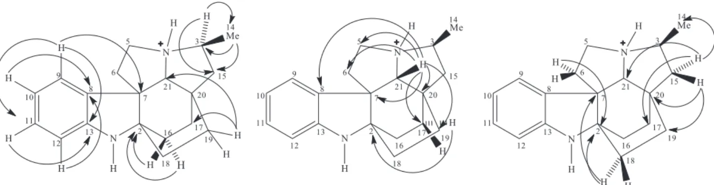
β-configuration for H-21 and the methyl (3H-14), as well as a boat conformation for both C and F rings (Figure 3). Thus, the sum of all spectroscopic data enabled the identification of compound 1 as (–)-(3S,7S,21R)-rel-(3αH)-15(14→ 3)-abeo-2,16,17,20,6,7-hexahydro-15H,8aH ,16a,20a-ethano-1H-indolizino[3,1-cd]carbazole, a new alkaloid that could be denominated either (3αH)-15(14→ 3)-abeo-aspidofractinine, or yet, according with the IUPAC rules,26 as 3,15-cyclo-14,15-seco-3α-aspidofractinine.
Compound 5 was obtained as an orange amorphous solid, m.p. 104.9-107.0 °C, [α]D
20 +31.5 ± 1 (c 0.15, MeOH) {lit.13 [α]
D23.5 +27 ± 8; c 0.251, CHCl3; lit.14 [α]D
25 +34 ± 15; c 0.042, MeOH}. Its HRESIMS showed a protonated molecule peak at m/z 323.2124 [M + H]+, compatible with the molecular formula C21H26N2O (calcd. 323.2123). The FT-IR spectrum exhibited a tertiary amide I band at 1646 cm-1, as well as bands at 1177 and 1124 cm-1 attributed to C–N bonds. Absorptions at 1481 and 1379 cm-1 were assigned to symmetrical bending of methylene and methyl groups, respectively. Analysis of the CPD, attached proton test (APT) and HSQC 13C NMR spectra (Table 3) allowed identification of 21 carbons, in agreement with the
suggested molecular formula, which could be correlated with six non-hydrogenated (3 sp2 and 3 sp3), five mono-hydrogenated (1 sp3 and 4 sp2), nine methylene and one methyl carbons. The signal at d 171.7 (C-22), typical of carbonyl, in addition to a signal of a methyl at d 25.6 (C-23), suggested the presence of an N-acetyl group, in agreement with the band at 1646 cm-1 in the IR spectrum. The 1H NMR spectrum (Table 3) exhibited three aromatic proton signals at d 7.59 (d, J 7.6 Hz, H-9), 7.28 (t, J 7.6 Hz, H-11) and 7.15 (t, J 7.6 Hz, H-10). Their multiplicities indicated the presence of a 1,2-disubstituted benzene ring, despite the non-detection of the fourth proton, even on the HSQC spectrum or by changing the solvent (see Supplementary Information). A singlet at d3.99 (H-21), a broad quartet at d 3.80 (J 10.0 Hz, H-5α), a triple doublet at Figure 2. Key long-range correlations for protonated compound 1 observed through the HMBC spectrum.
d3.68 (J 12.0, 10.0, 2.5 Hz, H-5β) and a multiplet for two protons at d 3.47-3.49 (H-3α and H-3β) were assigned to hydrogens attached to nitrogenated carbons. Other signals were viewed between 1.4 and 2.4 ppm, characteristic of hydrogens attached to sp3 carbons, with a singlet standing out at d2.37 (3H-23), attributed to the methyl of the acetyl group. The similarity of the spectral data with those of compounds 1 and 4 suggested the structure of the alkaloid
N-acetylaspidofractinine for compound 5, which was confirmed by the HMBC analysis. The N-acetyl group was confirmed by the coupling of the 3H-23 (d 2.37) protons with the carbonyl at d 171.7 (C-22). Some key correlations were observed between the protons H-10 (d7.15), H-21 (d3.99) and H-6β (d1.90) with the carbon C-8 (d139.9); H-9 (d7.59) and H-11 (d7.28) with the carbon C-13 (d 142.6); H-5β (d 3.68), H-3β (d 3.49), H-19a (d 1.76) and H-15β (1.66) with the carbon C-21 (d69.4); H-6α (d3.07) and H-18a (d2.02) with the carbon C-2 (d70.3); H-9, H-5β and H-18a with the carbon C-7 (d
58.8). Unequivocal assignment of the methylene carbons C-15 to C-19 was made through the correlations through 3J
CH of the protons H-21 (d3.99) and H-19a (d1.76) with C-17 (d29.9); H-3β (d 3.49) with C-15 (d33.6); and H-18a (d2.02) with C-20 (d31.3). Analysis of the NOESY spectrum showed dipolar couplings of H-21 (d 3.99) with H-3α (d 3.47); H-3αwith H-15α (d 1.48); H-5α(d 3.80) with H-17β (d 2.29) and H-14β (d 2.04); H-6α(d 3.07) with H-17βand H-16β (d 2.40); and H-17β with H-14β, permitting determination of the α-configuration for H-21, boat conformations for the C and F rings, and a chair conformation for the D ring. Comparison of the NMR data of the deprotonated compound (Table 3) showed considerable deshielding for the protons of the nitrogen-bearing carbons of the protonated form. Similarly to that already observed for compound 1, the C-8 carbon also exhibited a higher shielding in the N-protonated molecule. A significant shielding for C-2 and C-15, of approximately 3 ppm, was also observed.
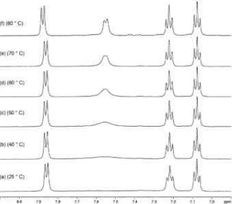
In order to explain the unexpected behavior of H-12 on the 1H NMR spectrum of compound 5, as noticed earlier, a series of variable-temperature 1H NMR experiments were performed. As can be noticed from Figure 4, the expected splitting pattern (two doublets and two triplets) of the four contiguous hydrogens of the aromatic system starts to rise up around 50 ºC (spectrum (a)), to be completely observed at 80 ºC (spectrum (f)). The integration of each absorption of the aromatic region at d 7.06-7.88 now reads one proton. The heteronuclear multiple quantum correlation (HMQC) spectrum (see Supplementary Information Figure S39) run at 70 ºC does show the correlation of the doublet at d7.55 with the carbon at d116.0, and the COSY spectrum (Figure
S38) now shows the complete coupling of the aromatic system. Thus, with these experiments it was proved that somehow an aromatic proton could not break through during a routine 1H NMR experiment.
Compound 2 was identified as (+)-aspidospermine, a plumeran alkaloid, [α]D20 +89.0 (c 0.19, CHCl3) {lit.6 [α]D +92}. Comparison of the
1H and 13C NMR data
(Table 2) with those published by Zèches-Hanrot et al.27 for the plumeran alkaloid aspidospermine showed that the chemical shifts of carbons C-8 (d 128.0) and C-13 (d141.0) should be reversed to C-8 (d143.7) and C-13 (d129.8), since C-13 is ortho to the methoxy group, and so should be more shielded than C-8, that is meta. Two other observed chemical shifts need to be emphasized. The
N-acetyl carbonyl in our case appeared at d172.1, which seems to be the most observed value, since in the literature it ranges between d 168.3 to 171.0.9,11,28-30 The value of
d 160.0, annotated for Zèches-Hanrot et al.,27 does not seem compatible, and no explanation for this difference is given. A similar strong shielding effect is observed for C-2, that in our case appears at d69.7 and in the literature27 at
d 64.0. We did not find any reasonable theoretical argument to explain this behavior, which may suggest that the NMR data, and their assignments, should be revised.
Compound 3 was characterized as the plumeran alkaloid (+)-demethoxyaspidospermine, otherwise named
N-acetylaspidospermidine,9 [α] D
20 +25.4 (c 0.11, MeOH) {lit.10 [α]
D +10; c 0.009, CHCl3}. 1H and 13C NMR data comparison (Table 2) with those from the literature9 revealed the chemical shift misassignments of both protons Figure 4. Partial 1H NMR (500 MHz, DMSO-d
6) spectra (d 7.0-8.0) (a)-(f),
and carbons-13 for the benzene carbons C-9 (d121.4; 7.12) and C-12 (d119.2; 8.18) for which the 3J
C,H correlation (observed only in the HMBC spectrum obtained in pyridine) of the proton H-9 (d7.34) with the carbon C-7 (d53.1) was fundamental for this assignment. Two other misassignments were done for C-6 (d37.4; 2.55/1.85) and C-17 (d21.9; 2.22/1.35), which positions were confirmed by long-range correlations seen in the HMBC spectrum in pyridine of the proton at d2.33 (H-6α), attached to the carbon at d39.1 (C-6), with the carbon at d 53.1 (C-7), and of the proton at d1.13 (H-15α) with C-17 (d 23.0; 2.18/1.06). Another mistake in the literature9 was the assignment of C-8, a substituted benzene carbon to which a value of d109.3 is annotated versus d134.9, experimentally observed, which is consistent with several other examples of plumeran alkaloids from the literature.28,30-33 Thus, the revision of the NMR data assignments is suggested.
Compound 4 was characterized as (–)-aspidofractinine, another plumeran alkaloid, [α]D20 –8.35 (c 0.23, MeOH) {lit.12 [α]
D –14; c 0.28, CHCl3}. Comparison of the 1H
and 13C NMR data (Table 2) with those reported in the literature11 showed that the slightly deshielded methylene at d 35.9, to which the protons at d 1.52/1.29 are attached (through HSQC analysis) is indeed C-15, not C-19 (d 35.3; 1.43/1.23) because a long-range correlation was observed in the HMBC spectrum of the deprotonated compound, in deuterated methanol, for the proton H-3a (d3.17) with the carbon C-15 (d 36.3), but not with C-19 (d36.4). The same is true for the methylenes C-17 (d29.5; 2.22/1.23) and C-16 (d26.6; 2.18/1.75) that are reversed in the literature, since the H-21 (d3.14) does show a 3J
C,H with the carbon at d29.5, but not with C-16 (d26.6). Once again the NMR data assignments need to be revised.
Compound 8 was identified as the (–)-enantiomer of N-methylakuammidine, [α]D20 –15.7 (c 0.43, MeOH) {lit.15 [α]
D
22 +15; c 0.43, H
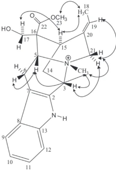
2O}. N-Methylakuammidine, sometimes referred to as macusine A,16 is a quaternary salt alkaloid, not yet reported for Aspidosperma pyrifolium. Its relative stereochemistry was deduced by the correlations observed in the NOESY spectrum between the protons H-5 (d 5.02), H-3 (d 4.92), H-21α (d 4.47) and H-21β(d 4.30) with the methyl attached to the nitrogen of the quaternary salt at d 3.24; H-5 (d 5.02) with the protons H-6β (d 3.86) and H-21β (d 4.30); H-3 (d 4.92) with the proton H-21α
(d 4.47); H-19 (d 5.50) with the diastereotopic methylene protons H-21α and H-21β; H-17α (d 3.74) with the protons H-15 (d 3.39) and H-14α (d 2.14); H-17β (d 3.64) with the proton H-14α (d 2.14); H-15 (d 3.39) with the proton H-14β (d 2.46); and 3H-23 (d 3.77) with the protons 3H-18 (d 1.70). All these dipolar couplings permitted to determine the β-configuration for protons H-3, H-5,
the N–CH3, and for the carboxyl methyl group, as well as the α-configuration for proton H-15. The E geometry of the double bond was confirmed by the dipolar interaction between the protons H-15 (d 3.39) and 3H-18 (d 1.70) (Figure 5). There are two reports about the 1H and 13C NMR data of N-methylakuammidine/macusine A that are found in the literature.15,16 Interestingly, the chemical shifts differ about 2 ppm, and no structure assignments have been done. Comparison of the current data (Table 2) with those from Hu, Zhu and Hesse15 showed a better compatibility.
The structures of the other known compounds, pirifoline (6),6,7 15-demethoxypirifoline (7),6,11 loganic acid (9),17-19 loganin (10)20 and 2-hydroxy-3-O-β -D-glucopyranosylbenzoic acid (11),19,21,22 were determined by comparison of their spectral data with those published in the literature. For those, no inconsistencies have been observed.
Antinociceptive and anti-inflammatory activities
In the formalin model of nociception test,35 the groups treated with different doses of APSE-Aq, indomethacin and morphine, significantly decreased the licking time during the early phase as compared with the control [control 44.88 ± 2.46 (n = 8); APSE-Aq 1 mg 36.38 ± 3.78 (n = 8); APSE-Aq 10 mg 25.33 ± 2.34 (n = 6); APSE-Aq 100 mg 20.00 ± 3.24 (n = 5); indomethacin 10 mg 20.00 ± 2.33 (n = 6); morphine 5 mg 19.25 ± 2.28 (n = 5)]. In contrast, in the late phase, only the groups treated with APSE-Aq 100 mg kg-1, indomethacin and morphine, significantly decreased licking time as compared with the control [control 22.40 ± 2.50 (n = 6); APSE-Aq 100 mg 6.00 ± 2.00 (n = 6); indomethacin 10 mg 2.80 ± 1.20 (n = 6); morphine 5 mg 5.25 ± 2.13 (n = 6)] (Figure 7). These results suggest that the aqueous fraction APSE-Aq presents possible central and peripheral effect.A similar result was found by Pereira et al.
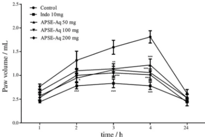
using the ethanol extract of other Aspidosperma species.36 In the paw edema induced by carrageenan test, the administration of APSE-Aq 100 mg kg-1 significantly reduced the carrageenan-induced paw edema two (p < 0.05), three (p < 0.01) and four hours (p < 0.001) after the administration of the stimulus compared to the animals treated with the vehicle. The APSE-Aq 50 mg kg-1 and 200 mg kg-1 groups reduced the paw edema three hours (APSE-Aq 50 mg p < 0.01; APSE-Aq 200 mg
p < 0.001) and four hours (APSE-Aq 50 mg p < 0.001; APSE-Aq 200 mg p < 0.001) after the administration of the stimulus compared to the animals with vehicle. The indomethacin reduced the paw edema from the second hour of administration of the stimulus (p < 0.001) (Figure 8). These results suggest that APSE-Aq presents a possible anti-inflammatory activity.
Figure 6. The effect of APSE-Aq on the abdominal writhing induced by acetic acid. Values are expressed as mean ± SEM of the number of observations. (a) vs. control (n = 6); (b) vs. APSE-Aq 50 mg kg-1
(n = 6); (c) vs. APSE-Aq 100 mg kg-1 (n = 5), respectively; at p < 0.0001
(one-way ANOVA followed by the Newman-Keuls pos hoc test). Indo: indomethacin.
Figure 7. The effect of APSE-Aq on the formalin test. The figure shows paw licking time (in seconds) at the early and late phases. Values are expressed as mean ± SEM of the number of observations. (a) vs. control; (b) vs. APSE-Aq 1 mg kg-1; (c) vs. APSE-Aq 10 mg kg-1, respectively; at
p < 0.0001 (one-way ANOVA followed by the Newman-Keuls pos hoc
test). Indo: indomethacin.
Conclusions
The phytochemical analysis of the ethanol extract from the seeds of Aspidosperma pyrifolium showed that the species is really a promising source of plumeran alkaloids. Among the 11 compounds obtained, the alkaloid (3αH )-15(14→3)-abeo-aspidofractinine (1) is being published by the first time in the literature and the compounds
N-methylakuammidine (8), loganic acid (9), loganin (10) and 2-hydroxy-3-O-β-D-glucopyranosylbenzoic acid (11) are being isolated and characterized for the first time from A. pyrifolium. Moreover, the alkaloid N-acetylaspidofractinine (5) was, for the first time, obtained as natural product and its 1H and 13C NMR data assignments are being reported for the first time in the literature. In addition, several chemical shift misassignments for aspidospermine (2), demethoxyaspidospermine (3) and aspidofractinine (4) have been revised.
The aqueous fraction obtained from the ethanol extract of A. pyrifolium showed a significant antinociceptive effect in the late phase of the formalin test, reducing the licking time compared to control for a similar value to morphine (5.25 ± 2.13), in addition to an anti-inflammatory effect causing reduction of the edema induced by carrageenan compared to control. Dose-response curves showed that the best doses of APSE-Aq were 100 mg kg-1 in the nociception induced by formalin [(20.0 ± 3.24) in the early phase, and (6.00 ± 2.00) in the late phase] and 200 mg kg-1 in the abdominal writhing induced by acetic acid (23.25 ± 2.56). These results permit to conclude that the aqueous fraction of the ethanol extract from seeds of A. pyrifolium presents antinociceptive and anti-inflammatory activities, contributing for the pharmacological knowledge of the plant.
Supplementary Information
Supplementary data are available free of charge at http://jbcs.sbq.org.br as PDF file.
Acknowledgements
The authors are grateful to CNPQ/PRONEX/CAPES/ FINEP/FUNCAP for the fellowships and financial support.
References
1. Macabeo, A. P. G.; Alejandro, G. J. D.; Hallare, A. V.; Vidar, W. S.; Villaflores, O. B.; Pharmacogn. Rev.2009, 3, 132. 2. Oliveira, V. B.; Freitas, M. S. M.; Mathias, L.; Braz-Filho, R.;
Vieira, I. J. C.; Rev. Bras. Plant. Med.2009, 11, 92.
3. Simões, C. M. O.; Schenkel, E. P.; Gosmann, G.; Mello, J. C. P.; Mentz, L. A.; Petrovick, P. R.; Farmacognosia: da Planta ao Medicamento, 5a ed.; Editora da UFSC: Brasil, 2004.
4. Pereira, M. M.; Jácome, R. L. R. P.; Alcântara, A. F. C.; Alves, R. B.; Raslan, D. S.; Quim. Nova2007, 30, 970.
5. Guimarães, H. A.; Braz-Filho, R.; Vieira, I. J. C.; Molecules
2012, 17, 3025.
6. Craveiro, A. A.; Matos, F. J. A.; Serur, L. M.; Phytochemistry
1983, 22, 1526.
7. Mitaine, A. C.; Mesbah, K.; Richard, B.; Petermann, C.; Arrazola, S.; Moretti, C.; Zèches-Hanrot, M.; Le Men-Olivier, L.;
Planta Med.1996, 62, 458.
8. Mitaine-Offer, A. C.; Sauvain, M.; Valentin, A.; Callapa, J.; Mallié, M.; Zèches-Hanrot, M.; Phytomedicine2002, 9, 142. 9. Atta-Ur-Rahman; Zaman, K.; Perveen, S.; Habib-Ur-Rehman;
Muzaffar, A.; Choudhary, M. I.; Pervin, A.; Phytochemistry
1991, 30, 1285.
10. França, O. O.; Brown, R. T.; Santos, C. A. M.; Fitoterapia 2000,
71, 208.
11. Araújo Jr., J. X.; Antheaume, C.; Trindade, R. C. P.; Schmitt, M.; Bourguignon, J.; Sant’ana, A. E. G.; Phytochem. Rev.2007, 6, 183.
12. Gagnon, D.; Spino, C.; J. Org. Chem.2009, 74, 6035. 13. Bycroft, B. W.; Schumann, D.; Patel, M. B.; Schmid, H.; Helv.
Chim. Acta1964, 47, 1147.
14. Guggisberg, A.; Gorman, A. A.; Bycroft, B. W.; Schmid, H.;
Helv. Chim. Acta1969, 52, 76.
15. Hu, W.-L.; Zhu, J.-P.; Hesse, M.; Planta Med.1989, 55, 463. 16. Yin, W.; Kabir, M. S.; Wang, Z.; Rallapalli, S. K.; Ma, J.; Cook,
J. M.; J. Org. Chem.2010, 75, 3339.
17. Di, L.; Li, N.; Zu, L.-B.; Wang, K.-J.; Zhao. Y.-X.; Wang, Z.;
Bull. Korean Chem. Soc. 2011, 32, 3251.
18. Zhang, X.; Xu, Q.; Xiao, H.; Liang, X.; Phytochemistry2003,
64, 1341.
19. Sunghwa, F.; Koketsu, M.; Nat. Prod. Res.2009, 23, 1408. 20. Lin, M.-H.; Liu, H.-K.; Huang, W.-J.; Huang, C.-C.; Wu, T.-H.;
Hsu, A.-L.; J. Agric. Food Chem. 2011,59, 7743.
21. Rashid, M. A.; Gustafson, K. R.; Cardellina II, J. H.; Boyd, M. R.;Phytochemistry 1996, 41, 1205.
22. Sakushima, A.; Coskun, M.; Maoka, T.; Phytochemistry 1995,
40, 257.
23. Koster, R.; Anderson, M.; Beer, E. J.; Fed. Proc. 1959, 18, 412. 24. Hunskaar, S.; Hole, K.; Pain1987, 30, 103.
25. Winter, C. A.; Risley, E. A.; Nuss, G. W.; Proc. Soc. Exp. Biol. Med.1962, 111, 544.
26. International Union of Pure and Applied Chemistry (IUPAC);
Preferred IUPAC Names, Provisional Recommendations; 2004, ch. 10.
27. Zèches-Hanrot, M.; Nuzillard, J.-M.; Richard, B.; Schaller, H.; Hadi, H. A.; Sévenet, T.; Men-Olivier, L. L.; Phytochemistry
28. Guimarães, H. A.; Vieira, I. J. C.; Braz-Filho, R.; Crotti, A. E. M.; Almeida, V. S.; Paula, R. C.; Helv. Chim. Acta2013,
96, 1793.
29. Mclean, S.; Reynolds, W. F.; Zhu, X.; Can. J. Chem.1987, 65, 200.
30. Brennan, J. P.; Saxton, J. E.; Tetrahedron1986, 42, 6719. 31. Ahond, A.; Janot, M. M.; Langlois, N.; Lukacs, G.; Potier, P.;
Rasoanaivo, P.; Sangare, M.; Neuss, N.; Plat, M.; Le Men, J.; Hagaman, E. W.; Wenkert, E.; J. Am. Chem. Soc.1974, 96, 633. 32. Wenkert, E.; Cochran, D. W.; Hagaman, E. W.; Schell, F. M.;
Neuss, N.; Katner, A. S.; Potier, P.; Kan, C.; Plat, M.; Koch, M.; Mehri, H.; Poisson, J.; Kunesch, N.; Rolland, Y.; J. Am. Chem. Soc. 1973, 95, 4990.
33. Liu, Y.-P.; Li, Y.; Cai, X.-H.; Li, X.-Y.; Kong, L.-M.; Cheng, G.-G.; Luo, X.-D.; J. Nat. Prod. 2012, 75, 220.
34. Rios, E. R. V.; Rocha, N. F. M.; Carvalho, A. M. R.; Vasconcelos, L. F.; Dias, M. L.; de Sousa, D. P.; de Sousa, F. C. F.; Fonteles, M. M. F.; Chem.-Biol. Interact.2013, 203, 573.
35. Melo, F. H. C.; Rios, E. R. V.; Rocha, N. F. M.; Citó, M. C. O.; Fernandes, M. L.; de Sousa, D. P.; de Vasconcelos, S. M. M.; de Sousa, F. C. F.; J. Pharm. Pharmacol.2012,64, 1722. 36. Pereira, M. M.; Souza Júnior, S. N.; Alcântara, A. F. C.;
Piló-Veloso, D.; Alves, R. B.; Machado, P. O.; Azevedo, A. O.; Moreira, F. H.; Castro, M. S. A.; Raslan, D. S.; Rev. Bras. Plant. Med.2006, 8, 1.