229
Imagens em Hematologia Rev. bras. hematol. hemoter. 2004;26(3):229-230
IMAGENS EM HEMATOLOGIA/IMAGES IN HEMATOLOGY
Gustavo J. Lourenço1
Rosemeire A. V. Bognone2
Maristela Zocca3
Marcia T. Delamain4
Afonso C. Vigorito5
Carmen S. P. Lima6
1
Pós-graduando em mestrado do Departamento de Clínica Médica da Faculdade de Ciências Médicas da Unicamp. 2
Bióloga responsável pelo Laboratório de Citogenética do Hemocentro da Unicamp.
3
Bióloga do Laboratório de Citogenética do Hemocentro da Unicamp.
4
Médica Assistente do Hemocentro da Unicamp. 5
Médico Assistente do Hemocentro da Unicamp. 6
Professora Doutora da Disciplina de Oncologia Clínica, do Departamento de Clínica Médica, da Faculdade de Ciências Médicas da Unicamp.
Morphologic, cytogenetic and molecular analyses in the identification of
acute promyelocytic leukaemia
Analise morfológica, citogenética e molecular na identificação da
leucemia promielocítica aguda
Correspondence to: Carmen Silvia Passos Lima, MD, PhD Hemocentro – Unicamp – Cidade Universitária “Zeferino Vaz” Caixa Postal 6198, Cep: 13083-970 – Campinas, SP – Brazil
Phone: + 55 19 3788-8740 – Fax: + 55 19 3788-8600 – e-mail: [email protected] The association of an exaggerated haemorrhagic
syndrome with certain leukaemias was described by Croizat & Favre-Gilli in 1949, 1 and in 1957 Hillstad 2 bestowed the appellation acute promyelocytic leukaemia (APL) upon this morphologic-clinical syndrome of acute myelogenous leukaemia (AML). This variant is called M3 in the FAB classification, and is characterised by the infiltration of bone marrow by promyelocytes. The haemorrhagic manifestations result, particularly, from procoagulant release by promyelocytes, and frequently determine fatal evolutions for these patients.
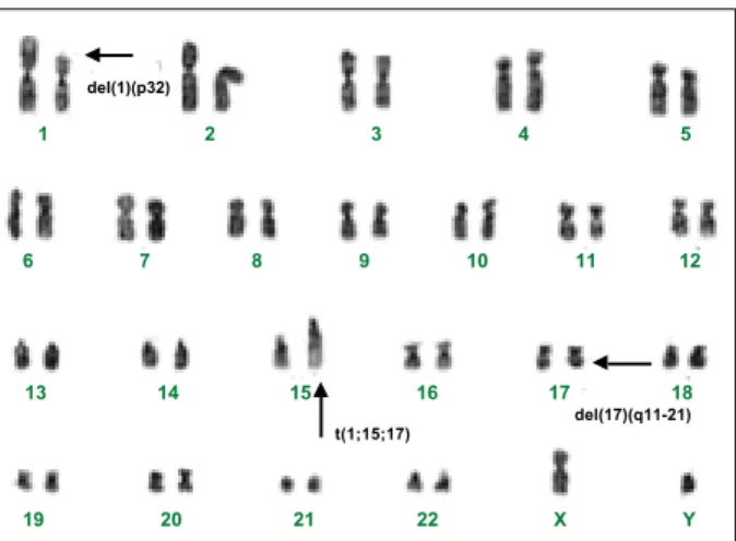
A translocation between chromosomes 15 and 17 t(15;17)(q22;q11-21) is present in most, maybe all, cases
Fig. 1 – Bone marrow smear obtained from the acute promyelocytic leukaemia at diagnosis predominantly showing promyelocytes (Romanovsky, 1000x)
Fig. 2 – G-banded karyotype obtained from the acute promyelocytic leukaemia patient at diagnosis: 46,XY,t(1;15;17)(p32;q22;q11-21). The arrows show the chromosomal abnormalities
1 2 3 4 5
6 7 8 9 10 11 12
13 14 15 16 17 18
19 20 21 22 X Y del(17)(q11-21) t(1;15;17)
230
Rev. bras. hematol. hemoter. 2004;26(3):229-230 Imagens em Hematologia
of APL 3 but this is not found in other AML subtypes. In addition, several descriptions exist of patients with seemingly classic APL but with variant translocations between 15q, 17q and a third chromosome.3,4
As a result of the translocation t(15;17) or its variants, the retinoic acid receptor gene (RARA) from chromosome 17 is moved to chromosome 15, where it is fused with a gene called PML to give rise to a new PML/RARA hybrid gene.3-6 A clinically intriguing correlate of the molecular genetic detection of a PML/RARA rearrangement in APL is the remarkable, albeit temporary, response to all-trans retinoic acid (ATRA) treatment. After an initial transient proliferation, the APL cells differentiate senesce and die. In consequence, a decrease in mortality by hemorrhagic manifestations has been seen in treated patients.
Herein, we present, for educational purposes, the images obtained from morphologic (Figure 1), cytogenetic (Figure 2), and molecular (Figure 3) analyses of a APL case seen at the Haematology and Haemotherapy Centre of the State University of Campinas.
It is important to comment that the patient was initially treated with ATRA attaining haematologic remission without significant haemorrhagic episodes. Another finding to be mentioned was the presence of the PML-RARA rearrangement in the variant translocation found in the case.
References
1. Croizat P, Favre-Gilli J. Les aspects du syndrome hemorrhagigue des leucémies. Sangre 1949;20:417.
2. Hillstad LK. Acute promyelocytic leukemia. Acta Med Scand 1957;159:189.
3. Heim S, Mitelman F. In: Acute myeloid leukemia. In: Heim S, Mitelman F. Cancer Cytogenetics. 2nd ed. New York: Willey-Liss 1995, p.69-140.
4. Grignani F, Fagioli M, Alcalay M et al. Acute promyelocytic leukemia: from genetics to treatment. Blood 1994;83:10-25.
5. Alcalay M, Zangrilli D, Pandolfi PP et al. Translocation breakpoint of acute promyelocytic leukemia lies within the retinoic acid receptor a locus. Proc Natl Acad Sci USA 1991;88:1.977-81.
6. Miller WH, Levine K, De Blassio A et al. Detection of minimal residual in acute promyelocytic leukemia by a reverse transcription polymerase chain reaction assay for the PML/ RARa fusion mRNA. Blood 1993;82:1.689-94.
Avaliação: Editor e dois revisores externos
Conflito de interesse: não declarado
Recebido: 17/04/2004 Aceito: 17/05/2004
Fig. 3 – Ethidium bromide-stained 2% agarose gel showing reverse transcriptase-polymerase chain reaction (RT-PCR) products of detection of PML/RARA fusion in the acute promyelocytic leukaemia patient. Fragments of 650 bp and 400 bp (Lane 1) expressing fusion mRNAs with breakpoints PML/RARA transcript and fragments of 370 bp corresponding to the control microglobulin b2. Lane 2 shows negative control and Lane 3 shows DNA size markers Ladder 100 bp
1 2 3
650 bp 400 bp