37
REV. HOSP. CLÍN. FAC. MED. S. PAULO 58(1):37-38, 2003From the Department of Medicine of the Federal University of Sergipe and São Lucas Hospital.
Received for publication on February 26, 2002.
FALCIFORM LIGAMENT ABSCESS:
REPORT OF A CASE
Valdinaldo Aragão de Melo, Gustavo Barreto de Melo, Renata Lemos Silva, João Fernandes Britto Aragão and José Eraldo Marques Rosa
MELO VA de et al. - Falciform ligament abscess: report of a case. Rev. Hosp. Clín. Fac. Med. S. Paulo 58(1):37-38, 2003.
Falciform ligament abscess is rare. We report a case of a 65-year-old man who presented with right upper quadrant abdominal pain, postprandial fullness, and fever. Computed tomography disclosed a cylindrical mass in the anterior abdomen that aroused suspicion of a hepatic abscess. At laparoscopic surgery, an abscess of the falciform ligament was found and drained. Two months later, the patient developed recurrence of the abscess secondary to acute calculous cholecystitis. Abscess drainage and cholecystectomy were performed. The presence of right uppper quadrant abdominal pain, epigastric tenderness, fever, leukocytosis, and a mass in the anterior abdomen should arouse suspicion of falciform ligament abscess. Its treatment consists of abscess drainage.
DESCRIPTORS: Falciform ligament. Abscess. Gallbladder. Drainage. Calculous cholecystitis.
INTRODUCTION
Few cases of falciform ligament abscess have been reported. This im-plies that the pathology of falciform ligament abscess is poorly understood, and many surgeons may encounter one without being able to identify it. Therefore, we report a case of falciform ligament abscess secondary to acute calculous cholecystitis.
CASE REPORT
A 65-year-old man presented with right upper quadrant abdominal pain and postprandial fullness without weight loss. These symptoms had ap-peared 2 months earlier.
At admission, his body temperature was 38.5°C. His heart beat was 82 systolic. Abdominal examination dis-closed a palpable mass that was slightly tender. Laboratory studies
were normal for gamma-GT (48 U/L), aspartate transaminase (40 U/L), and alanine transaminase (21 U/L). How-ever, his white blood cell count was 18,000 per cubic millimeter. Other laboratory tests were normal.
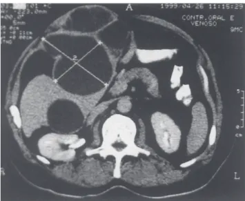
At abdominal ultrasound, the gall-bladder was enlarged, and a sonolucent mass that was interpreted as a hepatic abscess was found in the right upper quadrant. Computed tomography re-vealed a cylindrical mass in the ante-rior abdomen that aroused suspicion of a hepatic abscess. It extended along the course of the falciform ligament to the porta hepatis (Fig. 1).
Laparoscopic exploration con-firmed the presence of an abscess in the falciform ligament, which was drained percutaneously. The
postop-erative course was satisfactory. How-ever, 2 months later, the patient pre-sented with the same symptoms of the disease. A recurrence of the falciform ligament abscess and acute calculous cholecystitis were found. Therefore, cholecystectomy and another abscess drainage were performed. A follow-up ultrasound showed no evidence of in-tra-abdominal abscess.
DISCUSSION
be-38
Falciform ligament abscess: report of a case Melo VA et al.
REV. HOSP. CLÍN. FAC. MED. S. PAULO 58(1):37-38, 2003
falciform ligament due to umbilicus in-fection in children has been reported4.
On abdominal examination, right upper quadrant pain, distension, and
epigastric tenderness is common1,3,5.
Some cases may present spiking fevers and leukocytosis in laboratory studies3.
Ultrasound and computed tomog-raphy scans should be helpful in de-tecting the presence of an abscess and in evaluating the existence of chole-cystitis, even though it may be diffi-cult to properly visualize the gallblad-der. On computed tomography scan-ning, free air limited to the area sur-rounding the falciform ligament indi-cates the presence of an abscess1.
It is important to differentiate be-tween falciform ligament abscess and hepatic abscess because antimicrobials are efficient in treating the latter, while being almost useless to the former4.
Drainage by laparoscopic surgery is the treatment of choice for falciform ligament abscess.
Figure 1 - Falciform ligament abscess as a cylindrical mass in the anterior abdomen at computed tomography.
tween the gallbladder and the falciform ligament.
Clinically, the presence of an ab-scess is very uncommon. Few articles were found regarding it in the literature
1-5. The occurrence of falciform ligament
abscess secondary to
ventriculope-ritoneal shunt infection has been re-ported3. Three other reported cases were
caused by cholecystitis1,2,5.
Cholecysti-tis is considered the cause of the falciform ligament abscess presented here, either by direct or lymphatic spread. Additionally, abscess of the
REFERENCES
1 . BROCK JS, PACHTER HL, SCHREIBER J et al. - Surgical diseases of the falciform ligament. Am J Gastroenterol 1992; 87(6):757-758.
2 . DOSCHER W, CHARDAVOYNE R, TEICHER I - Abscess of falciform ligament. N Y State J Med 1980; 80(7 Pt 1):1131-1133.
3 . LAUCKS SS, BALLANTINE TVN, BOAL DK - Abscess of the falciform ligament in a child with a ventriculoperitoneal shunt. J Pediatr Surg 1986; 21(11):979-980.
4 . LIPINSKI JK, VEGA JM, CREMIN BJ - Falciform ligament abscess in the infant. J Pediatr Surg 1985; 20(5):556-558. 5 . SONES PJ, THOMAS BM, MASAND PP - Falciform ligament
abscess: appearance on computed tomography and sonography. Am J Roentgenol 1981; 137:161-162.
RESUMO
MELO VA de e col. - Abscesso de ligamento falciforme: relato de
caso. Rev. Hosp. Clín. Fac. Med.
S. Paulo 58(1):37-38, 2003.
Abscesso de ligamento falciforme é raro. É relatado um caso de um ho-mem de 65 anos que apresentou dor no quadrante superior direito do abdo-me, plenitude pós-prandial e febre. A
tomografia computadorizada revelou uma massa cilíndrica no abdome an-terior que causou suspeita de absces-so hepático. Na cirurgia laparoscópica, um abscesso de ligamento falciforme foi encontrado e drenado. Dois meses depois, o paciente desenvolveu recidi-va do abscesso secundário a colecistite aguda calculosa. Drenagem do absces-so e colecistectomia foram realizados.
A presença de dor no quadrante supe-rior direito, febre, leucocitose e abau-lamento no abdome ântero-superior deve causar suspeita dessa patologia. Seu tratamento consiste de drenagem do abscesso.
DESCRITORES: Ligamento