Case Report
299
MIRANDA LEC, CARVALHO E, LIMA DL. Giant schistosomal granuloma mimicking rectum neoplasia – case report. Rev bras Coloproct, 2011;31(3): 299-300.
AbstRACt: We report the case of a young man from an area where schistosomiasis is endemic, in the state of Pernambuco, who presented with hemorrhage. Initially diagnosed as rectum neoplasia, subsequent investigation demonstrated rectal giant schistosomal granuloma. the diagnoses and clinical aspects of the case are discussed in this study.
Keywords: schistosomiasis; granuloma; neoplasm; rectum; general surgery.
Giant schistosomal granuloma mimicking rectum neoplasia –
case report
LUIZ EDUARDO CORREIA MIRANDA1, EDUARDO CARVALHO2, DIEGO LAURENTINO LIMA3
1Adjunct Professor of Abdominal Surgert at Faculdade de Ciências Médicas of Universidade de Pernambuco (UPE) – Recife (PE),
Brazil. 2Doctor of the Endoscopy Service at Hospital Geral Octávio de Freitas – Recife (PE), Brazil. 3Medical student at Faculdade
de Ciências Médicas of UPE – Recife (PE), Brazil.
Study carried out at the University Hospital Oswaldo Cruz – Recife (PE), Brazil. Financing source: none.
Conlict of interest: nothing to declare.
Submitted on: 08/02/2010 Approved on: 19/09/2010
INtRODUCtION
Schistosomiasis mansoni is a chronic infection caused by the direct contact with fresh water that con-tains cercaria, the larval form of the parasite. The dis-ease is endemic in Brazil, being prevalent in the North-east region and spread to the West and South; there are serious consequences to the people who are infected1. Hyperplastic manifestations of schistosomiasis are un-common and may present in different clinical forms, including the pseudotumoral form. In this paper, we re-port the case of a rectal schistosomal pseudotumor.
CAsE REPORt
A 24-year-old man presented symptoms of con-stipation for a long period and rectal bleeding for one month. He denied using any type of medication, as well as weight loss, anorexia, hematemesis, jaundice or fever. He comes from an endemic area of schisto-somiasis, and did not present with acute suffering.
Phisical examination: abdomen was lat, not
tender, no masses, no hepatomegalies or
splenom-egalies. No clinical indings were related to hepatic insuficiency or portal hypertension. His blood tests
were normal, except for mild microcytic anemia and eosinophilia. Colonoscopy demonstrated a mass mea-suring from 3 to 4 cm, bleeding in the anterior rectal wall, 6 cm from the anal margin (Figure 1);
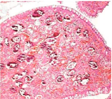
Pathological report showed chronic rectal
muco-sa inlammation and granulomas consisting of epithe -lioid cells and some nucleus Langhans giant cells, in-volving eggs with long lateral spine, which suggested rectal schistosomal granuloma (Figure 2). Patient was treated with praziquantel and colonoscopy after 90 days showed the clinical resolution of the granuloma.
DIsCUssION
Giant schistosomal granuloma mimicking rectum neoplasia – case report Luiz Eduardo Correia Miranda et al.
300 Journal of Coloproctology
July/september, 2011
Vol. 31 Nº 3
REFERÊNCIAs
1. Santana HJ, Lima CA. Pseudotumor esquistossomótico de cólon – Relato de um caso. Rev bras Coloproct 1985;5(1):17-21. 2. Kalil M, Battisti Netto O, Vieira LCA, Cintra LC. Forma
pseudotumoral intra-abdominal da esquistossomose mansônica. Ver Col Bras Cir [Internet] 2006 Mai-Jun; 33(3). Available from: http://www.scielo.br/rcbc
3. Prata A. Esquistossomose mansoni. In: Doenças infecciosas e parasitárias. 6ª ed. Rio de Janeiro: Guanabara-Koogan; 1976. 4. Neto JB. Manifestações hiperplásicas da esquistossomose
mansônica. J Bras Med 1983;45(5):37-40.
5. Lantsberg L, Khodadadi J, Krugliak P. Schitosomal granuloma mimicking adenocarcinoma of the rectum. J Clin Gastroenterol 1987;9(4):489-90.
Correspondence to:
Luiz Eduardo Correia Miranda
Serviço de Cirurgia Geral e Transplante de Fígado Rua Arnóbio Marques, 310
CEP: 50100-130 – Recife (PE), Brazil. E-mail: [email protected] with a granulomatous inlammatory reaction2. The
schistosomal granuloma is the most uncommon type of hyperplastic manifestation3. The incidence in the in-testinal form of schistosomiases are more frequent in the rectum, sigmoid and descending colon. Differential diagnosis of granuloma are adenocarcinoma, sarcoma, polyps, tuberculosis and lymphoma. The chronic
dis-ease is caused by a granulomatous inlammation that
occurs in response to the deposit of eggs in the tissue4. The clinical presentation of the intestinal form of the schistosomal granuloma may vary from
dyspepti-cal symptoms to schistosomal proctocolitis, abdominal pain, nausea, tenesmus, mucous-bloody diarrhea and transrectal bleeding2. Santana and Lima1 described a schistosomal granuloma of the colon in the descend-ing sigmoid junction, simulatdescend-ing malignant neoplasm. Lantsberg et al.5 described the rectal pseudotumor of an Ethiopian man who presented with rectal bleeding and received praziquantel after the disease was diagnosed by a rectal biopsy. The conclusion is that the diagnosis of the pseudotumoral form of schistosomiasis should be considered for patients who come from endemic areas for schistosomiasis with rectal mass. Praziquantel heals
60 to 90% of these patients, and endoscopic indings
may induce to a diagnostic error of rectal neoplasm.
Figure 1. Large and fragile lesion in the anterior rectal wall 6 cm from the anal margin.
Figure 2. Rectal giant schistosomal granuloma.
REsUmO: Nós relatamos o caso de um homem, jovem, proveniente de uma área endêmica para esquistomossomose, no Estado de Pernambuco, e que apresentou hematoquezia. Inicialmente diagnosticado como neoplasia do reto, a investigação subsequente demonstrou um granuloma esquistossomótico gigante do reto. O diagnóstico e os aspectos clínicos do caso são discutidos.