CESAR MAP, KLUG WA, ORTIZ JA, FANG CB, CAPELHUCHMIK P. What is the value of proctography for diagnostic of outlet obstruction? Rev bras Coloproct, 2011;31(3): 257-261.
AbstRACt: the diagnosis of constipation is complicated due to the multiplicity and complexity of the causes. Regarding diagnostic tests, proctography is the best choice because it provides information on functions and visualization of abnormalities. Objective: to measure the isolated value of proctography in patients with obstructed defecation. Method: We evaluated 40 constipated patients at the Coloproctology Clinic of Santa Casa de Misericórdia de São Paulo. the test was performed by introducing 120 mL of barium contrast in the rectum and by analyzing the different stages of evacuation. three x-rays were performed in the lateral position: rest, anal contraction and evacuation. Results: the diagnoses were: rectocele: 2 (5.0%); anismus: 8 (20.0%); perineal descent: 13 (32.5%); sigmoidocele: 6 (15.0%); internal invagination: 10 (25.0%); rectocele + sigmoidocele 9 (22.5%); rectocele + internal invagination 11 (27.5%); rectocele + anismus: 18 (45.0%). several patients presented multiple disorders. Conclusion: Constipation by obstructed defecation depends on multiple factors and it is important to have an accurate diagnosis. Proctography is essential, but insuficient as a single procedure. the other tests contribute with the diagnosis, therefore, they should be included in the investigation.
Keywords: constipation; defecation; defecography.
What is the value of proctography for diagnostic of outlet obstruction?
MARIA AUXILIADORA PROLUNGATTI CESAR1, WILMAR ARTUR KLUG2, JORGE ALBERTO ORTIZ3, CHIA BIN
FANG5, PERETZ CAPELHUCHMIK6
1Doctorate in Surgery at Faculdade de Ciências Médicas of Santa Casa de São Paulo – São Paulo (SP), Brazil; Assistant
Professor and Doctor of the Department of Medicine at Universidade de Taubaté – Taubaté (SP), Brazil. 2Professor of the Department of Surgery at Faculdade de Ciências Médicas of Santa Casa de São Paulo – São Paulo (SP), Brazil. 3Master’s degree in Surgery at Faculdade de Ciências Médicas of Santa Casa de São Paulo; Head of the anal physiology sector of the coloproctology área at Faculdade de Ciências Médicas of Santa Casa de São Paulo – São Paulo (SP), Brazil. 5Master’s
degree in Surgery at Faculdade de Ciências Médicas of Santa Casa de São Paulo; Adjunct Professor of the Department of Surgery at Faculdade de Ciências Médicas of Santa Casa de São Paulo – São Paulo (SP), Brazil. 6Professor of the
Department of Surgery at Faculdade de Ciências Médicas of Santa Casa de São Paulo – São Paulo (SP), Brazil.
Study carried out at the discipline of Coloproctology, Department of Surgery at Faculdade de Ciências Médicas of Santa Casa de São Paulo. Financing source: none.
Conlict of interest: nothing to declare.
Submitted on: 21/01/2011 Approved on: 30/06/2011
INtRODUCtION
Constipation is a disorder characterized by twice
or less bowel movements per week, having dificult
evacuation, hard stool and the sensation of incomplete evacuation1-6.
It is classified in two types: slow transit or colonic inertia and obstructed defecation. Iner-tia is the less common disorder, and it is caused by slower transit. The obstructed defecation is an
evacuation disorder such as the inability to evac-uate the rectal volume, the full rectum feeling, rectal pain, descent of the pelvic diaphragm and excessive effort1-6. The most common disorder in obstructed defecation is the non-relaxation of the puborectal muscle or anismus1-6.
puborectalis, rectocele, invagination, prolapse, recto-cele, hernia and perineal descen. It is also possible to measure perineal descent and anorectal angle at rest, contraction and evacuation1,3,4,7-10.
The objective of this study was to assess the im-portance of proctography diagnosing constipation by obstructed defecation.
PAtIENts AND MEtHOD
In this study, 40 proctographies of patients pre-senting with constipation at the Coloproctology clin-ic of Santa Casa de Misericórdia de São Paulo were analyzed. They were refractory to the treatment and diagnosed with obstructed defecation. Their proctog-raphies were compared to those of the asymptomatic control group, comprised of 20 patients.
Proctography consisted of the introduction of 120 mL of barium contrast in the rectum by a rectal probe 14, with the patient in supine, left lateral posi-tion. Afterwards, the contrast marker was placed with
the same barium contrast, ixated on the sacrum and
pubis.
After the introduction of the contrast, three x-rays in the lateral position were performed. The patient was sitting on the chair for the proctography: at rest, anal contraction and evacuation. The following guidelines were determined after the analysis of the x-rays:
a) pubococcygeus: between the upper pubis and the coccyx;
b) anal canal: from the anus to the anorectal junc-tion;
c) rectal axis: posterior rectum.
Afterwards, the following measures were
de-ined:
a) position of pelvic diaphragm: between the upper extremity of the anal canal and the pubococcy-geus muscles through a perpendicular line; b) perineal position: between the lower extremity
of the anal canal and the pubococcygeus muscles through a perpendicular line;
c) Length of the anal canal;
d) Anorectal angle: between the rectum axis and the anal canal at the intersection of lines.
Data were analyzed by the Student’s t test, with
signiicance of 0.05%.
REsULts
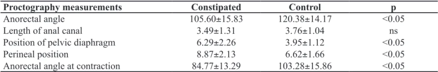
Forty proctographies of patients who were con-stipated due to obstructed defecation were compared with 20 proctographies of patients in the control group. The measurements of the proctographies in the positions at rest, contraction and evacuation are dem-onstrated in Tables 1 to 3, respectively. At rest, pelvic diaphragm was lower, as well as the anorectal angle
Proctography measurements Constipated Control p
Anorectal angle 105.60±15.83 120.38±14.17 <0.05
Length of anal canal 3.49±1.31 3.76±1.04 ns
Position of pelvic diaphragm 6.29±2.26 3.95±1.12 <0.05 Perineal position 8.87±2.13 6.62±1.66 <0.05 Anorectal angle at contraction 84.77±13.29 103.28±15.86 <0.05 Table 1. Proctography measurements at rest in constipated patients, compared with the control group.
ns: not signiicant.
Proctography measurements Constipated Control p
Anorectal angle 84.77±13.29 103.28±15.86 <0.05 Length of the anal canal 6.42±4.67 4.71±0.96 ns Position of pelvic diaphragm 4.13±1.62 3.05±1.12 ns Perineal position 7.46±1.51 6.19±1.63 <0.05
Table 2. Proctography measurements at contraction position in constipated patients, compared with the control group.
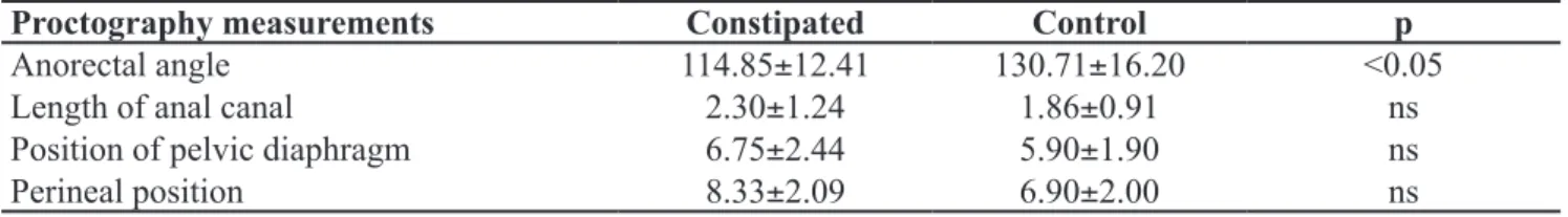
and the perineal position; at contraction, the anorectal angle was lower, as well as the perineal position; at evacuation, anorectal angle was lower. The other mea-surements were not statistically different.
The differences between proctographies of
pa-tients with different speciic diagnoses were compared
to those of the patients in the control group at rest, con-traction and evacuations. These differences are demon-strated in Tables 4 to 6. There were many differences, marked with *, except as to the length of the anal canal, since there was no variation between the groups.
In relation to diagnostics, many patients present-ed more than one diagnosis at proctography, with the following rates:
a) rectocele: 2 (5.0%);
b) puborectal paradoxal contraction: 8 (20.0%); c) perineal descent: 13 (32.5%);
d sigmoidocele: 6 (15.0%);
e) internal invagination: 10 (25.0%); f) rectocele + sigmoidocele: 9 (22.5%); g) rectocele + invagination: 11 (27.5%);
h) rectocele + paradoxal contraction: 18 (45.0%).
DIsCUssION
Constipation caused by inertia or obstructed defeca-tion is complex and little understood. It is multifactorial and includes factors regarding diet, age, gender, general
Proctography measurements Constipated Control p
Anorectal angle 114.85±12.41 130.71±16.20 <0.05
Length of anal canal 2.30±1.24 1.86±0.91 ns
Position of pelvic diaphragm 6.75±2.44 5.90±1.90 ns
Perineal position 8.33±2.09 6.90±2.00 ns
Table 3. Proctography measurements at evacuation position in constipated patients, compared with the control group.
ns: not signiicant.
Rectocele
Puborectal paradoxal contraction
Invagination sigmoidocele Control p
Anorectal angle 102.40±12.51* 100.70±1.08* 105.80±15.89* 100.20±9.64* 120.38±14.17 <0.05 Length of anal
canal 3.16±1.43 3.79±1.09 2.91±1.22 2.75±1.21 3.76±1.04 ns Position of
pelvic diaphragm 6.56±2.27* 6.22±2.31* 7.16±2.33* 5.93±2.04 3.95±1.12 <0.05 Perineal position 9.19±2.30* 8.96±2.32* 9.01±2.44* 7.85±2.25 6.62±1.66 <0.05 Table 4. Proctography measurements at rest and diagnoses of causes for constipation.
ns: not signiicant.
Rectocele
Puborectal paradoxal contraction
Invagination sigmoidocele Control p
Anorectal angle 79.83±11.24* 79.74±0.26* 86.90±9.62* 84.00±7.01* 103.28±15.86 <0.05 Length of anal
canal 6.63±6.48 5.59±2.22 5.42±2.18 5.05±1.99 4.71±0.96 ns Position of
pelvic diaphragm 4.36±1.59* 4.32±1.49* 4.97±1.45* 4.47±0.97 3.05±1.12 <0.05 Perineal position 7.82±1.30* 7.50±0.97* 8.00±1.89* 7.08±2.43 6.19±1.63 <0.05 Table 5. Proctography measurements at contraction and diagnoses of causes for constipation.
Rectocele
Puborectal paradoxal contraction
Invagination sigmoidocele Control p
Anorectal angle 111.17±2.88* 111.74±9.70* 117.40±14.15* 114.33±16.47* 130.71±16.20 <0.05 Length of anal
canal 1.88±0.99 2.28±0.76 2.16±1.64 1.70±1.02 1.86±0.91 ns Position of
pelvic diaphragm 8.08±2.16* 6.41±2.39 7.84±2.74 7.58±3.36 5.90±1.90 <0.05 Perineal position 9.21±2.02* 7.81±1.92 9.31±2.51* 8.70±3.20 6.90±2.00 <0.05 Table 6. Proctography measurements at evacuation and diagnoses of causes for constipation.
ns: not signiicant.
conditions, hormones and intestinal polypeptides, parity, neurological lesions and physiology of pelvic organs1-6.
Pelvic functional phenomena related to evacuation and analyzed by anal physiology tests are prevalent for obstructed defecation. Clinical diagnoses are based on history and markers, manometry, proctography, electro-myography, and latency of the pudendal nerve1-6.
Rectocele is a common diagnosis, being present in almost all constipated patients. It varies as to dimension and is usually associated with other alterations. Proctogra-phy images are clear and a good way to diagnose1-4,7-12.
In our sample, diagnoses were based on the asso-ciation of clinical, manometric and radiological data, as well as electromyography in selected patients after the correction of eating and hygiene habits and the ex-clusion of associated diseases. The previous selection of patients excluded those who were constipated due to colonic inertia. Among the tests, we separated the results obtained by proctography with the objective to assess its diagnostic potential in an isolated way. It was clear that, because of the multiplicity and associa-tion of causes, the approach to these patients required the use of different physiological methods.
We believe that proctography is useful to ana-lyze constipation. The method should be investigat-ed due to its importance, because it not only enables current diagnoses, but also a more detailed analy-sis of the pelvic diaphragm. In spite of that, when we perform this test on asymptomatic patients,
nor-mal indings may occur9. The possible alterations in young asymptomatic patients are perineal descent, invagination and rectocele, and their importance is not clear at the proctography9.
In our sample, the measurements of anorectal an-gles in constipated patients were lower than the
con-trol group in all phases of the test, and the differences
were signiicant. At rest, we observed that the values
of the constipated patients were lower than the control group, as well as at contraction and evacuation. The length of the anal canal increases at the moment of contraction, and decreases at evacuation, with no sta-tistical differences as to the control group. At rest, the
position of the pelvic diaphragm presented signiicant
higher values for the constipated patients, because they were located at a lower position; however, there were no differences during contraction and effort.
For those who have rectocele, sigmoidocele and invagination, radiographic evidence is essential and
conirms the diagnosis; however, at puborectal para -doxal contraction, we observed perineal descent and lower anorectal angle. This difference may be impor-tant, because the other possible way to diagnose this disorder is electromyography. For sigmoidocele, there
were no signiicant differences in proctography mea -surements in comparison to those who do not have this condition, except for the radiographic evidence of the presence of colon loop, which presses the rectum.
In relation to the position of the pelvic diaphragm and the perineal position, we observed perineal de-scent. This is in accordance with the usually accepted idea that the efforts made by constipated patients lead to alterations in the position of the pelvic diaphragm. Factors such as age, gender, parity, associated condi-tions and obstetric trauma certainly interfere in the re-sults, but they were not considered for not being the objective of this study. On the other hand, when series of patients are investigated, the mean values positive-ly contribute to the interpretation of the disorders.
Proctography has demonstrated many indings
important to remember that patients with refractory constipation at clinical treatment should be fully as-sessed, because only one examination may lead to a wrong diagnosis of the cause of constipation. A very important example is the presence of rectocele (fre-quent diagnosis at proctography), associated with pu-borectal paradoxal contraction. The former would be surgically treatable, but the outcomes could be nega-tive in case there was associated puborectal paradoxal contraction. The latter is clinically treatable, and proc-tography is not gold standard for this diagnosis.
Proctography usually inds signs of perineal descent,
because this may suggest that the patient may present in-nervation compromise of the pelvic diaphragm; in this case, it is recommended to investigate fecal continence.
CONCLUsION
Proctography proved to be important to assess constipation through diagnoses and measurements, and it is useful as an examination associated with the full evaluation of the constipated patient.
REsUMO: O diagnóstico da constipação é difícil pela multiplicidade e complexidade das causas. Dos exames diagnósticos, a proctograia é preferida, fornecendo informações da função e visualização de anormalidades. Objetivo: Medir o valor isolado da proctograia, em pacientes com diagnóstico de defecação obstruída. Método: Avaliamos 40 pacientes com constipação intestinal do Ambulatório de Colo-proctologia da santa Casa de Misericórdia de são Paulo. O exame foi feito introduzindo-se 120 mL de contraste no reto e analisando-se as diferentes fases da evacuação. Foram realizadas três radiograias na posição lateral: repouso, contração anal e evacuação. Resultados: Os diagnósticos foram: retocele: 2 (5,0%); contração paradoxal do puborretal: 8 (20,0%); descida perineal: 13 (32,5%); sigmoidocele: 6 (15,0%); invaginação interna: 10 (25,0%); retocele + sigmoidocele: 9 (22,5%); retocele + invaginação: 11 (27,5%); retocele + contração paradoxal: 18 (45,0%). Vários pacientes apresentaram distúrbios múltiplos. Conclusão: Constipação por defecação obstruída depende de múltiplos fatores e é importante o diagnóstico preciso. A proctograia é essencial, mas insuiciente como procedimento isolado. Os outros exames são importante contribuição para irmar o diagnóstico, devendo ser incluídos na investigação.
Palavras-chave: constipação intestinal; defecação; defecograia.
REFERENCEs
1. Cesar MAP, Klug WA, Aguida HAC, Ortiz JA, Bin FC, Kapelhuchnik P. A presença de retocele interfere nos
resultados de exames de isiologia anal? Rev bras Coloproct
2008;28(3):329-33.
2. Cesar MAP, Klug WA, Aguida HAC, Ortiz JA, Fang CB, Capelhuchnik P. Alterações das pressões anais em pacientes constipados por defecação obstruída. Rev bras Coloproct 2008;28(4):402-8.
3. Cesar MAP, Klug WA, Ortiz JA, BIN FC, Kapelhuchnik P.
Diagnóstico do anismus através dos exames de isiologia
anal. Rev bras Coloproct 2009;29(2):192-6.
4. Cesar MAP, Klug WA. Fisiologia Anorretal e cirurgia. Investigação dos distúrbios de evacuação (Constipação intestinal e incontinência fecal). In Speranzini MB, Deutsch CR, Yagi OK. Manual de diagnóstico e tratamento para o residente de cirurgia. 2009(2):1465-72.
5. Cesar MAP, Oliveira CC. Existe importância na utilização da manometria anal no diagnóstico da síndrome do intestino irritável? Rev bras Coloproct 2009;29(3):358-62.
6. Vieira EP, Pupo Neto J, Lacombe DLP. Contribuição da manometria ano retal na avaliação da constipação intestinal crônica. Rev bras Coloproct 2005;25(4):348-60.
7. Mellgren A. Diagnosis and treatment of constipation Eur J Surg 1995;161(9):623-34.
8. Karasick S, Ehrlich SM. Is constipation a disorder of defecation or impaired motility? Distinction based on defecography and colonic transit studies. AJR 1996;166(1):63-6.
9. Fang CB, Peixoto VCS, Klug WK, Ortiz JA, Capelhuchnik P. Esvaziamento retal em voluntários assintomáticos através da
proctograia. Rev bras Coloproct 1997;17(3):175-9.
10. Hiltunen KM, Kolehmainen H, Matikainen M. Does defecography help in diagnosis and clinical decision-making in defecation disorders? Abdom Imaging 1994;19(4):355-8. 11. Sobrado Jr. CV, Pires CEF, Araújo SEA, Amaro Jr. E,
Habr-Gama A, Kiss DR. Avaliação computadorizada do esvaziamento retal em voluntários assintomáticos. Rev bras Coloproct 2003;23(1):5-8.
12. Sentovich SM, Rivela LJ, Thorson AG, Christensen MA, Blatchford GJ. Simultaneous dynamic proctography and
peritoneography for pelvic loor disorders. Dis Colon Rectum
1995;38(9):912-5.
Correspondence to:
Maria Auxiliadora Prolungatti Cesar, Serviço de Clínica Cirúrgica do Hospital Universitário de Taubaté