393
INTRODUCTION
Revista da Sociedade Brasileira de Medicina Tropical 43(4):393-395, jul-ago, 2010
Article/Artigo
Cytokine expression in the duodenal mucosa of patients with visceral
leishmaniasis
Expressão de citocinas na mucosa duodenal de pacientes com leishmaniose visceral
Kleber Giovanni Luz
1, Felipe Francisco Tuon
2, Maria Irma Seixas Duarte
3, Guilherme Mariz Maia
4,
Paulo Matos
5, Ana Maria de Oliveira Ramos
6and Antônio Carlos Nicodemo
21. Department of Infectious Diseases, Federal University of Rio Grande do Norte, Natal, RN, Brazil. 2. Department of Infectious Diseases, Medical School, University of São Paulo, São Paulo, SP, Brazil. 3. Laboratory of Infectious Diseases Pathology, Pathology Department, School of Medicine, University of São Paulo, São Paulo, SP, Brazil. 4. Hospital Infantil Varela Santiago, Natal, RN, Brazil. 5. Medical Laboratory of Pathology Getúlio Sales, Natal, RN, Brazil. 6. Hospital of Pediatrics, Federal University of Rio Grande do Norte, Natal, RN, Brazil.
Address To: Dr. Kleber Giovanni Luz. R. Desembargador Túlio Bezerra de Melo 3631/1000, Candelária, 59065-200 Natal, RN, Brasil.
Phone: 55 11 3069-6530; Fax: 55 11 3069-7508 e-mail: [email protected]
Received in 08/12/2009
Accepted in 29/04/2010
ABSTACT
Introduction: Visceral leishmaniasis (VL) is a neglected tropical disease with a complex immune response in diferent organs. his patern of organ-speciic immune response has never been evaluated in the gastrointestinal tract. he aim of this study was to determine the in situ
immune response in duodenal biopsies on patients with VL. Methods: A case-control study was conducted on 13 patients with VL in comparison with nine controls. he immune response was evaluated using immunohistochemistry, for CD4, CD8, CD68, IL-4, IFN-γ, TNF-α and IL-10. Histological indings from the villi, crypts and inlammatory process were analyzed.
Results: All the cases of VL presented Leishmania antigens. No antigen was detected in the control group. he villus size was greater in the VL patients (p < 0.05). CD68 (macrophages) and CD4 levels were higher in the VL patients (p < 0.05). No diferences in the expression of CD8, TNF-α, IL-10 or IL-4 were demonstrated. he number of cells expressing IFN-γ was lower in the VL patients (p < 0.05). Conclusions: Low levels of cytokines were found in the gastrointestinal tract of patients with VL. his patern was not found in other organs afected by the disease. Immunotolerance of this tissue against Leishmania could explain these indings, as occurs with intestinal bacteria.
Key-words: Visceral leishmaniasis. Leishmaniasis. Leishmania.
RESUMO
Introdução: Leishmaniose visceral (LV) é uma doença tropical negligenciada com uma resposta imune complexa em diferentes órgãos. Este padrão de resposta imune órgão-especíica nunca foi avaliada no trato gastrointestinal. O objetivo deste estudo foi determinar a resposta imune
in situ em biópsias duodenais de pacientes com LV. Métodos: Um estudo de caso controle com 13 pacientes com LV foi comparado com 9 controles. A resposta imune foi avaliada por imunohistoquímica para CD4, CD8, CD68, IL-4, IFN-γ, TNF-α e IL-10. Achados histológicos nos vilos, criptas e processo inlamatório foram analisados. Resultados: Todos os casos de LV apresentaram antígenos de Leishmania. Nenhum antígeno foi encontrado no grupo controle. O tamanho do vilo foi maior em pacientes com LV (p < 0,05). CD68 (macrófagos) e CD4 estavam aumentados em pacientes com LV (p < 0,05). Nenhuma diferença foi demonstrada na expressão de CD8, TNF-α, IL-10 e IL-4. O número de células expressando IFN-γ foi mais baixo que no grupo controle (p < 0,05). Conclusões: Baixos níveis de citocinas foram encontrados no trato gastrointestinal de pacientes com LV. Este padrão não foi encontrado em outros órgãos acometidos pela doença. Uma imunotolerância do tecido contra Leishmania
poderia explicar estes achados, como ocorre com as bactérias entéricas.
Palavras-chaves: Leishmaniose visceral. Leishmaniose. Leishmania.
Leishmaniasis is a neglected tropical disease occurring in more than 80 countries throughout the world with increasing numbers of cases in some regions. he disease usually presents in two clinical forms: visceral and cutaneous. he later is recognized as localized hard-to-treat ulcers. he visceral form is a chronic disease leading to hepatosplenomegaly with pancytopenia1. he immune patern of visceral
leishmaniasis (VL) associated with cytopenia is responsible for recurrent bacterial infection, which is a common cause of death in these patients.
Opportunistic bacteria gain access through the respiratory, the urinary and, especially, the digestive tract. The mechanism through which bacterial translocation occurs is generally explained by cytopenia. Nevertheless, a recent study showed that local immune factors may be the causes of these complications, including speciic T cell depletion with cell deactivation and further cytokine imbalance2. his
previous study demonstrated the existence of immune dissociation in the lungs. However, no studies have evaluated local immune conditions in the digestive tract, which is considered to be the organ most associated with bacterial translocation, because of the area and the major bacterial load. Furthermore, one study has revealed that 12% of patients with VL show gastrointestinal symptoms3, while other studies have
demonstrated impairment of vitamin A absorption among VL patients4 and colitis in dogs5.
he aim of the present study was to determine the in situ immune response in the gastrointestinal tract of patients with VL, by means of cell/cytokine evaluation by means of histopathological indings.
METHODS
Cases
394
Luz KG et al - Visceral leishmaniasis and cytokine expression in the duodenum
RESULTS
DISCUSSION
VL is caused by Leishmania infantum chagasi, and all cases included in this study were from the city of Natal, in northeastern Brazil, and were recruited between January 2003 and January 2004. All the patients underwent endoscopy with duodenal biopsy by means of Watson’s capsule, before the treatment for VL. The control group included children who underwent endoscopy because of gastrointestinal symptoms but who showed normal examinations for VL.
Histology
he duodenal tissue was ixed in 10% neutral-bufered formalin, embedded in parain, sectioned to micron thicknesses and stained with hematoxylin and eosin. Semi-quantitative methods were used to evaluate the size of the villi (0 = normal to 3 = atrophic), crypt characteristics (0 = normal to 3 = hyperplasia with hypertrophy four times normal values) and the presence of an inlammatory process (0 = normal to 3 = signiicant inlammatory process). he villus/ crypt ratio was also analyzed.
Immunohistochemical detection of cells, inflammation phenotype and cytokines
he in situ immune response was studied through cytokine analysis and immunohistochemical study of phenotypic markers. he parain-embedded duodenal biopsies were resected and 5μm sections were mounted on sylane-treated slides. he sections were subjected to immunohistochemical processing with monoclonal antibodies, using the streptavidin-biotin peroxidase method with an endogenous biotin blocking system (Dako, Carpinteria, CA, USA) with modiications as described elsewhere6. Positive and
negative control biopsies were used to avoid bias. he following monoclonal antibodies for cell phenotypes and cytokines were used in this process: CD4 (M834/Dako, Denmark), CD8 (M7103/Dako, Denmark), CD68 (M786/Dako, Denmark), IL-4 (AB204/R&D Systems, Minneapolis, MN, USA), IL-10 (MAB 217/R&D Systems), TNF-α (IP300/Genzyme, UK) and IFN-γ (IP500/Genzyme, UK).
he presence of Leishmania in the tissue was determined by means of antigen detection using immunohistochemistry as previous described2. We classiied the results as positive or negative. No
quantitative method was used.
Data analysis
Quantitative estimates of the diferent cell subsets in each biopsy were analyzed according to the density of the labeled cells for each stain (immunohistochemistry), using a grid scale, with 10 × 10 subdivisions in an area of 10mm2, to count ields
under high magniication (×400) in at least 10 ields, as described elsewhere7.
he cytokine and immunolabeled cell medians in the control and VL tissues were compared by means of the nonparametric Mann-Whitney test. We took into consideration all diferences in which the likelihood of similarity (p < 0.05) was signiicant. he chi-square test or the Fisher exact test was used, as appropriate.
Ethical
his study was approved by the Ethics Commitee of Hospital das Clinicas, Federal University of Rio Grande do Norte. Cases with immunodeficiency, comorbidities and previous treatment were excluded.
hirteen patients with VL were evaluated and nine patients were included in the control group. he median age was 3.14 years (range: 1-9) in the VL group and 7.6 years (range: 3-12) in the control group (p < 0.05). Leishmania antigens were detected in all patients with VL and none in the control group. No complication occurred following the endoscopic biopsy.
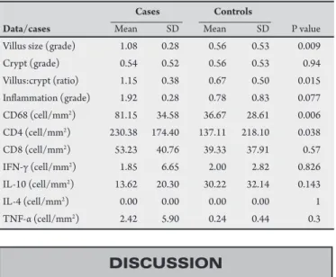
he results relating to villus size, crypts, villus/crypt ratio and inlammation are described in Table 1. he villus size was greater in the VL patients (p < 0.05).
he number of cells expressing CD68 (macrophages) and CD4 was higher in the patients with VL (p < 0.05). No diference in CD8 expression was demonstrated. he number of cells expressing cytokines was also evaluated, but no diferences in IFN-γ, TNF-α, IL-10 or IL-4 expression were found.
he number of cases expressing IFN-γ was signiicantly lower among the patients with VL (p = 0.023). he number of cells expressing IL-10 in the VL group was similar to the number in the control group, and this was also found in relation to TNF-α and IL-4.
TABLE 1 - Case control study of patients with visceral leishmaniasis, evaluating the in situ immune response.
Cases Controls
Data/cases Mean SD Mean SD P value
Villus size (grade) 1.08 0.28 0.56 0.53 0.009 Crypt (grade) 0.54 0.52 0.56 0.53 0.94 Villus:crypt (ratio) 1.15 0.38 0.67 0.50 0.015 Inlammation (grade) 1.92 0.28 0.78 0.83 0.077 CD68 (cell/mm2) 81.15 34.58 36.67 28.61 0.006
CD4 (cell/mm2) 230.38 174.40 137.11 218.10 0.038
CD8 (cell/mm2) 53.23 40.76 39.33 37.91 0.57
IFN-γ (cell/mm2) 1.85 6.65 2.00 2.82 0.826
IL-10 (cell/mm2) 13.62 20.30 30.22 32.14 0.143
IL-4 (cell/mm2) 0.00 0.00 0.00 0.00 1
TNF-α (cell/mm2) 2.42 5.90 0.24 0.44 0.3
The presence of Leishmania in intestinal tissue has been recognized since the time of the irst cases of this disease and Muigai et al described the pathological characteristics very well4. Here, we
described some in situ aspects of the disease that had not been shown previously. he increased numbers of macrophages in the intestinal tissue was not a new discovery, nor was the lymphoid iniltration (CD4 and CD8 cells). he morphological indings regarding villi, crypts and inflammation were also compatible with previous studies. he villi may be atrophic or hypertrophic, but are found to be normal in most cases, including in animal studies8. hese data
explain why chronic diarrhea and malabsorption are uncommon in immunocompetent patients with visceral leishmaniasis9.
395
Rev Soc Bras Med Trop 43(4):393-395, jul-ago, 2010IL-10 with lower IFN-gamma is associated with an old concept of h2 patern of immune response. Nevertheless, localized cutaneous leishmaniasis caused by Leishmania (Viannia) braziliensis is the best concept of h1 patern. Despite these theories, the lack of diference in IL-4, IL-10 and TNF-α expression associated with lower levels of IFN-γ could be explained as an immunotolerant patern, previously called a state of anergy11.
he gut is considered to be an organ with an immune privilege, because of the confusing interplay between microorganisms, antigens and the intestinal epithelial barrier. he intestinal barrier can secrete immunoregulatory mediators that promote the generation of tolerogenic antigen-presenting cells, phagocytic innate immune cells and regulatory cells of the adaptive immune system. his complex interaction maintain the gut homeostasis12.
he presence of Leishmania did not change this microsystem and the tolerance of the immune response enabled survival of this parasite for long periods without any inflammatory process. A detailed evaluation of the innate immune response is necessary to conirm this theory. NK cell, CD4CD25fox3+ cell and apoptosis evaluation are simple analyses that could bring out new concepts regarding immunotolerance in visceral leishmaniasis, with diferences in relation to other organs.
Our study showed some limitations. he number of patients was too small to determine diferences in some variables in relation to cytokine expression. he immune study was restricted to the duodenal mucosa. Studies in diferent parts of the gastrointestinal tract, such as the large intestine, should be performed because of their speciic histological characteristics.
Dysregulation of the immunoregulatory network would lead to colitis, which has been described in dogs5. his would increase the
percentage of intestinal signs and symptoms, thereby increasing the risk of translocation and further septicemia in such patients.
Cytokine responses in the intestine (in this case, the duodenum) were largely absent and only small changes in villus/ crypt structures could be observed, thus indicating that apart from iniltration of macrophages, the structure was largely normal. he only conclusion that can be drawn from the results presented is that duodenal biopsies from VL patients are not very diferent from those of controls, with the exception of cell iniltration and, possibly, increased villus size.
CONFLICT OF INTEREST
REFERENCES
he authors are not part of any associations or commercial relationships that might represent conlicts of interest in the writing of this study (e.g., pharmaceuticalstock ownership, consultancy, advisoryboard membership, relevant patents,or research funding).
1. Guerin PJ, Olliaro P, Sundar S, Boelaert M, Crot SL, Desjeux P, et al. Visceral leishmaniasis: current status of control, diagnosis, and treatment, and a proposed research and development agenda. Lancet Infect Dis 2002; 2:494-501. 2. Tuon FF, Guedes F, Fernandes ER, Pagliari C, Amato VS, Seixas Duarte MI.
In situ immune responses to interstitial pneumonitis in human visceral leishmaniasis. Parasite Immunol 2009; 31:98-103.
3. Barati M, Sharii I, Daie PM, Fasihi HM. Bacterial infections in children with visceral leishmaniasis: observations made in Kerman Province, Southern Iran, between 1997 and 2007. Ann Trop Med Parasitol 2008; 102: 635-641. 4. Muigai R, Gatei DG, Shaunak S, Wozniak A, Bryceson AD. Jejunal function and
pathology in visceral leishmaniasis. Lancet 1983; 27:476-479.
5. Adamama-moraitou KK, Rallis TS, Koytinas AF, Tontis D, Plevraki K, Kritsepi M. Asymptomatic colitis in naturally infected dogs with Leishmania infantum: a prospective study. Am J Trop Med Hyg 2007; 76:53-57.
6. Moussallem TM, Guedes F, Fernandes ER, Pagliari C, Lancellotti CL, de Andrade Jr HF, et al. Lung involvement in childhood measles: severe immune dysfunction revealed by quantitative immunohistochemistry. Hum Pathol 2007; 38:1239-1247.
7. Guedes F, de Andrade Jr HF, Fernandes ER, Tuon FF, Brasil A, Pagliari C, et al. he efects of human herpesvirus 8 infection and interferon-gamma response in cutaneous lesions of kaposi sarcoma difer among human immunodeiciency virus-infected and uninfected individuals. Br J Dermatol 2008;159:839-846. 8. Gonzalez JL, Insa F, Novoa C, Pizarro M. Intestinal amyloidosis in hamsters with
visceral leishmaniasis. Br J Exp Pathol 1986; 67:353-360.
9. Baba CS, Makharia GK, Mathur P, Ray R , Gupta SD, Samantaray JC. Chronic diarrhea and malabsorption caused by leishmania donovani. Indian J Gastroenterol 2006; 25:309-310.
10. Duarte MI, Corbet CE. Histopathological paterns of the liver involvement in visceral leishmaniasis. Rev Inst Med Trop Sao Paulo 1987; 29:131-136. 11. Xu D, Liu H, Komai-Koma M, Campbell C, Mcsharry C, Alexander J, et al.
Cd4+cd25+ regulatory t cells suppress diferentiation and functions of th1 and th2 cells, leishmania major infection, and colitis in mice. J Immunol 2003; 170: 394-399.