INTRODUCTION
1. Núcleo de Doenças de Transmissão Vetorial, Instituto Adolfo Lutz, São Paulo, SP. 2. Laboratório Regional de Ribeirão Preto, Superintendência de Controle de Endemias, Ribeirão Preto, SP. 3. Centro de Vigilância Epidemiológica “Prof. Alexandre Vranjac”, Secretaria do Estado da Saúde de São Paulo, São Paulo, SP. 4. Grupo de Vigilância Epidemiológica, São José do Rio Preto, SP. 5. Centro de Controle de Zoonoses, Prefeitura Municipal de Ribeirão Preto, Ribeirão Preto, SP.
Address to: Dra. Akemi Suzuki. Núcleo Doenças Transmissão Vetorial/IAL. Av. Doutor Arnaldo 355, 01246-902 São Paulo, SP, Brasil.
Phone: 55 11 3068-2901 e-mail: [email protected] Received in 27/10/2010 Accepted in 04/02/2011
Reemergence of yellow fever: detection of transmission in the State
of São Paulo, Brazil, 2008
Reemergência de febre amarela: detecção de transmissão no Estado de São Paulo, Brasil, 2008
Eduardo Stramandinoli Moreno
1, Iray Maria Rocco
1, Eduardo Sterlino Bergo
2, Roosecelis Araujo Brasil
1,
Melissa Mascherati Siciliano
3,
Akemi Suzuki
1, Vivian Regina Silveira
1, Ivani Bisordi
1, Renato Pereira de
Souza
1, and Yellow Fever Working Group
1,2,3,4,5ABSTACT
Introduction: Following yellow fever virus (YFV) isolation in monkeys from the São José do Rio Preto region and two fatal human autochthonous cases from the Ribeirão Preto region, State of São Paulo, Brazil, two expeditions for entomological research and eco-epidemiological evaluation were conducted. Methods: A total of577 samples from humans, 108 from monkeys and 3,049 mosquitoes were analyzed by one or more methods: virus isolation, ELISA-IgM, RT-PCR, histopathology and immunohistochemical. Results: Of the 577 human samples, 531 were tested by ELISA-IgM, with 3 positives, and 235 were inoculated into mice and 199 in cell culture, resulting in one virus isolation. One sample was positive by histopathology and immunohistochemical. Using RT-PCR, 25 samples were processed with 4 positive reactions. A total of 108 specimens of monkeys were examined, 108 were inoculated into mice and 45 in cell culture. Four virus strains were isolated from Alouata caraya. A total of 931 mosquitoes were captured in Sao Jose do Rio Preto and 2,118 in Ribeirão Preto and separated into batches. A single isolation of YFV was derived from a batch of 9 mosquitoes Psorophora ferox, collected in Urupês, Ribeirão Preto region. A serological survey was conducted with 128 samples from the municipalities of São Carlos, Rincão and Ribeirão Preto and 10 samples from contacts of patients from Ribeirão Preto. All samples were negative by ELISA-IgM for YFV. Conclusions:
he results conirm the circulation of yellow fever, even though sporadic, in the Sao Paulo State and reinforce the importance of vaccination against yellow fever in areas considered at risk.
Keywords: Yellow fever. Flavivirus. Epizooties. Entomological surveillance.
RESUMO
Introdução: A partir do isolamento do vírus febre amarela (VFA), de macacos, da região de São José do Rio Preto e de dois casos humanos autóctones fatais, da região de Ribeirão Preto, Estado de São Paulo, foram realizadas duas expedições para pesquisa entomológica e avaliação ecoepidemiológica. Métodos: Um total de 577 amostras de humanos, 108 de macacos e 3.049 mosquitos foram analisados por um ou mais métodos: isolamento viral, ELISA-IgM, RT-PCR, histopatologia e imunohistoquímica. Resultados: De 577 amostras humanas, 531 foram testadas por ELISA-IgM, sendo 3 positivas, 235 foram inoculadas em camundongos, 199 em cultura de células, obtendo-se 1 isolamento viral. Uma amostra foi positiva por histopatologia e imunohistoquímica. Por RT-PCR foram processadas 25 amostras com 4 reações positivas. Os 108 espécimes de macacos foram inoculados em camundongos, 45 em cultura de células, obtendo-se 4 isolamentos de VFA, de Alouata caraya. Um total de 931 mosquitos foram capturados em São José do Rio Preto e 2.118 em Ribeirão Preto e separados em lotes. Um único isolamento de VFA foi derivado de um lote de 9 mosquitos Psorophora ferox, coletados em Urupês, região de Ribeirão Preto. Um inquérito sorológico foi realizado com 128 amostras dos municípios de São Carlos, Rincão e Ribeirão Preto e mais 10 amostras de contactantes de pacientes de Ribeirão Preto. Todas as amostras foram negativas por ELISA-IgM para VFA.
Conclusões: Os resultados conirmam a circulação, mesmo que esporádica, do VFA no Estado de São Paulo e reforça a importância da vacinação antiamarílica nas áreas consideradas de risco.
Palavras-chaves: Febre amarela. Flavivírus. Epizotias. Vigilância entomológica.
Yellow fever (YF) is an infectious disease, endemic in the tropical forests of Africa and Central and South America1. Clinical manifestations in
humans range from asymptomatic to mild and severe forms. Yellow fever virus (YFV) has two transmission patterns in the Americas: sylvatic yellow fever (SYF) and urban yellow fever (UYF)1,
but both lead to the same clinical disease. The sylvatic cycle involves mosquitoes and monkeys. Some monkeys are highly susceptible to YFV, such as the howler monkey (genus Alouata), while other species show strong resistance to the virus, such as the capuchin monkey (genus Cebus)2. he sylvatic
yellow fever cycle in the Americas includes vectors from the genus Haemagogus and Sabethes, and the main species are Hg. janthinomys, Hg. albomaculatus,
Hg. leucocelaenus, Sa. chloropterus, Sa. Soperi and Sa. Cyaneus2,3. Urban yellow fever involves humans and
Aedes aegypti mosquitoes2-4.
Yellow fever virus is the prototype member of the genus Flavivirus, family Flaviviridae1. he genome is
a single strand, positive sense RNA, approximately 11kb in length. he complete genome has 10,862 nucleotides that encode 3,411 aminoacids1,4-8.
Genetic studies of YFV strains have revealed some variations; the YFV strains isolated in South America and Africa are genetically distinct and are associated with diferent geographic regions. hus, in Africa ive genotypes have been identiied: West Africa I, West Africa II, East Africa, Central and East Africa and Angola8,9. In South America two genotypes have
been identiied: South America I, which involves strains identiied from Brazil, Panama, Colombia, Ecuador, Venezuela and Trinidad, and genotype South America II from Peru8,11.
Yellow fever usually occurs as outbreaks in cycles of 7 to 10 years, alternating with periods with lower numbers of cases4. Epizooties are usually registered
before human cases are detected.
Brazil has the most YF enzootic areas in continental America. Yellow fever is endemic in the
Article/Artigo
METHODS
northern region and appears sporadically as epidemic/endemic in the central-western region12. Since 2000, SYF has been spreading
progressively, transcending its usual boundaries and reaching other areas formerly known as enzootic13-15. The last urban outbreak
in Brazil occurred in 19424. he last autochthonous cases were
reported in the State of São Paulo 47 years ago15, but in 2000, two
autochthonous SYF cases were notiied along the border of the State of Minas Gerais14.
Ater YF laboratory conirmation in four monkeys from the region of São José do Rio Preto and two conirmed fatal human autochthonous cases in the region of Ribeirão Preto, two expeditions for entomological studies and eco-epidemiological assessment of the likely sites of infection (LSI) were conducted. his study presents the results of laboratory analysis performed with blood/serum and tissues of humans and monkeys and mosquitoes collected in these regions from January to July 2008.
Biological samples from humans
A total of 577 biological samples from individuals with suspected yellow fever infection were processed from January to July 2008. hese samples came from networks of private and public healthcare in the State of São Paulo. he human autochthonous cases occurred in April and May 2008.
Serosurvey
Serological surveys to determine the prevalence of IgM antibodies against YFV were conducted in the municipalities of São Carlos, Rincão and Ribeirão Preto, in areas close to the LSI, following the conirmation of human YF cases.
Biological samples-monkeys
Monkeys that were found dead, though still in a suitable state for storage, were submited to necropsy to remove the liver, spleen, kidneys, heart, lungs and occasionally blood. he samples were sent, in liquid nitrogen, for virus research and in 10% formalin for immunohistochemical investigation of viral antigen.
Vectors
Expeditions to the region of São José do Rio Preto and Ribeirão Preto were conducted in April and June 2008, respectively.
To catch vectors, mobile collection was used, with stops for 15-20min, with the aid of capturing oral suction and dip nets at times of highest light intensity between 9am and 4pm. he expeditions lasted four days. he mosquitoes collected were stored in cryo-resistant tubes, transported in a liquid nitrogen lask and stored at -70°C in the laboratory. Identiication of mosquitoes was performed on a cold table and the mosquitoes were separated into batches of the same species or genus.
Entomological area research
São José do Rio Preto region: entomological surveys were conducted in the municipalities of Nova Aliança, Mendonça and Urupês, in areas where monkeys were found dead. These environments consist of small remaining fragments of forest, mostly gallery forests, surrounded by areas of agriculture and pasture. he local vegetation consists of heavily modiied savannah sensu strictu, forming small groves of varying density, depending on soil and water availability. he area presents a mesothermal humid climate: a dry
and mild winter, with average temperatures during the coldest month above 18°C and an annual average of 25.3°C. he average rainfall during the driest month is less than 60mm16
Ribeirão Preto region: an entomological survey was conducted close to the likely sites where the two humans were infected. hese areas are located in rural/wild areas in the municipalities of Luiz Antônio, São Carlos and Rincão, surrounding the Jataí Farm Forest Reserve located in Luiz Antônio. he Farm Forest Reserve has an extension of 4,532 hectares, with vegetation consisting primarily of
cerradosensu latu, with variations related to forests covering water bodies: riparian forest, gallery forest, semideciduous forest and transition forest riparian/savannah, as well as dry forests: high altitude savannah, high altitude open savannah and low altitude savannah17.
Adjacent areas are used for technified agriculture linked to industrial complexes, with a predominance of cultures of sugar cane and citrus plantations, as well as eucalyptus and pine, and pasture areas18.
he study areas are located in a climate zone that has two well deined seasons; hot and rainy in the period from October to April, and another cold and relatively dry in the period from May to September. he total estimated average annual rainfall is 1,400mm, and in the dry period, the monthly average is below 20mm. he average annual temperature is 22°C, with an average maximum of 29°C and a minimum of 16°C19.
he State of São Paulo Environmental Department20 describes
the presence of species of the following monkeys Callicebus personatus nigrirons, Alouata caraya e Cebus apella in these regions.
Laboratory diagnosis
Depending on whether the samples were acute or convalescent, they were analyzed by one or more of the following methods: virus isolation (in mice and/or cell culture), capture ELISA IgM antibodies, indirect immunoluorescence, RT-PCR and sequencing. he samples from necropsy were analyzed by histopathology and immunohistochemical.
Capture ELISA IgM
The tests were performed in accordance with the protocol described by Kuno et al21. The samples were processed at 1:40
dilution in PBS containing 0.5% bovine albumin. he system used for revelation of the test consisted of a conjugate (anti-lavivirus labeled peroxidase) and substrate (ABTS). Samples with optical density reading of greater than 0.2 were considered positive. Positive and negative controls were added to all assays.
Isolation of virus in mice
To isolate the virus, samples of blood, serum, tissues from the autopsies of monkeys and humans, and mosquitoes were inoculated in 1-3 day-old Swiss mice. Samples of liver and brain of human and monkeys were inoculated separately, and the other viscera were processed in a mixed suspension. The tissues were macerated, suspended in a solution of bovine albumin, 0.75%, with antibiotics (100μg/mL of streptomycin and 100UI/mL of penicillin) and centrifuged at 6,000rpm for 20min. Each suspension was inoculated by the intracerebral route in 6 suckling mice at a dose of 0.02ml/mouse. Blood samples or serum were inoculated separately, pure or diluted (50%) in albumin solution. he animals were observed for 21 days.
RESULTS
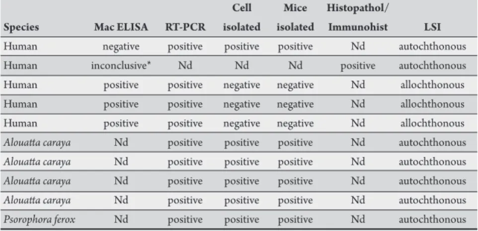
TABLE 1 - Laboratorial tests for yellow fever virus in the State of São Paulo, from January to July 2008.
Cell Mice Histopathol/
Species Mac ELISA RT-PCR isolated isolated Immunohist LSI
Human negative positive positive positive Nd autochthonous
Human inconclusive* Nd Nd Nd positive autochthonous
Human positive positive negative negative Nd allochthonous
Human positive positive negative negative Nd allochthonous
Human positive positive negative negative Nd allochthonous
Alouata caraya Nd positive positive positive Nd autochthonous Alouata caraya Nd positive positive positive Nd autochthonous Alouata caraya Nd positive positive positive Nd autochthonous Alouata caraya Nd positive positive positive Nd autochthonous Psorophora ferox Nd positive positive positive Nd autochthonous Mac ELISA: IgM antibody capture Enzyme-linked immunosorbent assay, RT-PCR: Reverse transcription polymerase chain reaction ,Histopathol/Immunohist: Histopathology/Immunohistochemistry, LSI: Likely site of infection, Nd: not done. *Absorbance reading next to the value of the cutof test
cases in which the deinition of species was not possible, in batches of the same genus. Mosquitoes were macerated and suspended in a solution 1.8% of bovine albumin and antibiotics (100μg/mL of streptomycin and 100UI/mL of penicillin) and centrifuged at 10,000rpm for 30min. Each supernatant was inoculated into mice in the same manner described above. he mice were observed for 21 days and those showing signs of disease had their brains harvested and subjected to subsequent passages to adapt the virus22,23.
Virus isolation in cell culture
he same suspensions of tissues from human and monkey tissues, prepared for inoculation in mice, in addition to blood and serum, were also subjected to virus isolation in cell culture. An aliquot of 20µl was inoculated into tubes seeded with cultured cells of Aedes albopictus, clone C6/3624. he tubes were incubated for 9 days at 28°C and then
agitated and centrifuged at 1,500rpm for 5min. he supernatants were stored at -70ºC, the pellets of cells were resuspended in PBS pH 7.5 and placed on slides with 12 parallel holes, for the indirect immunoluorescence assay (IFA) following the technique standardized by Gubler et al25. Polyclonal anti-yellow fever antibodies
prepared in mice and anti-mouse immunoglobulin conjugated marked with luorescein isothiocyanate (Sigma) were used. he positive samples were identified by IFA with YFV monoclonal antibodies (Center for Disease Control and Prevention, USA).
RNA extraction and RT-PCR
Total RNA was extracted using commercial kits. For tissue
fragments, the QIAamp® RNA Blood (Qiagen Inc., Ontario, CA)
was utilized and for serum, the QIAamp® Viral RNA Kit (Qiagen Inc., Ontario, CA) was used, in accordance with the manufacturer’s instructions.
Ampliication of viral RNA was performed by one-step RT-PCR, followed by a second ampliication (semi-nested)26 of the products
of the irst reaction, diluted at 1:50. he ampliied products were visualized by electrophoresis in 1.5% agarose gel stained with ethidium bromide. Positive samples were those that had a band compatible with the expected.
Sequencing
Positive samples were sequenced directly in an ABI 377 sequencer by the method of dideoxy chain-termination cycle sequencing using the BigDye terminator sequencing kit v.3.1 (Applied Biosystems,
Monkeys
A total of 108 specimens of monkeys were analyzed: 76 (70.5%)
Callithrixpenicillata, 13 (12%) Alouata caraya,13 (12%), Cebus apella and 6 (5.6%) other species. Tissue samples and/or blood of 108 monkeys were inoculated in mice and 45 in cell culture. YFV isolation was obtained from four Alouata caraya, two were collected in Mendonça, one in Urupês and the other in Nova Aliança on January 14, 2008, using mice and cell culture. Two of them were found dead in Mendonça and Nova Aliança. he third one, a sick infant, was found with its dead mother in Mendonça, and the fourth was found dead in Urupês, on February 14, 2008. All isolates were analyzed by RT-PCR, with positive results for SYF (Table 1).
Humans
A total of 577 samples from humans with clinical suspicion of YF infection were analyzed. Among them, ive were positive (two autochthonous and three allochthonous) by at least one of the techniques (Table 1). he serological survey was performed with 128 human samples from São Carlos (70), Rincão (22) and Ribeirão Preto (36). Besides these, another 10 serum samples from individuals who had been in contact with the patient from Ribeirão Preto were analyzed. All samples presented negative ELISA-IgM for YFV. Foster City, CA), in accordance with the manufacturer’s instructions, with the same pair of primers of the one step RT-PCR.
For the edition of the nucleotide sequences, the Chromas Lite v.2.01 (Technelysian Pty Ltd.) was used, excluding the sequences of primers.
Histopathology and immunohistochemistry
A total of 235 human tissue fragments and blood/serum, submited for virus isolation in mice, resulted in the isolation of one strain of YFV, later characterized by RT-PCR and sequencing. he same isolation was obtained in cell culture among 194 samples inoculated. Among the 25 samples tested by RT-PCR, four were positive, one autochthonous and three allochthonous.
Entomological research
A total of 3,049 mosquitoes were captured, 931 assembled in 148 lots, collected in the São Jose do Rio Preto region and 2,118 assembled in 172 lots, collected in the Ribeirão Preto region. Species richness is presented in Tables 2 and 3. A single isolation of YFV was derived from a batch of nine samples of Psorophora ferox collected in Urupês, Ribeirão Preto region, on March 14, 2008. Viral isolation was achieved in mice and identiication was achieved by RT-PCR and sequencing.
Histopathological analysis
Histopathological findings in both humans and monkeys consisted in lesions, predominantly mediozonal, sometimes extending from the hepatic parenchyma. Abundant apoptosis in hepatocytes, focal necrosis, steatosis and micro macrogoticular, hyperplasia and hypertrophy of Kupfer cells and space-port with mild lymphocytic iniltrate and no evidence of lesion interface were found (Figure 1).
TABLE 2 - Mosquito species collected in the São José do Rio Preto region, State of São Paulo, March 2008.
Pools Females
Species n n %
Aedes serratus group 14 295 31.7
Psorophora ferox 18 145 47.3
Ochlerotatus scapularis 27 117 12.6
Culex (Melanoconion) spp. 14 96 10.3
Aedeomyia squamipennis 1 39 4.2
Psorophora albigenu 7 39 4.2
Culex declarator ainis 6 38 4.1
Sabethes chloropterus 10 28 3.0
Sabethes tridentatus 5 21 2.3
Psorophora discrucians 5 19 2.0
Aedes terrens 3 18 1.9
Culex (Culex) spp. 6 18 1.9
Haemagogus leucocelaenus 7 16 1.7
Coquilletidia albicosta 3 7 0.8
Uranotaenia geometrica 1 6 0.6
Anopheles triannulatus 3 5 0.5
Howardina argyrothorax 2 3 0.3
Coquilletidia juxtamansonia 2 3 0.3
Haemagogus janthinomys/capricornii 3 3 0.3
Sabethes shannoni 1 3 0.3
Culex bidens 1 2 0.2
Limatus durhamii 2 2 0.2
Culex ameliae ainis 1 2 0.2
Anopheles parvus 1 1 0.1
Culex declaratory 1 1 0.1
Culex (Microculex) spp. 1 1 0.1
Wyeomyia aporonoma 1 2 0.1
Aedes albopictus 1 1 0.1
Total 148 931 100.0
TABLE 3 - Mosquito species collected in the Ribeirão Preto region, State of São Paulo, March/2008
Pools Females
Species n n %
Aedes serratus group 34 1,298 61.3
Psorophora ferox 19 359 16.9
Ochlerotatus scapularis 22 249 11.8
Culex (Melanoconion) spp. 9 40 1.9
Culex (Culex) spp. 8 25 1.2
Haemagogus leucocelaenus 5 17 0.8
Sabethes chloropterus 4 14 0.7
Aedes albopictus 8 14 0.7
Coquilletidia juxtamansonia 7 11 0.5
Ochlerotatus fulvus 7 9 0.4
Sabethes intermedius 1 9 0.4
Wyeomyia spp. 3 8 0.4
Anopheles triannulatus 5 7 0.3
Mansonia titillans 4 6 0.3
Howardina fulvithorax 4 6 0.3
Culex restuans/declarator 2 4 0.2
Mansonia wilsoni 2 4 0.2
Psorophora albigenu 3 4 0.2
Sabethes quasicyaneus 3 4 0.2
Sabethes undosus 2 4 0.2
Sabethes identicus 1 4 0.2
Anopheles albitarsis s.l. 3 3 0.1
Anopheles mediopunctatus group 1 3 0.1
Culex habilitator ainis 3 3 0.1
Haemagogus janthinomys/capricornii 2 2 0.1
Sabethes belisarioi 1 2 0.1
Sabethes tridentatus 2 2 0.1
Aedes terrens 1 1 0.0
Anopheles galvaoi 1 1 0.0
Culex bidens 1 1 0.0
Culex coronator 1 1 0.0
Sabethes glaucodaemon 1 1 0.0
Psorophora lanei ainis 1 1 0.0
Total 172 2,118 100.0
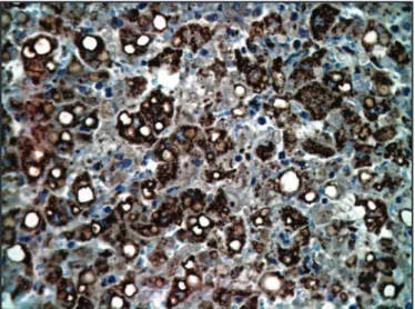
Immunohistochemistry
he presence of viral antigen was veriied by a brownish color, present in the cytoplasm of hepatocytes and Kuppfer cells (Figure 2).
FIGURE 2 - Hepatocytes positively imunostained with anti-anti-antigen of yellow fever virus amplified in reaction with enzyme-conjugated polymer (X400).
DISCUSSION
Monkey deaths have been registered in the area of transition to YF in the State of São Paulo since January 2008.
All the monkeys from which YFV was isolated belonged to the species Alouata caraya, which originated from cities outside the region of Sao José do Rio Preto. Due to their high susceptibility to YF, when found dead, infection in the Alouata caraya monkey species can indicate the emergence of YF12. Continuous monitoring
of these communities makes it possible to detect viral activity in the environment. he most abundant species, among the 108 monkeys found sick or dead, was Callithrix penicillata (over 70%).
The presence of three principal species of YFV vector in the region of São José do Rio Preto was observed: Haemagogus janthinomys, Haemagogus leucocelaenus and Sabethes chloropterus. However, these mosquitoes were identified in small numbers, which could be explained by the fact that the collections were made at ground level and these species favor the upper strata of trees29.
However, it should be noted that the forests studied did not have the same exuberance observed in the Amazon rainforest.
his is the irst report of the isolation of YFV from Psorophora ferox, although it had previously been found naturally infected by the arbovirus Ilheus30 andRocio31. However, these findings do
not necessarily mean that the species is a vector for these viruses.
Psorophora ferox was the second most abundantly caught species, corresponding to 47.3% in the region of São José do Rio Preto (Table 2) and 16.9% in the region of Ribeirão Preto (Table 3) from the total number of female specimens. Virus isolation could be facilitated by this abundance, since a dense population of mosquitoes is more susceptible to entering in contact with a circulating arbovirus and more easily accessed. Ps. ferox females are persistent and painful biters and they are not likely to be overlooked in biting collections. his aggressive behavior is the primary reason for its abundance in the collection. he virus isolation in a mosquito captured in the ield is an indication that this species could be naturally infected, but this
occurs only in certain circumstances and, up to now, no evidence of transmission associated with Psorophora had been veriied. Additional studies of vector competence and capacity are required to evaluate the possibility of this species acting as a vector, but it is most probable that the YF infection in this species is accidental. Most isolations of YF from mosquitoes come from Haemagogus or Sabethes, with occasional evidence of other species infected with YFV, such as
Aedes fulvus, Psorophora, Aedesscapularis and Psorophoraalbipes, each presenting one isolation2.
Atempts to isolate the virus in Aedes albopictus, collected in the Ribeirão Preto region, were all negative. his species is considered a potential vector of YF32or a potential link between urban and
rural YF33, since it is disseminated in the periurban, rural and sylvan
environments.
he Jataí Farm Forest Reserve and fragments of forests in the region of São José do Rio Preto, in principle, are ideal habitats for the circulation of YFV, since susceptible host monkeys and species of mosquitoes that are vectors of YF were found in this area. However, the forested environment is deteriorated and largely fragmenting this area, which may not be suicient for the development of a large group of monkeys. As a result, the region does not favor the concentration of vector species in the dry season in order to maintain the cycles of YFV for long periods. In addition to the collection points located within the Jataí Forest Reserve Farm, mosquitoes were also collected in rain forest areas near the Anhanguera Highway, a forest habitat rather modiied by human action. Nevertheless, it was possible to capture species known to be vectors of YF, oten found in preserved forested environments21. his suggests possible diferences in the
dynamics of YF in fragmented landscapes due to adaptations to their hosts and reservoirs, speciic to each region.
Further research is needed to assess the vector ability of other species of mosquitoes present in the region to identify possible alternative vectors and, in such cases, to determine the importance of these vectors in the maintenance of YFV in the environment.
In the expedition to the region of Ribeirão Preto, Haemagogus leucocelaenus and Sabethes chloropterus, known vectors of YF, were captured. Several reports exist of YFV isolation from Haemagogus lecocelaenus mosquitoes in the Amazon region34. More recently, in
2003, these mosquitoes have been found naturally infected with YFV in Rio Grande do Sul33.
No monkey positive for YFV was detected in the Ribeirão Preto region. hrough information from local people, the members of the expedition learned that groups of monkeys that had been previously seen or heard of in the region were no longer observed, suggesting the death of most or all of these groups.
Prompt laboratory diagnosis and the investigation of cases, whether autochthonous or allochthonous, is very important because of the environmental conditions favorable for the spread and establishment of the transmission of the agent in unafected areas of the state. his determines the implementation of control measures related to urban vectors (preventing the risk of urban YF) or of blockage by anti-yellow fever vaccination. he importance of highways that connect the Amazon region to other states of Brazil should be highlighted, since these help disseminate diferent strains of YFV33.
four following the onset of symptoms. he RT semi-nested PCR permited conirmation of 50% of SYF cases in this period. hrough necropsy, it was possible to correlate morphological indings and the immunopathology of infection, conirmed by immunohistochemical research positive for viral antigen.
Another important aspect concerns the role that the human population exercises in the maintenance of YFV in fragmented landscapes. Within a given location, for each case diagnosed as typical SYF, numerous asymptomatic cases also exist that usually go unnoticed. In Brazil, serological investigations have shown that 90% of infections are asymptomatic34. hus, in primary infections of
wild YF, the symptoms are atypical, requiring the existence of a cycle
Haemagogus - Man - Haemagogus for the appearance of secondary or tertiary cases, the only ones likely to be diagnosed clinically34. It
is important to assess whether the dynamics of YF is inluenced by human populations living and working near forest fragments with small populations of monkeys.
he present study indings conirm the circulation of YFV, even if sporadic, in the State of São Paulo and reinforce the importance of maintaining vaccination coverage for YF in areas considered at risk. he high mobility of the human population to transmission areas, individuals in the course of viremia to areas still without established transmission, associated with high rates of infestation by Aedes aegypti
in Brazil represent a risk for the reemergence of YF.
he indings indicate the need for eco-epidemiological studies, with a multidisciplinary focus, to elucidate the dynamics of transmission and geographical distribution of YFV in the state. Equally relevant is the impact of disease on wildlife endangered species.
ACKNOWLEDGMENTS
he authors declare that there is no conlict of interest.
CONFLICT OF INTEREST
REFERENCES
he authors are grateful to Miriam S. A. Marques for supplying the cell cultures and Lucy T.A. dos Santos and Regina C.W.F. Mozetic for supplying the suckling mice. Yellow Fever Working Group: Adriana Yurika Maeda, Rosa M Tubaki, Terezinha L. M. Coimbra, Selma Petrella, Luiz E. Pereira, Fernanda G. Silva, Cecília L. S. Santos, Regiane T. Menezes, Rubens P. Cardoso Jr, Mônica R. Bocchi, José J. N. Martins, Cilea H. Tengan and Cristina Kanamura.
1. Barnet ED. Yellow fever: Epidemiology and control. Clin Infect Dis 2004; 44:850-856.
2. Vasconcelos PFC. Febre amarela. Rev Soc Bras Med Trop 2003; 36:275-293. 3. Dégallier N, Travassos da Rosa APA, Vasconcelos PFC, Travassos da Rosa ES,
Rodrigues SG, Sá Filho GC, et al. New entomological and virological data on the vectors of sylvatic yellow fever in Brazil. Braz J Assoc Adv Sci 1992; 44:136-142. 4. Monath TP. In: Monath TP, editor. he Arboviruses: ecology and epidemiology.
Yellow fever, Vol V. Boca Raton (FL): CRC Press; 1988. p. 139-241.
5. Westaway EG, Briton MA, Gaidamovich SY, Horzinek MC, Igarashi A, Kaariainen L, et al. Flaviviridae. Intervirology 1985; 24:183-192.
6. Rice CM, LenchesEM, Eddy SR, Shin SH, Strauss JH. Nucleotide sequence of yellow fever virus: implications for lavivirus gene expression and evolution. Science 1985; 229:726-733.
7. Chambers TJ, Hahn CS, Galler R, Rice CM. Flavivirus genome organization, expression, and replication. Ann Rev Microbiol 1990; 44:649-688.
8. Wang E, Weaver SC, Shope RE, Tesh RB, Wats DM, Barret ADT. Genetic variation in yellow fever virus: duplication in the 3’ noncoding region of strains from Africa. Virology1996; 225:274-281.
9. Zanoto PMA, Gould EA, Gao GF, Harvey PH, Holmes EC. Population dynamics of laviviruses revealed by molecular phylogenies. Proc Nat Acad Sci 1996; 93:548-553.
10. Mutebi JP, Wang H, Li L, Bryant JE, Barret ADT. Phylogenetic and evolutionary relationships among yellow fever virus isolates in Africa. J Virol 2001;75:6999-7008.
11. Briant JE, Holmes EC, Barret ADT. Out of Africa: A Molecular Perspective on the Introduction of Yellow Fever Virus into the Americas. PLoS Pathogens 2007; 3:Issue 5 e 75.
12. Ministério da Saúde. Secretaria de Vigilância em Saúde. Guia de vigilância epidemiológica. 6a ed. Brasília: Ministério da Saúde; 2005.
13. Fundação Nacional de Saúde. Manual de Febre Amarela. Brasília: Fundação Nacional de Saúde; 2005.
14. Rocco IM, Katz G, Tubaki RM, Suzuki A, Coimbra TLM, Ferreira IB, et al. Febre amarela silvestre no Estado de São Paulo: casos humanos autóctones. Rev Inst Adolfo Lutz 2003; 62:201-206.
15. Filippis AMB, Nogueira RMR, Schatzmayr HG, Tavares DS, Jabor AV, Diniz SCM, et al. Outbreak of jaundice and hemorrhagic fever in the Southeast of Brazil in 2001: detection and molecular characterization of yellow fever virus. J Med Virol 2002; 68:620-627.
16. Teixeira LA. Da transmissão hídrica a culicidiana: a febre amarela na sociedade de medicina e cirurgia de São Paulo. Rev bras Hist 2001; 21: 217-241. 17. Instituto Brasileiro de Geografia e Estatística. Recursos Naturais e Meio
Ambiente: Uma visão do Brasil. Rio de Janeiro: Departamento de Recursos Naturais e Estudos Ambientais; 1992.
18. Pires JSR. Análise ambiental voltada ao planejamento a gerenciamento do ambiente rural: abordagem metodológica aplicada ao Município de Luiz Antônio - SP. [Tese Doutorado]. [São Carlos]: Universidade Federal de São Carlos; 1995. p.192.
19. Pires AMZCR, Santos JE, Pires JSR. Caracterização e diagnóstico ambiental de uma unidade da paisagem. Estudo de caso: Estação Ecológica de Jataí e Estação Experimental de Luiz Antônio. In: JE Santos, JSR Pires, editores. Estação Ecológica de Jataí, vol. 1. Estudos Integrados em Ecossistemas. São Carlos: Editora Rima; 2000; p. 346.
20. Secretaria do Meio Ambiente do Governo do Estado de São Paulo. Fauna Ameaçada no Estado de São Paulo. Série Documentos Ambientais. São Paulo, SP: SMA/CED. PROBIO; 1998.
21. Kuno G, Gomez I, Gubler DJ. Detecting artiicial antidengue IgM complexes using a enzyme linked immunosorbent assay. Am J Trop Med Hyg 1987; 36:153-159. 22. Beaty B, Calisher CH, Shope RE. Arboviruses. In: Schmidt NJ, Emmons RW,
editors. Diagnostic Procedures for Viral, Ricketsial and Chlamydial Infections, 6th ed. Washington: American Public Health Association; 1989. p. 797-855.
23. Travassos da Rosa APA, Travassos da Rosa ES, Travassos da Rosa JFS, Dégallier N, Vasconcelos PFC, Rodrigues SG, et al. Os arbovirus no Brasil: Generalidades, Métodos e Técnicas de Estudo. Documento Técnico n° 2, Belém, PA, Instituto Evandro Chagas. Fundação Nacional da Saúde. Ministério da Saúde; 1994. 24. Igarashi A. Isolation of Singh´s Aedes albopictus cell line clone sensitive to dengue
and chikungunya virus. J Gen Virol 1978; 40:531-544.
25. Gubler DJ, Kuno G, Sather GE, Velez M, Oliver A. Mosquito cell culture and speciic monoclonal antibodies in surveillance for dengue viruses. Am J Trop Med Hyg 1984; 33:158-165.
26. Deubel V, Huerre M, Cathomas G, Drouet MT, Wuscher N, Le Guenno B, et al. Molecular detection and characterization of yellow fever virus in blood and liver specimens of a non-vaccinated fatal human case. J Med Virol 1997; 53:212-217. 27. Brito T, Siqueira AS, Santos RT, Nasser ES, Coimbra TL, Alves VA. Human fatal yellow fever. Immunohistochemical detection of viral antigens in liver, kidney and heart. Pathol Res Pract 1992; 188:177-181.
28. Sabbatini E, Bisgaard K, Poggi S, Piccioli M, Ceccarelli C, Pieri F, et al. he EnvisionTM + System: a new immunohistochemical method for diagnostis and
research. Critical comparison with the APAAP, ChemMateTM, CSA, LABC and
29. Pinheiro FP, Travassos da Rosa APA, Freitas RB, Travassos da Rosa JFS, Vasconcelos PFC. Arboviroses - Aspectos clínicos epidemiológico. In: Instituto Evandro Chagas, organizador. 50 anos de contribuição às Ciências Biológicas e à Medicina Tropical. Belém: Fundação de Serviço de Saúde Pùblica; 1986. Vol 1. p. 375-408.
30. Travassos da Rosa JFS, Travassos da Rosa APA, Vasconcelos PFC, Pinheiro FPI, Rodrigues SG, Travassos da Rosa ES, et al. An overview of arbovirology in Brazil and neighbouring coutries. Belém: Instituto Evandro Chagas; 1998. 31. Lopes OS, Saccheta L, Francy DB, Jakob WL, Calisher CH. Emergence of a new
arbovirus disease in Brazil. III Isolation of Rocio virus from a Psorophora ferox (Humbodt, 1819). Amer J Epidemiol 1981; 113:122-125.
32. Foratini OP. Culicidologia médica. São Paulo: Editora da Universidade de São Paulo; 2002.
33. Vasconcelos PFC, Sperb AF, Monteiro HAO, Torres MAN, Souza MRS, Vasconcelos HB et al. Isolations of yellow fever virus from Haemagogus leucocelaenus in Rio Grande do Sul State, Brazil. Trans Royal Soc Trop Med Hyg 2003; 97:60-62.