Uterine artery embolization with spherical pva-pvac particles as
Uterine artery embolization with spherical pva-pvac particles as
Uterine artery embolization with spherical pva-pvac particles as
Uterine artery embolization with spherical pva-pvac particles as
Uterine artery embolization with spherical pva-pvac particles as
preparation for surgical resection of miyomas
preparation for surgical resection of miyomas
preparation for surgical resection of miyomas
preparation for surgical resection of miyomas
preparation for surgical resection of miyomas
Embolização das artérias uterinas com partículas de PVA-PVAC esférico como
Embolização das artérias uterinas com partículas de PVA-PVAC esférico como
Embolização das artérias uterinas com partículas de PVA-PVAC esférico como
Embolização das artérias uterinas com partículas de PVA-PVAC esférico como
Embolização das artérias uterinas com partículas de PVA-PVAC esférico como
preparo para posterior ressecção cirúrgica de miomas
preparo para posterior ressecção cirúrgica de miomas
preparo para posterior ressecção cirúrgica de miomas
preparo para posterior ressecção cirúrgica de miomas
preparo para posterior ressecção cirúrgica de miomas
JURACI GHIARONI1; GAUDENCIO ESPINOSA LOPEZ,TCBC-RJ2; ANTONIO CARLOS COUTINHO JUNIOR3; ALBERTO SCHANAIDER,TCBC-RJ4
A B S T R A C T A B S T R A C T A B S T R A C T A B S T R A C T A B S T R A C T
Objective Objective Objective Objective
Objective: To evaluate the use of a new spherical particle of polyvinyl alcohol and polyvinyl acetate (PVA-PVAc) for uterine artery embolization in patients with myoma with surgical indication. MethodsMethodsMethodsMethods: twelve patients underwentMethods uterine myoma embolization with PVA-PVAc particles. Three to nine months later, they were submitted to laparotomy with myomectomy. We analyzed the following parameters: volume of the uterus and of the biggest myoma; concentrations of follicle stimulating hormone and hemoglobin; menstrual bleeding (number of days and used absorbents), signs and symptoms before treatment, after embolization and after myomectomy. ResultsResultsResultsResultsResults: The mean age was 37 years and mean uterine volume prior to treatment, 939.3 cc. Three years after embolization, there was reduction in uterine volume (p = 0.0005), increase in hemoglobin concentration after embolization (p = 0.0004), without variation after the myomectomy. There was no significant variation of the follicle stimulating hormone (p = 0.17). There was no case of ovarian failure, but one of the patients had endometrial atrophy. Two patients became pregnant, with good obstetric indicators. Signs and symptoms improved after embolization, and remained after myomectomy. ConclusionConclusionConclusionConclusionConclusion: Arterial embolization with spherical PVA-PVAc particles is promising in the preparation for myoma surgery, since it was associated with a reduction in uterine volume, decrease in intraoperative bleeding and made possible the use of smaller incisions, increasing the chance of preserving the uterus.
Key words Key words Key words Key words
Key words: Patients. Leiomyoma. Uterine artery embolization. Polyvinyls. Treatment outcome
Work performed at the Clementino Fraga Filho University Hospital – HUCFF, Federal University of Rio de Janeiro –UFRJ, Rio de Janeiro State – RJ, Brazil.
1. Associate Professor, Department of Obstetrics and Gynecology, UFRJ; 2. Associate Professor, Department of Surgery, UFRJ; 3. Radiologist, Diagnostic Imaging Clinic (CDPI); 4. Professor, Department of Surgery, Faculty of Medicine, UFRJ.
INTRODUCTION
INTRODUCTION
INTRODUCTION
INTRODUCTION
INTRODUCTION
T
he use of embolic materials for the treatment of tumors dates back to 1904, but their application specifically for the treatment of uterine myomas occurred only decades after 1. The first series of cases in which occlusion of theuterine artery with embolic materials proved effective was reported by Professor Ravina in 1995 2. In the last decade,
arterial embolization has established itself as an alternative for the treatment of myomas 3,4.
In 2004, the American College of Obstetrics and Gynecology recommended treatment with uterine artery embolization (UAE), though with reservations for women wishing to preserve fertility. In this circumstance, myomectomy remains the best approach5. Another report
from that institution 6 reaffirms arterial embolization is a
safe, valid and effective option for the management of myomas.
Currently, in the treatment of uterine myomas basically three types of materials are used: non-spherical, flaked polyvinyl alcohol (PVA), gelatin sponge (Gelfoam), and trisacryl gelatin microspheres (Embospheras).
PVA has advantageous features such as adequate biocompatibility and good elasticity, with great compression capacity and memory, resuming the original shape when in contact with aqueous solutions, such as blood. It also presents good chemical resistance to acids, alkalis and detergents.
However, currently available PVA particles have irregular shapes and sizes, as well as a tendency to aggregate, so that occlusion occurs in many cases before the desired point.
METHODS
METHODS
METHODS
METHODS
METHODS
Twelve patients underwent uterine artery embolization with PVA-PVAc particles so we could evaluate its clinical use.
The criteria for inclusion of patients were: having myomas with indication for treatment and desire to preser-ve the uterus. In all subjects there were indications for vo-lume reduction of myomas before surgery.
Before the procedure, patients underwent a study of pelvic magnetic resonance imaging (MRI) in order to rule out other pelvic diseases and provide an accurate assessment of the volumes of the uterus and of the biggest myoma.
The technique of embolization consisted of puncture of the right femoral artery and aortoiliac angiography with a Simmons-1 5-F catheter (Cordis) supported on a Roadrunner 0.035 hydrophilic guide wire (Cook), followed by selective catheterization of the hypogastric arteries. After verifying the uterine volume and the presence of neovascularization (myomas), we proceeded to the distal embolization of the uterine arteries with spherical PVA-PVAc, manufactured by the Alberto Luiz Coimbra Institute of Postgraduate Studies and Research in Engineering of the Federal University of Rio de Janeiro (COPPE-UFRJ), yet commercially available. We also performed a control angiography after the procedure, which had an average duration of 60 minutes.
After embolization, another MRI of the pelvis was requested in order to quantify the volume reduction of the uterus and of the biggest myoma.
In an interval ranging from three to nine months after embolization, the patient underwent myomectomy by laparotomy with a transverse suprapubic incision. The suture of the uterine wall was made with 0 or 2-0 poliglactin sutures. The myomas were fixed in formalin and sent for histopathological examination.
The following signs and symptoms were evaluated at three moments (baseline, UAE and post-surgery): number of days of menstrual flow (less than or greater than four); number of absorbent pads used per day (less than or greater than three); presence of dysmenorrhea, pollakiuria and increased abdominal size.
We also measured the serum concentrations of hemoglobin and follicle-stimulating hormone (FSH) between the third and fifth days of the menstrual cycle to assess ovarian function. Both examinations were performed befor to and three months after embolization, and after myomectomy. At the end of treatment (after the operation) the uterine volume was measured by ultrasonography.
For statistical analysis, we performed the Wilcoxon test for the presence of significant variation in the volume of the uterus and of the biggest myoma between pre to post-treatment with UAE. We used the ANOVA test to analyze the hormonal function (follicle stimulating hormone
and hemoglobin) over three time points (baseline, post-UAE and post-surgery). We also applied the Bonferroni test for identifying times that differed from each other, and the corrected McNemar test for analyzing the variation in signs and symptoms during treatment. The criterion for determining significance level was 5%. We used the SAS® statistical software for composition and interpretation of results.
The study was approved by the Ethics in Research Committee of the Clementino Fraga Filho University Hos-pital (HUCFF) under No 313/09.
RESULTS
RESULTS
RESULTS
RESULTS
RESULTS
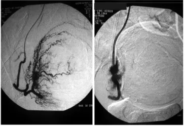
The age of patients ranged between 23 and 44 years. In all cases, the control after embolization showed good results, with bilateral distal occlusion of uterine arteries (Figure 1).
The hospital stay after embolization was 24 hours in all cases, and after myomectomy it ranged from two to three days. The duration of surgery in cases of myomectomy varied between 60 and 120 minutes. No patients required blood transfusion.
Histopathological analysis of removed myomas showed the presence of necrosis, hyalinization and visualization of PVA-PVAc particles.
After uterine artery embolization, patients were followed on average for three years (33 and 45 months). There was a significant variation in the uterine volume and in the volume of biggest myoma between pre- and post-UAE (Figures 2 and 3).
Three years after the embolization, and considering myomectomy, total uterine volume reduced significantly, approximately 460 mm3 (p = 0.0005), which
corresponded on average to 64.9% (Table 1).
Figure 1 Figure 1 Figure 1 Figure 1
Figure 1 - Arteriography showing absence of vascularized branches of the uterine artery after arterial embolization.
There was no variation of the follicle stimulating hormone over the three moments studied (baseline, post-UAE and post-myomectomy) in our series (p = 0.17).
Hemoglobin levels rose after embolization (p = 0.0004) and remained stable in the long term (three years). The number of myomas removed from 12 patients ranged from one to 18. In histopathology we found extensive inflammatory reaction, thrombosis and necrosis areas.
We observed a significant reduction of all pre-embolization (baseline) signs and symptoms when compared with post-UAE and after myomectomy (p <0.01). There was no significant variation between post - UAE and postoperative times in the sample. (Figure 4).
DISCUSSION
DISCUSSION
DISCUSSION
DISCUSSION
DISCUSSION
Myomectomy, especially for multiple and/or large tumors, has always been considered an intervention capable of serious complications because of perioperative bleeding. Therefore, many techniques are proposed to reduce the bleeding, whether by the use of mechanical tourniquets in uterine vessels and pelvic infundibulum, or with chemical tourniquets, such as vasopressin and epinephrine 7. In this
regard, UAE also present advantages as a preoperative procedure, since as well as progressively reducing the size of myomas, it decreases vascularization of tumors and hence bleeding during surgery 8.
We studied the application of a more selective method for the treatment of uterine myomas with the use of a new, domestically-manufactured, spherical shape embolic particle of PVA-PVAc. With this, the aim was to obtain evidence of able to substantiate their use in preparation for a safer and less morbid myomectomy.
Figure 4 Figure 4Figure 4
Figure 4Figure 4 - Percentage of signs and symptoms during treatment.
Figure 3 Figure 3Figure 3 Figure 3
Figure 3 - Volume of the biggest myoma pre- and post-UAE. There was significant reduction in the volume of the biggest myoma, on average 31.1% and amounted to 124.9 mm3 (± 50.4 – p = 0.006).
Figure 2 Figure 2Figure 2 Figure 2
Figure 2 - Uterine volume before and after UAE. We observed a significant reduction in uterine volume, average 32.3%, which corresponded to 304.9 mm3 (± 76.3 – p
= 0.001).
Table 1 Table 1Table 1 Table 1
Table 1 - Uterine volume after UAE and myomectomy.
Uterine volume (mm Uterine volume (mmUterine volume (mm Uterine volume (mm
Uterine volume (mm33333))))) M e a nM e a nM e a nM e a nM e a n S DS DS DS DS D M e d i a nM e d i a nM e d i a nM e d i a nM e d i a n M i n i m u mM i n i m u mM i n i m u mM i n i m u mM i n i m u m M a x i m u mM a x i m u mM a x i m u mM a x i m u mM a x i m u m
Post-UAE 634.4 347.3 639 109 1347
After three years 173.1 141.4 114.75 55 490
Moreover, if proven their effectiveness, there is the added advantage of a less invasive treatment, with the possibility of uterine preservation and pregnancies.
In the presented series, the improvement in hemoglobin concentration was significant after embolization, remaining stable after removal of myomas. This finding reflects the impact of treatment in decreasing menstrual bleeding and is in agreement with the data reported by other investigators 8.
Similar to the findings of this study, Brunereau et al., observed a 23% decrease in the volume of the biggest myoma in a series of 58 patients who underwent UAE 9.
There is report of a reduction of up to 70% of the volume after a one year follow-up 10. It is noteworthy that the rate
of decrease in the size of the uterus did not necessarily correlate with the improvement of the patient’s symptoms, especially menstrual flow and dysmenorrhea.
The choice for GnRH analogues, the only drugs approved by the U.S. Food and Drug Administration (FDA) to reduce the size of myomas preoperatively, has some limitations. It is to be used for a maximum six months due to side effects, and it is capable of causing reduction of smaller myomas, making them undetectable during surgery, but retains the risk of progressive regrowth once when treatment is discontinued 11.
The decrease in myoma volume in this study may also have been instrumental in the success of myomectomy, considering that in no patient required blood transfusion and in all myomectomy was successful. The positive aspects of UAE as a procedure prior to myomectomy are therefore evident.
Nevertheless, UAE is not without complications. Goodwin and Spies, two of the leading scholars of the procedure, published an article in 2009 with a series of 3,160 women and showed that major complications (uterine necrosis, fulminant sepsis) occurred in 4.8% of cases within the first month after the procedure, but the most common was persistent or recurrent pain12.
The currently marketed embolic particles have some undesirable features. The PVA particle, like the gelatin sponge, is a water-soluble polymer, hence susceptible to biodegradation, which theoretically could facilitate recanalization of the vascular bed treated over time. The particles features, such as shape, flocculation and highly irregular surface, with a wide variation in their grain size, facilitates their aggregation, resulting in an occlusion proximal to the desirable location, in addition to hindering the passage through the angiographic catheter13. Finally,
this product is not manufactured in Brazil, which entails the need to importation, at a very high cost.
To circumvent the negative aspects inherent to imported microparticles, several trials were performed at COPPE -UFRJ to test new mechanisms of synthesis of PVA for application in vascular embolization. This work has resulted in a new, Brazilian-technology particle, comprising of a surface of polyvinyl alcohol (PVA) and a core of polyvinyl acetate (PVAc), forming a spherical, more resistant to
degradation (by not being water soluble) material, available in suitable sizes and at a significantly lower manufacturing cost – the Spherus®®®®®. The PVAc is a toxic, non-carcinogenic polymer used in capsules that serve as containers acid pharmaceutical substances, which require pH-dependent release. This structure, PVA-PVAc shell-core, has the advantage of little swelling when in contact with aqueous solutions. The particle used has a size between 300 and 600 ìm, but its manufacturing process allows it to be made in other sizes if necessary. The new particles were experimentally tested and the results published by Mendes et al. 14.
Two spherical particles for embolization currently in commercial use, Embosphere Microsphere®®®®® and Contour SE®®®®®, differ from Spherus®®®® in that the first is® composed of gelatin Trisacryl, and the second consists of PVA, not containing PVAc in its structure and having a different manufacturing process.
Chua et al.15 published a series of 17
embolizations performed as preoperative preparation for myomectomy, comparing the use of PVA and flaked embospheres (microspheres). They observed that the microspheres penetrated deeper into the myoma and allowed a terminal embolization, minimizing ischemic lesions in normal myometrium and ovaries.
Pelage showed that the use of spherical particles is more accurate, with correlation between the particle size and the extent of necrosis generated 16.
In this series the improvement of clinical complaints happened after embolization and the degree of satisfaction of patients undergoing UAE was high. Two patients became pregnant, one had a cesarean delivery at term, the other was in the third trimester of pregnancy at the end of the evaluation.
For a patient with multiple myomas who wants to gestate, it is advantageous to reduce to a minimum the number of incisions in the uterus and this situation is facilitated by UAE.
The most important issue in UAE indication in patients who wish to maintain reproductive function is the possibility of secondary amenorrhea, whether by ovarian failure or by endometrium atrophy. One case has evolved to amenorrhea due to the latter. We emphasize the study of ovarian function prior to UAE since the failure of this organ is more common in women who have already had some function impairment. There is to date no test that can predict which patients are prone to this rare complication.
The indication of arterial embolization with PVA-PVAc spherical particles showed promise in preparation for surgery with removal of myomas, therefore associated with a reduction in uterine volume and decrease in intraoperative bleeding, and made the use of incisions smaller possible, with increased chance of preserving the uterus.
The studies should continue, with the expansion of research in this area of knowledge, in order to substantiate the proposed use of this new particle, which may become a safe and most economical option among the existing embolic agents.
R E S U M O R E S U M O R E S U M O R E S U M O R E S U M O
Objetivo Objetivo Objetivo Objetivo
Objetivo: avaliar a utilização de uma nova partícula de polivinil álcool e polivinil acetato (PVA-PVAc) esférica, para embolização das artérias uterinas, em pacientes portadoras de mioma, com indicação cirúrgica. MétodosMétodosMétodosMétodosMétodos: doze pacientes foram subme-tidas à embolização de miomas uterinos com partículas de PVA-PVAc. Três a nove meses depois, realizou-se uma laparotomia com miomectomia. Analisaram-se os seguintes parâmetros: volume do útero e do maior mioma; concentrações do hormônio folículo estimulante e de hemoglobina; sangramento menstrual (número de dias e de absorventes utilizados), sinais e sintomas antes do tratamento, após a embolização e após a miomectomia. ResultadosResultadosResultadosResultadosResultados: a média de idade foi 37 anos e a média do volume uterino, previamente ao tratamento, de 939,3cc. Três anos após a embolização, observou-se diminuição do volume uterino (p=0,0005). Houve melhora na concentração de hemoglobina (p= 0,0004), com elevação após a embolização, sem variação subsequente à miomectomia. Não ocorreu variação significante do hormônio folículo estimulante, (p=0,17). Não foi constatado nenhum caso de falência ovariana, mas uma das pacientes apresentou atrofia de endométrio. Duas pacientes engravidaram, com bons indicadores obstétricos. Quanto aos sinais e sintomas, houve melhora após a embolização, que se manteve após a miomectomia. ConclusãoConclusãoConclusãoConclusãoConclusão: a embolização arterial com partículas de PVA-PVAc esférico mostrou-se promis-sora no preparo para uma intervenção cirúrgica com retirada dos miomas, pois, associou-se à redução do volume uterino, à diminuição do sangramento operatório e tornou possível a utilização de incisões menores, aumentando a chance de preserva-ção do útero.
Descritores: Descritores: Descritores: Descritores:
Descritores: Pacientes. Leiomioma. Embolização de artéria uterina.. Polivinil. Resultado de tratamento.
REFERENCES
REFERENCES
REFERENCES
REFERENCES
REFERENCES
1. Dawbain RHM. The starvation operation for malignancy in the external carotid area. JAMA. 1904;17:792-5.
2. Ravina JH, Herbreteau D, Ciraru-Vigneron N, Bouret JM, Houdart E, Aymard A, et al. Arterial embolisation to treat uterine myomata. Lancet. 1995;346(8976):671-2.
3. Goodwin SC, Vedanthan S, McLucas B, Forno AE, Perella R. Preliminary experience with uterine artery embolization for uterine fibroids. J Vasc Interv Radiol. 1997;8(4):517-26. Erratum in: J Vasc Interv Radiol. 1999;10(7):991.
4. Worthington-Kirsch RL, Popky GL, Hutchins FL Jr. Uterine artery embolization for the management of leiomyomas: quality-of-life assessment and clinical response. Radiology, 1998;208(3):625-9. 5. Committee on Gynecologic Practice, american College of
Obstetricians and Gynecologists. ACOG Committee Opinion. Uterine artery embolization. Obstet Gynecol. 2004;103(2):403-4. 6. American College of Obstetricians and Gynecologists. ACOG
practice bulletin. Alternatives to hysterectomy in the management of leiomyomas. Obstet Gynecol. 2008;112 (2 Pt 1):387-400. 7. Taylor A, Sharma M, Tsirkas P, Di Spiezo Sardo A, Setchell M,
Magos A. Reducing blood loss at open myomectomy using triple tourniquets: a randomised controlled trial. BJOG. 2005;112(3):340-5.
8. Ngeh N, Belli AM, Morgan R, Manyonda I. Pre-myomectomy uterine artery embolisation minimises operative blood loss. BJOG 2004;111(10):1139-40.
9. Brunereau L, Herbreteau D, Gallas S, Cottier JP, Lebrun JL, Tranquart F, et al. Uterine artery embolization in the primary treatment of uterine leiomyomas: technical features and prospective follow-up with clinical and sonographic examinations in 58 patients. AJR Am J Roentgenol. 2000;175(5):1267-72.
10. Walker WJ, Pelage JP. Uterine artery embolisation for symptomatic fibroids: clinical results in 400 women with imaging follow-up. BJOG. 2002;109(11):1262-72.
11. Letterie G, Coddington CC, Winkel CA, Shawker TH, Loriaux DL, Collins RL. Efficacy of a gonadotropin-releasing hormone agonist in the treatment of uterine leiomyomata: long term follow-up. 1989;51(6):951-6.
12. Goodwin SC, Spies JB. Uterine fibroid embolization. N Engl J Med. 2009;361(7):690-7.
13. Laurent A, Wassef M, Namur J, Martal J, Labarre D, Pelage JP. Recanalization and particle exclusion after embolization of uterine arteries in sheep: a long-term study. Fertil Steril. 2009;91(3):884-92.
14. Mendes WDS, Chagas VLA, Pinto JC, Caldas JG, Espinosa G. Estudo comparativo da reação inflamatória renal entre álcool de polivinil-flocular e álcool de polivinil+acetato de polivinil-es-férico: estudo experimental. Rev Col Bras Cir. 2007;32(3):120-6.
15. Chua GC, Wilsher M, Young MP, Manyonda I, Morgan R, Belli AM. Comparison of particle penetration with non-spherical polyvinyl alcohol versus trisacryl gelatin microspheres in women undergoing premyomectomy uterine artery embolization. Clin Radiol. 2005;60(1):116-22.
16. Pelage JP. Polyvinyl alcohol particles versus tris-acryl gelatin microspheres for uterine artery embolization for leiomyomas. J Vasc Interv Radiol. 2004;15(8):789-91.
Received on 25/08/2012
Accepted for publication 28/10/2012 Conflict of interest: none
Source of funding: none
How to cite this article: How to cite this article: How to cite this article: How to cite this article: How to cite this article:
Ghiaroni J, Lopez GE, Coutinho Júnior AC, Schanaider A. Uterine artery embolization with spherical pva-pvac particles as preparation for
surgical resection of miyomas. Rev Col Bras Cir. [periódico na Internet] 2013;40(5). Disponível em URL: http://www.scielo.br/rcbc
Address correspondence to: Address correspondence to: Address correspondence to: Address correspondence to: Address correspondence to: Juraci Ghiaroni