Correlation between doppler of the right hepatic vein with
Correlation between doppler of the right hepatic vein with
Correlation between doppler of the right hepatic vein with
Correlation between doppler of the right hepatic vein with
Correlation between doppler of the right hepatic vein with
ultrasound transcutaneous guided biopsy in liver diseases
ultrasound transcutaneous guided biopsy in liver diseases
ultrasound transcutaneous guided biopsy in liver diseases
ultrasound transcutaneous guided biopsy in liver diseases
ultrasound transcutaneous guided biopsy in liver diseases
Correlação entre o doppler da veia hepática direita com a biópsia transcutânea
Correlação entre o doppler da veia hepática direita com a biópsia transcutânea
Correlação entre o doppler da veia hepática direita com a biópsia transcutânea
Correlação entre o doppler da veia hepática direita com a biópsia transcutânea
Correlação entre o doppler da veia hepática direita com a biópsia transcutânea
guiada pela ultrassonografia em hepatopatias
guiada pela ultrassonografia em hepatopatias
guiada pela ultrassonografia em hepatopatias
guiada pela ultrassonografia em hepatopatias
guiada pela ultrassonografia em hepatopatias
GLEIM DIASDE SOUZA1; LUCIANA RODRIGUES QUEIROZ2; CARMEN AUSTRALIA PAREDES MARCONDES RIBAS3; MARCELO MAZZADO NASCIMENTO3; THELMA LAROCCA SKARE3; RONALDO MAFIA CUENCA, TCBC-DF3; GUSTAVO HENRIQUE SOARES TAKANO4
A B S T R A C T A B S T R A C T A B S T R A C T A B S T R A C T A B S T R A C T
Objective Objective Objective Objective
Objective: To correlate chronic liver diseases diagnosed by ultrasound-guided transcutaneous liver biopsy with B-mode and Doppler ultrasound findings of the right hepatic vein; to compare the wave patterns between the study group and the control group; to compare the right hepatic vein Doppler findings with histopathology findings as a possible marker of chronic liver disease. MethodsMethodsMethodsMethods:Methods Were studied 38 patients with chronic liver disease diagnosed by biopsy and serology (study group) and 10 persons without serologic liver disease (control group), who were assessed by B-mode and Doppler ultrasound. The criteria were based on histology classification of the Brazilian Society for Pathology of chronic hepatitis. Chi-square, Fisher’s exact and Student t tests were used. ResultsResultsResultsResultsResults: The B-mode and Doppler ultrasound were useful in inferring the differentiation between individuals with chronic liver disease from normal ones. There were significant differences between the study group and the controls when comparing the histopathology findings, B-mode and Doppler ultrasound in relationship to the wave patterns of the right hepatic vein. Conclusion:Conclusion:Conclusion:Conclusion:Conclusion: The correlation of liver biopsies with B-mode and Doppler ultrasound of hepatic vein was possible; individuals with liver disease showed alterations in the flow of the right hepatic vein and normal subjects did not, the wave pattern in normal controls being triphasic, and in patients with chronic liver disease, monophasic or biphasic; Doppler of the right hepatic vein is a useful marker for chronic liver disease.
Key words: Key words: Key words: Key words:
Key words: Ultrasonography, Doppler. Hepatic veins. Biopsy. Liver cirrhosis. Hepatitis C.
From the Post-Graduation Program of Principles of Surgery of the Evangelical Faculty of Paraná/Evangelical University Hospital, Curitiba, Paranà State – PR, and Digimed Diagnostic Imaging Clinic Ltd, Brasília, Federal District – DF, Bra7. Master, profesor at the Universidade de Brasília – DF, Brazil.
1. PhD, Post-Graduattion Program in Principles of Surgery, Medical Research Institute, Evangelical Faculty of Paraná / Evangelical University Hospital of Curitiba.; 2. Master’s Degree; Preceptress, Residency in Radiology, Hospital de Base do Distrito Federal; 3. PhD, Permanent Professor, Post-Graduation Program in Principles of Surgery, Medical Research Institute, Evangelical Faculty of Paraná; Coordinator, Service of Metabolic and Bariatric Surgery, Evangelical University Hospital of Curitiba; 4. PhD, Permanent Professor, Post-Graduation Program in Principles of Surgery, Evangelical Faculty of Paraná / Evangelical University Hospital of Curitiba; 5.PhD, Permanent Professor, Post-Graduation Program in Principles of Surgery, Medical Research Institute, Evangelical Faculty of Paraná / Evangelical University Hospital of Curitiba; 6. Master’s Degree; Assistant Professor, University de Brasília – DF.
INTRODUCTION
INTRODUCTION
INTRODUCTION
INTRODUCTION
INTRODUCTION
H
epatic veins drain little pressure blood from hepatic sinusoids to the inferior cava vein1. The normal andabnormal wave features of the greater hepatic vessels (hepatic veins, hepatic artery and portal vein) are already described2. At duplex ultrasound, the flow in the hepatic
veins is usually pulsatile due to pressure changes in the right atrium. The normal liver is easily adaptable to changes in pressure3, and the triphasic pattern in the right hepatic
vein occurs due to reversal flow in the inferior vena cava during systole1.
The passage of blood through the vessels is changed by differential pressure between the vessel end and the resistance of its wall4. A key factor pertaining to
cardiac function is the relative position of the veins in the
circulatory system, which depend on the physiological state of the vessels and the demand for blood. Each vessel in a normal human body has a characteristic flow pattern that is represented by its wave aspect of the observed spectral Doppler sonography, which reflects both the anatomical position of the veins as the physiological need of the body in relation to the vessels4. Cardiac function can exert the
same effect on the flow through all veins, and the main determinants of the flow characteristics of a vein are vascular resistance - which varies according to physiological need of the body – and the distance of the vessel to the heart.
Vascular resistance can be changed by pathological conditions or physiological differences5. The
predominantly hepatopetal (toward the liver)7. The wave
pattern in Doppler of hepatic vein in healthy people is triphasic8. The characteristic triphasic spectrum in Doppler
of the hepatic veins consists of a peak at systolic and diastolic anterograde flow followed by a short flow reversal peak resulting from pressure variations during cardiac cycle2,9,10. The Doppler normal triphasic wave patterns of
hepatic veins can also be defined as two opposites anterograde flow peaks toward the heart during diastole and ventricular contraction, followed by a short period of peak reverse flow towards the liver during atrial contraction11,12. Parenchymal liver alterations can impair the
structure of the walls of the veins and result in slowdown9
in Doppler waves, described in patients with liver cirrhosis. This damping of waves can be attributed to the hardness of the parenchyma of patients with various hepatic diseases9.
This study aims to evaluate the usefulness of Doppler hepatic vein as a marker of chronic liver disease, through: 1) the correlation of ultrasound-guided transcutaneous liver biopsy with data from B-mode Doppler ultrasounds of the right hepatic vein; and 2) the comparison of wave patterns between the study (liver disease) and control (healthy) groups.
METHODS
METHODS
METHODS
METHODS
METHODS
We conducted a prospective study with 48 individuals divided into two groups: 1) study group composed of 38 liver patients with an indication for ultrasound-guided transcutaneous biopsy for staging of disease, and 2) control group consisted of ten healthy young persons.
The sole inclusion criterion was the diagnosis of liver disease clinically done with positive serological tests. Exclusion criteria were patients: 1) submitted to hepatic surgical procedures; 2) under 18 years of age; 3) with decompensated nephropathy or heart disease; 4) with morbid obesity.
Ultrasound evaluation Ultrasound evaluation Ultrasound evaluation Ultrasound evaluation Ultrasound evaluation
All individuals underwent B-mode liver ultrasound and Doppler of the right hepatic vein. In the study group an ultrasound-guided percutaneous biopsy was also carried out. The control group did not undergo biopsy.
For correlation we used the liver B-mode ultrasound, Doppler of the right hepatic vein and the histological classifications defined by the Brazilian Society of Pathology (SBP).
The study was held at Digimed Clinic Diagnostic Imaging Ltd., Brasília, Federal District – DF, Brazil with Toshiba® brand appliances, Xario model year 2009 and Aplio, 2010 with 3-9mHz probes. All sonographic examinations followed the same protocol, with quantification and classification of parenchymal abnormalities. The ultrasound exams covered the entire
analysis of liver echotexture, which was subsequently compared with the renal echogenicity. We characterized the changes of chronic diseases based on visual criteria of hepatic steatosis graded as mild, moderate and severe. In mild steatosis, there was only increased echogenicity of the liver parenchyma; in moderate, hyperechogenicity was associated with attenuation of the ultrasound and subsequent plans; when intense, it was characterized by the above mentioned changes and loss of definition of the vascular structures of the liver and diaphragm.
After the analysis, patients were classified into subgroups formed by individuals with and without sonographic changes, which were comparing to histopathological alterations, correlating the findings.
Hepatic vein Doppler Hepatic vein Doppler Hepatic vein Doppler Hepatic vein Doppler Hepatic vein Doppler
The patients were examined in the morning, fasting, in supine position, with arms outstretched to the side of the head. All segments of the liver were examined and the focal parenchymal lesions identified.
The color Doppler mode was done on the right hepatic vein, identifying the pulsatility pattern and flow direction. The evaluation of the right hepatic vein was performed at the 10th or 11th right intercostal space during
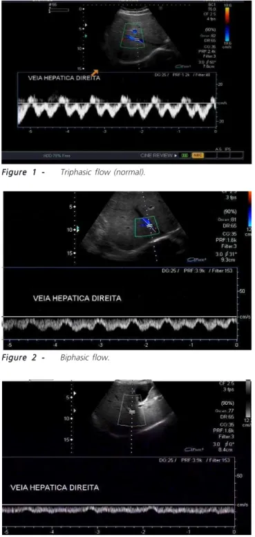
a short period of apnea, 2.0cm distal from the confluence of the hepatic veins, to avoid artifacts in the wave pattern. The analysis was recorded for at least eight cardiac cycles (about 10 seconds). The angulation of the transducer was 30° to 60°. The wave patterns found were classified as follows: 1) normal triphasic phase (Figure 1); 2) biphasic short reverse flow (Figure 2); 3) no reverse flow but with fluctuations of more than 10% of the mean amplitude and monophasic (Figure 3) or flat with wave oscillation less than 10% of the mean amplitude. Due to changes in vessel diameter of up to 2mm per cardiac cycle during systole and diastole and to flow in different directions, we did not calculate hepatic veins velocimetry.
Histopathologic evaluation Histopathologic evaluation Histopathologic evaluation Histopathologic evaluation Histopathologic evaluation
Ultrasound-guided liver biopsies were performed with Histo® automatic pistol and thick caliber needles -18G. We collected three fragments of 20 x 3mm from the right lobe of the liver.
The material was sent to the laboratory Micra of Cytology and Histology, being fixed in 10% formalin and subsequently subjected to histological processing with conventional successive baths in absolute ethanol, xylene and paraffin inclusion for stainings with hematoxiline and eosin, PAS, Gomori trichrome, reticulin and Perls technique for ferric deposits. Slides were analyzed by the same pathologist using a Nikon® optical microscope (100 to 1000x magnification).
(steatosis, lymphoid aggregates, hemorrhagic necrosis, glassy inclusions and iron deposits) absent / present 0-4.
Statistical methodology Statistical methodology Statistical methodology Statistical methodology Statistical methodology
To evaluate the association between qualitative variables we used the chi-square or Fisher exact tests. To evaluate the homogeneity of the groups in terms of age we applied the Student t test for independent samples. P values less than 0.05 indicated statistical significance. Considering each individual as alterated or not, we tested the null hypothesis of the altered probability among patients
be equal to the one of controls, versus the alternative hypothesis of different probabilities. In relation to gender, we tested the null hypothesis of equal distribution of gender between patients and controls, versus the alternative hypothesis of different distributions. We also tested the null hypothesis of equal mean age between patients and controls, versus the alternative hypothesis of different means.
RESULTS
RESULTS
RESULTS
RESULTS
RESULTS
B mode ultrasound evaluation B mode ultrasound evaluation B mode ultrasound evaluation B mode ultrasound evaluation B mode ultrasound evaluation
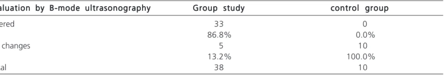
The B-mode ultrasonography assessment was peformed in 48 people in the two groups, ie, group study with 38 patients with liver disease and a control group of ten healthy young men. The control group was 100% nor-mal. In the study group there was no change in five (13.2%) individuals, three (7.9%) had grade III steatosis and / or diffuse hyperechogenicity grade III, six (15.8%) had chronic liver disease and cirrhosis, ten (26.3%) displayed diffuse hyperechogenicity grade I, and 14 (36.8%), diffuse hyperechogenicity grade II.
All patients from both groups underwent liver conventional ultrasound (B-mode) and, based on the results, they were grouped into two groups: altered and normal. We tested the null hypothesis probability of the altered group being equal to the one of the normal group, versus the alternative hypothesis of different probabilities. The results showed that they are different, with statistical significance (p=0.001).
Hepatic vein Doppler Hepatic vein Doppler Hepatic vein Doppler Hepatic vein Doppler Hepatic vein Doppler
The evaluation was conducted in both groups (48 subjects). Standard triphasic pattern was observed in six (15.8%) individuals from the study group and in nine (90%)from the control group. The monophasic wave was seen in six (15.8%) patients from the study group and none from the control. The biphasic pattern was seen in 26 (68.4%) subjects from the study group and in only one person from the control group (10.0%).
Considering each individual classified according to the wave pattern, we tested the null hypothesis of same distribution between patients and controls versus the alternative hypothesis of different distributions. We observed a statistical significant difference (p<0.001) in favor of the study group that had abnormal echogenicity of the liver parenchyma and changes in the right hepatic vein flow (Table 1).
Histopathological results correlation with Histopathological results correlation with Histopathological results correlation with Histopathological results correlation with Histopathological results correlation with Doppler of the right hepatic vein
Doppler of the right hepatic vein Doppler of the right hepatic vein Doppler of the right hepatic vein Doppler of the right hepatic vein
All SBP criteria for chronic liver disease (structural changes 0-4; portal inflammatory infiltrate 0-4, periportal activity of 0-4, parenchymal (lobular) activity of 0-4, etiological markers (steatosis, lymphoid aggregates, hemorrhagic necrosis, glassy inclusions and iron deposits)
Figure 1 Figure 1 Figure 1 Figure 1
-Figure 1 - Triphasic flow (normal).
Figure 2 Figure 2 Figure 2 Figure 2
-Figure 2 - Biphasic flow.
Figure 3 Figure 3 Figure 3 Figure 3
absent / present 0-4), were separately correlated with the wave patterns found in the evaluation by Doppler.
Structural changes Structural changes Structural changes Structural changes Structural changes
We performed the correlation between the distribution (semiquantification) of the structural changes (histopathological classification of chronic liver disease) and the wave patterns found on Doppler evaluation of right hepatic vein. The results were: from the 38 patients analyzed, 26 showed a biphasic waveform: 13 grade 0 (50%), ten grade 1 (38.5%), two grade 2 (7.7%), and one grade 3 (3.8%). Six individuals showed monophasic flow: 0 grade 3 (60%), one grade 1 (20%), one grade 2 (20%) and none grade 3 (0.0%). Six patients had triphasic flow, five grade 0 (83.3%), one grade 1 (16.7%), none grade 2 or 3 (0%).
By grouping the results regarding distributions (semiquantifications) of histopathological evaluation of structural changes in correlation with Doppler of the right hepatic vein, two groups were formed: group 0-1 (absent or minimal changes) and group 2-3 (moderate or severe). We tested the null hypothesis of probability of group 2/3 being equal to the control group, versus the alternative hypothesis of different probabilities. The result was p=0.592, indicating a non-significant statistical trend in favor of individuals in the study group, who had histopathological changes and had also changes in the flow of the right hepatic vein.
Inflammatory portal infiltrate Inflammatory portal infiltrate Inflammatory portal infiltrate Inflammatory portal infiltrate Inflammatory portal infiltrate
We performed correlation between the distribution (semiquantification) of portal inflammatory infiltrate and right hepatic vein Doppler wave patterns. The results were: from the 38 patients, 26 showed biphasic waveform, with varying portal inflammatory infiltrates: one (3.8%) patient was characterized as absent, 17 (65.4%) as mild, seven (26.9%) moderate and one (3.8%) as severe. Five individuals displayed a monophasic flow, all of whom (100.0%) had portal inflammatory infiltrates characterized as mild; six subjectes had triphasic wave flow, five (83.3%) with mild portal inflammatory infiltrates and one (16.7%) with moderate.
By grouping the results, two groups were formed: group 1 of mild or minimal changes and group 2 of moderate or intense changes. We then tested the null hypothesis that the probability of the study group 2 was equal to one of
control group 2, versus the alternative hypothesis of different probabilities. The result was p=1, indicating no statistical correlation between this histopathologic finding and the evaluation by Doppler of the right hepatic vein.
Periportal activity Periportal activity Periportal activity Periportal activity Periportal activity
We performed the correlation between the distribution (semiquantification) of periportal activity and the wave patterns found on Doppler evaluation of right hepatic vein. The following results were found: from the 38 patients we observed that the flow was biphasic in 26. From these, 14 (53.8) were categorized as having absent periportal inflammation, seven (26.9%) as mild, four (15.4%) as moderate and one (3.8%) as intense. The flow was monophasic in five, four (80.0%) with absent periportal activity and one (20.0%) with moderate. The flow was triphasic in six people, four (66.7%) with absent periportal activity, one (16.7%) with mild and one (16.7%) moderate; there were no cases of severe periportal inflammatory activity.
By grouping the results, two groups were formed: group 1 with absent or mild activity and group 2 with moderate or intense. We tested the null hypothesis that the probability of study group 2 was equal to the one of control group 2, versus the alternative hypothesis of different probabilities. The p=1 indicated no statistical correlation between this histopathologic finding and the evaluation by Doppler of the right hepatic vein.
Parenchymal activity Parenchymal activity Parenchymal activity Parenchymal activity Parenchymal activity
We performed the correlation between the distribution (semiquantification) of parenchymal activity and wave patterns found on Doppler evaluation of the right hepatic vein. The results were: from the 38 patients analyzed in the study group, 26 had biphasic flow: four (15.4%) had absent parenchymal activity, 16 (61.5%) mild, four (15.4%) minimum and two (7.7%) moderate. Five showed monophasic Doppler flow and they all (100%) were classified as mild activity. Six had triphasic flow, one (16.7%) with absent activity, three (50%) with mild and two (33.3%) with minimal.
By grouping the results, two groups were formed: group 1 with absent or mild parenchymal activity and group 2, with moderate or intense. We tested the null hypothesis that the probability of study group 2 was equal to the probability of control group 2, versus the alternative
Table 1 -Table 1 -Table 1 Table 1
-Table 1 - Frequencies of the altered and normal groups as for the pattern of ultrasound echogenicity.
Evaluation by B-mode ultrasonography Evaluation by B-mode ultrasonographyEvaluation by B-mode ultrasonography Evaluation by B-mode ultrasonography
Evaluation by B-mode ultrasonography Group studyGroup studyGroup studyGroup studyGroup study control groupcontrol groupcontrol groupcontrol groupcontrol group
Altered 33 0
86.8% 0.0%
No changes 5 10
13.2% 100.0%
hypothesis of different probabilities. The p=1 indicated that there was no statistical correlation between this histopathologic finding and the evaluation by Doppler of the right hepatic vein.
Etiologic markers Etiologic markers Etiologic markers Etiologic markers Etiologic markers
Following the criteria of SBP, we analyzed all histopathological etiological markers (steatosis, lymphoid aggregates, hemorrhagic necrosis, glassy inclusions and iron deposits); however, only steatosis and iron deposit were found. Consequently, only these two etiological markers were correlated with wave patterns found by Doppler evaluation.
Hepatic steatosis Hepatic steatosis Hepatic steatosis Hepatic steatosis Hepatic steatosis
We performed the correlation between the distribution (semiquantification) of steatosis and wave patterns found on Doppler evaluation. The results were: from the 38 patients analyzed, the flow was biphasic in 26. From this group, four (15.4%) had absent steatosis, nine (34.6%) had steatosis 1, three (11.5%) steatosis 2 and one (3.8%) steatosis 3. Nine patients in this group (34.6%) did not have steatosis. In assessing the monophasic wave pattern three individuals (60%) were classified as steatosis 1 and two (40%) and steatosis 2. In the triphasic wave pattern, three subjects (50%) were categorized as absent, one (16.7%) as steatosis 1 and in two (33.3%) steatosis did not occur.
By grouping the results of the histopathological evaluation of steatosis in correlation with Doppler, two groups were formed: group 1 without alterations and group 2 with steatosis 1, 2 and 3. We tested the null hypothesis that the probability of study group 2 was equal to the probability of control group 2, versus the alternative hypothesis of different probabilities. Results returned p=0.047, indicating statistical significance inferring that individuals in the study group who had exhibited steatosis also had altered flow of the right hepatic vein.
Iron deposits Iron deposits Iron deposits Iron deposits Iron deposits
We performed the correlation between the distribution (semiquantification) of iron deposits (etiologic marker) and the wave patterns found on Doppler. The results were: from the 38 patients analyzed in the study group, the flow was biphasic in 25. Of these, 23 (92%) were categorized as absent, one (4%) as mild and one (4%) as minimal. All five people (100.0%) with monophasic waveform were classified as absent. Of the six individuals with triphasic waveforms, five (83.3%) had absent iron deposits, and one (16.7%) as moderate.
By grouping the results of histopathological evaluation of iron deposits in correlation with Doppler study, two groups were formed: group 1 with absent or mild deposits and group 2 with moderate or intense ones. We tested with the null hypothesis that the probability of the study group 2 was equal to the probability of control group
2, versus the alternative hypothesis of different probabilities. The result was p=0.431, indicating that there was no statistical correlation between this histopathologic finding and the evaluation by Doppler of the right hepatic vein.
DISCUSSION
DISCUSSION
DISCUSSION
DISCUSSION
DISCUSSION
It is controversial on the literature whether the abnormalities in the liver parenchyma can be diagnosed by Doppler of the hepatic veins. Some authors say that the Doppler is insufficient, while others argue that it has high sensitivity and specificity in the diagnosis of cirrhosis10. In
the correlation between histopathological findings and hepatic vein Doppler, the wave pattern was altered in patients with fibrotic changes, predominantly with a biphasic pattern.
The definition and classification of chronic liver disease depends on adequate biopsy and histology observation. The diagnosis of cirrhosis is of particularly value in prognosis and requires histological evidence of diffuse fibrosis associated with abnormal architecture. The normalizations made by the SBP facilitated characterization of the degree of fibrosis in the evolution process.
In some studies the ultrasound diagnosis of cirrhosis is relatively accurate. Objective signs, such as surface roughness or increased volume of the caudate lobe, are considered of great value. The changes detected in this study failed to safely characterize the degree of involvement of the hepatic parenchyma, but separated with good accuracy the normal people from the ones with liver disease. Recent studies suggest that the flattening of the phasic oscillations of the hepatic veins pulse is significantly associated with liver cirrhosis. These signals are very sensitive (75%) in the diagnosis of cirrhosis 13. In Doppler (duplex) of
the right hepatic vein, we could clearly separate the nor-mal individuals from the ones with liver diseases.
The compression of the hepatic veins in cirrhosis by regenerative nodules may explain changes in phasic oscillation. The abnormal wave pattern of hepatic veins is correlated with parenchymal fibrosis around the hepatic veins. Other factors are indeterminate and may also play a role. The inflammation and necrosis seem not to affect the wave pattern of the hepatic veins13,14..
Ultrasound is commonly the first imaging test in the clinical study of patients with hepatitis. Some studies in chronic liver disease have shown increased values in the hepatic artery resistance, associated with degeneration of the architecture of the liver affected by the disease. Thus, ultrasound, being noninvasive, is very useful for detection of chronic diseases in the liver. The increase in resistance is correlated with high rates of fibrosis15. But till
REFERENCES
REFERENCES
REFERENCES
REFERENCES
REFERENCES
1. Jequier S, Jequier JC, Hanquinet S, Gong J, Le Coultre C, Belli DC. Doppler waveform of hepatic veins in healthy children. AJR Am J Roentgenol. 2000;175(1):85-90.
2. Abu-Yousef MM. Duplex Doppler sonography of the hepatic vein in tricuspid regurgitation. AJR Am J Roentgenol. 1991;156(1):79-83.
3. Abu-Yousef MM, Mufid M, Woods KT, Brown BP, Barloon TJ. Normal lower limb venous Doppler flow phasicity: is it cardiac or respiratory? AJR Am J Roentgenol. 1997;169(6):1721-5. 4. Farrant P, Meire HB. Hepatic vein pulsatility assessment on spectral
Doppler ultrasound. Br J Radiol. 1997;70(836):829-32.
5. Nelson TR, Pretorius DH. The Doppler signal: where does it come from and what does it mean? AJR Am J Roentgenol. 1988;151(3):439-47.
6. Taylor KJ, Burns PN. Duplex Doppler scanning in the pelvis and abdomen. Ultrasound Med Biol. 1985;11(4):643-58.
7. Taylor KJ, Holland S. Doppler US. Part I. Basic principles, instrumentation, and pitfalls. Radiology. 1990;174(2):297-307.
R E S U M O R E S U M O R E S U M O R E S U M O R E S U M O
Objetivos: Objetivos: Objetivos: Objetivos:
Objetivos: Correlacionar os achados da biópsia transcutânea hepática guiada por ultrassonografia com os dados ultrassonográficos modo B e Doppler da veia hepática direita; comparar os padrões de onda entre os grupos de estudo (hepatopatas) e controle (sadios); e avaliar se o Doppler da veia hepática direita serve como marcador de hepatopatia crônica. Métodos: Métodos: Métodos: Métodos: Métodos: Foram estudados 38 pacientes portadores de hepatopatia crônica comprovada por sorologia e biópsia (grupo de estudo) e dez pacientes sem hepatopatia sorológica (grupo controle), avaliados pela ultrassonografia modo B e Doppler. Os critérios histológicos foram a classifi-cação da Sociedade Brasileira de Patologia de Hepatite Crônica. Resultados: Resultados: Resultados: Resultados: Resultados: A ultrassonografia modo B e o Doppler diferenciaram os indivíduos portadores de hepatopatia crônica dos normais (p=0,047). Houve diferença significativa entre o grupo de estudo e o controle na comparação entre os achados histopatológicos, ultrassonográficos modo B e o Doppler nos padrões de onda da veia hepática direita (p=0,001). Conclusão: Conclusão: Conclusão: Conclusão: Conclusão: Foi possível correlacionar a biópsia hepática com a ultrassonografia modo B e o Doppler da veia hepática direita; os hepatopatas apresentaram alteração no fluxo da veia hepática direita e os normais não, sendo que o padrão de onda nos controles saudáveis foi trifásico e nos hepatopatas bifásico ou monofásico; e o Doppler da veia hepática direita serviu como marcador de hepatopatia crônica.
Descritores: Descritores: Descritores: Descritores:
Descritores: Doppler. Veia hepática direita. Biópsia. Fibrose hepática. Hepatite C.
It suggested that resistance within the intrahepatic branch was opposite to the main artery. In 43 patients with hepatitis C, Doppler was needed to estimate the extent of fibrosis, but the liver vasculature was not valid as a cirrhosis marker14. Another study concluded that the
hepatic flow is not related to the severity in liver histology in hepatitis C16.
The color Doppler is fast and simple and shows retrograde flow in the hepatic veins. A short flash of color change in the hepatic veins can prove the reverse flow, so the recording of the spectral curve can be reserved for exams without color17. Several studies
suggest that healthy individuals almost always have triphasic pattern of Doppler curves. It seems that abnormal curves in healthy people may indicate some degree of abnormality in liver18-23. These same studies
showed a high prevalence of normal individuals showing triphasic waves on Doppler.
A study with 139 patients submitted to ultrasound described 43 of them with abnormal Doppler curves, 26 being explained by liver disease. In the 17 remaining subjects, 12% of the tests showed no liver problem. These studies confirmed that the pattern flow in the hepatic veins can be seen in hepatic disorders. Therefore, it was suggested that abnormal Doppler waves reflect an increased stiffness of the parenchyma around the hepatic veins and can be nonspecific indicators of abnormalities in individuals with normal biochemical tests19.
In conclusion, the correlation of liver biopsies with B-mode ultrasound and Doppler of the hepatic vein was possible, individuals with liver disease showed alterations in the flow of the right hepatic vein and normal subjects did not, the wave pattern being triphasic in normal individuals and monophasic or biphasic in patients with chronic liver disease, and the Doppler of the right hepatic vein is a useful marker for chronic liver disease.
8. Burns PN. Hemodynamics. In: Taylor KJW, Burns PN, Wells PTN, editors. Clinical applicatios of Doppler ultrasoud. New York: Raven; 1988. p. 46-75.
9. Colli A, Fraquelli M, Andreoletti M, Marino B, Zuccoli E, Conte D. Severe liver fibrosis or cirrhosis: accuracy of US for detection— analysis of 300 cases. Radiology. 2003;227(1):89-94.
10. Murat A, Akarsu S, Cihangiroðlu M, Yildirim H, Serhatlioðlu S, Kalender O. Assessment of Doppler waveform patterns and flow velocities of hepatic veins in children with acute viral hepatitis. Diagn Interv Radiol. 2006;12(2):85-9.
11. Paltiel HJ. Pediatric abdominal applications of color Doppler ultrasonography. Ultrasound Q. 2002;18(3):161-85.
12. Meyer RJ, Goldberg SJ, Donnerstein RL. Superior vena cava and hepatic vein velocity patterns in normal children. Am J Cardiol. 1993;72(2):238-40.
13. Colli A, Cocciolo M, Riva C, Martinez E, Prisco A, Pirola M, et al. Abnormalities of Doppler waveform of the hepatic veins in patients with chronic liver disease: correlation with histologic findings. AJR Am J Roentgenol. 1994;162(4):833-7.
15. Piscaglia F, Nolsøe C, Dietrich CF, Cosgrove DO, Gilja OH, Bachmann Nielsen M, et al. The EFSUMB Guidelines and Recommendations on the Clinical Practice of Contrast Enhanced Ultrasound (CEUS): update 2011 on non-hepatic applications. Ultraschall Med. 2012;33(1):33-59.
16. Coulden RA, Lomas DJ, Farman P, Britton PD. Doppler ultrasound of the hepatic veins: normal appearances. Clin Radiol. 1992;45(4):223-7.
17. Bolondi L, Li Bassi S, Gaiani S, Zironi G, Benzi G, Santi V, et al. Liver cirrhosis: changes of Doppler waveform of hepatic veins. Radiology. 1991;178(2):513-6.
18. Shapiro RS, Winsberg F, Maldjian C, Stancato-Pasik A. Variability of hepatic vein Doppler tracings in normal subjects. J Ultrasound Med. 1993;12:701-3.
19. Pedersen JF, Dakhil AZ, Jensen DB, Søndergaard B, Bytzer P. Abnormal hepatic vein Doppler waveform in patients without liver disease. Br J Radiol. 2005;78(927):242-4.
20. von Herbay A, Vogt C, Häussinger D. New methods in abdominal ultrasound: do they have a clinical value? Panoramic imaging, harmonic imaging technologies and contrast medium enhanced ultrasound. Z Gastroenterol. 2001;39(4):295-304.
21. Arda K, Ofelli M, Calikoglu U, Olçer T, Cumhur T. Hepatic vein Doppler waveform changes in early stage (Child-Pugh A) chronic parenchymal liver disease. J Clin Ultrasound. 1997;25(1):15-9.
22. Teichgräber UK, Ehrenstein T, Lemke M, Liebig T, Stobbe H, Hosten N, et al. Automated speech recognition for the generation of medical records in computed tomography. Rofo. 1999;171(5):396-9.
23. O’Donohue J, Ng C, Catnach S, Farrant P, Williams R. Diagnostic value of Doppler assessment of the hepatic and portal vessels and ultrasound of the spleen in liver disease. Eur J Gastroenterol Hepatol. 2004;16(2):147-55.
Received on 01/06/2012
Accepted for publication 27/07/2012 Conflict of interest: none
Source of funding: no
How to cite this article: How to cite this article: How to cite this article: How to cite this article: How to cite this article:
Souza GD, Queiroz LR, Ribas CAPM, Nascimento MM, Skare TL, Cuenca RM, Takano GHS. Correlação entre o Doppler da veia hepática direita com a biópsia transcutânea guiada pela ultrassonografia em hepatopatias. Rev Col Bras Cir. [periódico na Internet] 2012; 39(6). Disponível em URL: http://www.scielo.br/rcbc
Address for correspondence: Address for correspondence: Address for correspondence: Address for correspondence: Address for correspondence: Gleim Dias de Souza