Amorim Amorim Amorim Amorim Amorim
Spirometry evaluation in patient with tuberculosis sequelae treated by lobectomy 117
Rev. Col. Bras. Cir. 2013; 40(2): 117-120
Original Article
Original Article
Original Article
Original Article
Original Article
Spirometry evaluation in patient with tuberculosis sequelae
Spirometry evaluation in patient with tuberculosis sequelae
Spirometry evaluation in patient with tuberculosis sequelae
Spirometry evaluation in patient with tuberculosis sequelae
Spirometry evaluation in patient with tuberculosis sequelae
treated by lobectomy
treated by lobectomy
treated by lobectomy
treated by lobectomy
treated by lobectomy
Avaliação espirométrica de doentes com sequela de tuberculose submetidos à
Avaliação espirométrica de doentes com sequela de tuberculose submetidos à
Avaliação espirométrica de doentes com sequela de tuberculose submetidos à
Avaliação espirométrica de doentes com sequela de tuberculose submetidos à
Avaliação espirométrica de doentes com sequela de tuberculose submetidos à
lobectomia
lobectomia
lobectomia
lobectomia
lobectomia
E
LIASA
MORIM1; R
OBERTOS
AADJ
UNIOR, TCBC-SP
2; R
OBERTOS
TIRBULOV3A B S T R A C T
A B S T R A C T
A B S T R A C T
A B S T R A C T
A B S T R A C T
Objective Objective Objective Objective
Objective: To evaluate pre and post-operative spirometry in patients with tuberculosis sequelae undergoing lobectomy. MethodsMethodsMethodsMethodsMethods: We selected 20 patients, aged between 15 and 56 years, of both genders, with a history of tuberculosis treatment, with repeated infections or hemoptysis and indication of pulmonary lobectomy. The tuberculosis treatment time was six months, and onset of symptoms, between one and 32. We evaluated and compared vital capacity (VC), forced vital capacity (FVC), forced expiratory volume (FEV1), the FEV1/FVC, forced expiratory flow (FEF) and peak expiratory flow (PEF) preoperatively and after the first, third and sixth postoperative months (POM). The significance level (á) used in all tests was 5%, ie, it was considered significant when p <0.05. ResultsResultsResultsResultsResults: The averages found were: Vital Capacity (VC) – Preoperative: 2.83; 1st POM: 2.12; 3rd POM: 2.31; 6th POM: 2.43. Forced Vital Capacity (FVC) – Preoperative: 2.97; 1st POM: 2.21; 3rd POM: 2.35; 6th POM: 2.53. Expiratory Volume in 1 second (FEV1) – Preoperative: 2.23; 1st POM: 1.75; 3rd POM: 1.81; 6th POM 1.97. There was marked decrease in lung function in the first month after surgery, but there was an improvement of the parameters from the third month, with gradual increase up to the sixth month. ConclusionConclusionConclusionConclusion: There was no recovery of preoperative spirometric parameters at six months postoperatively in patients withConclusion sequelae of tuberculosis submitted to lobectomy.
Key words: Key words: Key words: Key words:
Key words: Pneumonectomy. Tuberculosis. Spirometry. Preoperative care. Postoperative care.
Work performed at the University Hospital of the Federal University of Maranhão at Presidente Dutra, Maranhao State, Brazil.
1. Staff Physician, University Hospital, Federal University of Maranhão; 2. Professor, Thoracic Surgery, Department of Surgery, Faculty of Medical Sciences of the São Paulo Wholly Home; 3. Associate Professor, Pulmonology, Department of Internal Medicine, Faculty of Medical Sciences of the São Paulo Wholly Home.
INTRODUCTION
INTRODUCTION
INTRODUCTION
INTRODUCTION
INTRODUCTION
T
uberculosis (TB) caused by Mycobacterium tuberculosis,
or M. tuberculosis, is still common in countries like Brazil
1.
It reaches the 21st century as a public health problem and
remains unsolved, with significant morbidity and mortality
2.
It is the most common infectious disease in humans
3, killing
nearly three million people worldwide each year
4.
Tuberculosis is perhaps the oldest disease known to
mankind, with records of injuries found in vertebrae of
mummies from about four thousand years ago
1,5-10.
Modern chemotherapy promoted remarkable
reduction of tuberculosis. The specific treatment, initiated
in 1944 with the discovery of streptomycin, followed in 1952
with isoniazid
11,12.
The most common sequelae are destroyed lung,
bronchiectasis, fungal ball, and tracheal stenosis
13-15.
The pulmonary function and ventilation test, or
spirometry, is very important for the indications of operative
treatment. It is used to evaluate the conditions of mortality
and anesthetic procedures.
There are few references in the literature
regarding changes in pulmonary function after parenchymal
resection in tuberculosis.
The Faculty of Medical Sciences of the São Paulo
Wholly Home (FCMSCSP), pursuant to on of its lines of
research, is developing studies on pulmonary function, both
pre-operative and postoperative. Many of these works have
been completed
16-19.
The aim of this study was to evaluate the results
of spirometry in patients undergoing thoracotomy for
lobectomy as treatment of sequelae of tuberculosis.
METHODS
METHODS
METHODS
METHODS
METHODS
This study was approved by the Research Ethics
Committee of the University Hospital, Federal University of
Maranhão at Presidente Dutra County.
118
Rev. Col. Bras. Cir. 2013; 40(2): 117-120
Amorim AmorimAmorim Amorim Amorim Spirometry evaluation in patient with tuberculosis sequelae treated by lobectomy
who sought the Clinic of Thoracic Surgery, University Hospital
of Presidente Dutra, Federal University of Maranhão, From
September 2007 to February 2010.
The age of patients ranged from 15 to 56 years,
the treatment time was six months, and the onset of
symptoms, between one and 32 years after treatment.
After medical interview, with reports of recurrent
hemoptysis and a history of tuberculosis treated, were
requested chest radiography and computed tomography (CT).
With the radiological diagnosis of sequel, the
following preoperative tests were then performed:
perfusion-ventilation scintigraphy, research of alcohol-acid resistant
bacilli (AFB) in sputum, red blood cells, cardiac evaluation,
and pulmonary function test.
The volume-time curve obtained by forced
spirometry was performed in compliance with the criteria
established by the American Thoracic Society
20and the best
three acceptable curves were chosen. From there, we
obtained the values of forced vital capacity (FVC), forced
expiratory volume in one second (FEV1), FEV1/FVC, forced
expiratory flow between 25% and 75% of FVC (FEF
25-75%), peak forced expiratory flow (PEF). The normal
reference values used for all curves were proposed by
Pereira et al.
21.
The evaluation of the effect of lobectomy in the
variables (VC, FVC, FEV1, FEV1/FVC, FEF and PEF) after
the first, third and sixth months compared to preoperative
values was performed using the paired t test. The
significance level (á) used in all tests was 5%, ie, it was
considered significant when p <0.05.
RESULTS
RESULTS
RESULTS
RESULTS
RESULTS
Imaging tests (chest radiography and CT)
confirmed the diagnosis of fungal ball and bronchiectasis
of the segments to be operated.
The ventilation-perfusion scintigraphy showed
pulmonary perfusion deficit in the diseased segments,
researches for AFB were negative, given that this was an
exclusion criterion. Cardiac evaluation, blood counts and
pulmonary function tests were all consistent with the
performance of surgery.
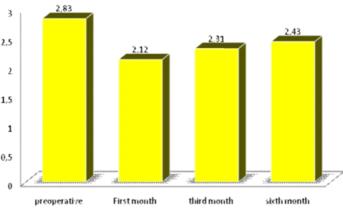
Figure 1 depicts the mean values of VC with the
standard deviations and calculated values of p for
comparison between pre-and postoperative periods. It is
observed that in all three comparisons (pre and first month,
pre and third month, and pre and sixth month) there were
significant differences (p <0.05) between the means of the
two moments.
Figure 2 shows the mean values of FVC with
comparison between the same three pairs of moments.
There were statistically significant differences when
comparing the four moments (p <0.05).
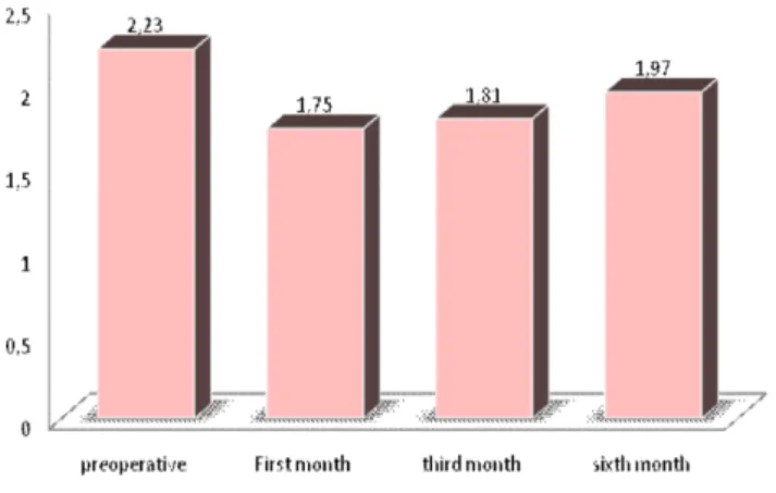
As for VEF1, There was no statistically significant
difference (p> 0.05) between the preoperative and
six-month postoperative periods, the opposite occurring for
the other comparisons (pre and first month and pre and
third month - p <0.050), with progressive recovery values
(Figure 3).
In Figure 4 we find the average of PEF of the
four periods: preoperative and the three postoperative.
There was a statistically significant difference (p <0.05)
among all comparisons with baseline PEF, indicating that
by the sixth month there was full recovery of variable PEF.
DISCUSSION
DISCUSSION
DISCUSSION
DISCUSSION
DISCUSSION
Spirometry involves carrying out measures of lung
volumes and flows that are obtained during the movements
of inspiration and forced expiration; the device then
transforms them into numbers and graphs. It is essential in
cases of surgical resection, especially in lung operations, as
it can help predict the risk of postoperative complications.
In this work, we have chosen spirometry due to
it being the mostly used method in surgical practice, its
availability at the hospital, and the difficulty of getting other
methods. The disease incidence is still high in the state of
Maranhão, with large numbers of symptomatic sequelae.
Figure 2 Figure 2 Figure 2
Figure 2 Figure 2 - Mean forced vital capacity of the four times studied (p <0,05).
Figure 1 Figure 1Figure 1
Amorim Amorim Amorim Amorim Amorim
Spirometry evaluation in patient with tuberculosis sequelae treated by lobectomy 119
Rev. Col. Bras. Cir. 2013; 40(2): 117-120
FVC is considered the most important measure of
spirometry because it represents the maximum flow of air
exhaled after maximal inspiration, held at the shortest interval
of time. Any process that alters the dynamic pulmonary also
alters the maximum flow, being the measurement most
sensitive to variation. In restrictive ventilatory disorders, FVC
displays values †below the reference ones.
FEV1 is the volume obtained in one second
expiratory FVC; it is the most representative spirometric
measurement of clinical alterations and restrictive ventilatory
disturbances; it may be normal or reduced.
Perin
19compared spirometric evaluation of
patients who underwent abdominoplasty in the immediate
preoperative and 19 and 36 postoperative months and
showed no significant difference with the correction of
rectus abdominis muscles.
This work showed marked decrease in lung
function in the first month after surgery, attributed to the
great difficulty of the patient to perform the maneuvers
due to the trauma of the chest wall muscles and the pain
associated with respiratory effort.
Helene Junior
18assessed respiratory function in
patients who underwent abdominoplasty in the preoperative
period and in the fourth POD, and observed reduced respiratory
function on the fourth POD, with normalization at the 30th day.
According to Tercan et al.
22, VC presents
significant postoperative improvement until the 30th day in
abdominoplasty, though not reaching preoperative values.
In this study, the improvement in lung function
was observed from the third postoperative month on due
to the absence of pain and improved lung compliance, with
progressive recovery of the parameters in the sixth month.
This study sought to assess whether there was
total recovery of spirometric parameters within 180 days
after lobectomy. The results did not show significant recovery
of parameters, reaching the preoperative average. This
occurred only in measures of FEV1/FVC and FEF.
The present study is the first to investigate the
profile of patients with sequelae of tuberculosis by evaluating
spirometry in the pre and postoperative periods of
lobectomy. Further studies should be performed in longer
periods, of a year or more, to verify whether the reason for
this change is due to the specific lung segment operated.
Thus, we conclude that, after six postoperative
months, patients with tuberculosis sequelae submitted to
lobectomy did not present with spirometric parameters
recovery when compared to preoperative values.
Figure 4 Figure 4 Figure 4 Figure 4
Figure 4 - Mean peak forced expiratory flow of the four times studied (p <0,05).
Figure 3 Figure 3 Figure 3 Figure 3
Figure 3 - Mean forced expiratory volume in one second of the four times studied (p <0,05).
R E S U M O
R E S U M O
R E S U M O
R E S U M O
R E S U M O
Objetivo: Objetivo: Objetivo: Objetivo:
Objetivo: Avaliar a espirometria no pré e pós-operatório de doentes com sequela de tuberculose, submetidos à lobectomia. Métodos:Métodos:Métodos:Métodos:Métodos: Foram selecionados 20 doentes, com idade entre 15 e 56 anos, de ambos os sexos, com história pregressa de tratamento de tuberculose, apresentando infecção de repetição ou hemoptises. Foram submetidos à lobectomia pulmonar. O tempo de tratamento da tuberculose foi seis meses e o aparecimento dos sintomas entre um e 32 anos. Foram avaliadas a capacidade vital (CV), a capacidade vital forçada (CVF), o volume expiratório forçado (VEF1), o VEF1/CVF, o fluxo expiratório forçado (FEF) e o pico de fluxo expiratório (PFE) após o primeiro, terceiro e sexto meses em relação ao pré-operatório. O nível de significância (á) aplicado em todos os testes foi 5%, ou seja, considerou-se significativo quando p<0,05. Resultados:Resultados:Resultados:Resultados:Resultados: As Médias encontradas foram as seguintes: Capacidade Vital (CV) Pré-operatória-2,83 ; 1º PO 2,12; 3º PO 2,31; 6º PO 2,43. Capacidade Vital Forçada (CVF) Pré-operatória- 2,97; 1º PO 2,21; 3º PO 2,35; 6º PO 2,53. Volume Expiratório no 1º Segundo (VEF1) Pré-operatório 2,23; 1º PO 1,75; 3º PO 1,81; 6º PO 1,97. Houve diminuição acentuada das funções respiratórias no primeiro mês de pós-operatório, porém houve melhora dos parâmetros a partir do terceiro mês, com progressivo aumento até o sexto mês de pós-operatório.Conclusão: Conclusão: Conclusão: Conclusão: Conclusão: Não houve recuperação dos parâmetros espirométricos, comparados aos do pré operatório, após seis meses de pós-operatório nos pacientes com sequela de tuberculose submetidos à lobectomia.
Descritores: Descritores: Descritores: Descritores:
120
Rev. Col. Bras. Cir. 2013; 40(2): 117-120
Amorim AmorimAmorim Amorim Amorim Spirometry evaluation in patient with tuberculosis sequelae treated by lobectomy
REFERENCES
REFERENCES
REFERENCES
REFERENCES
REFERENCES
1. Campos CA, Marchiori E, Rodrigues R. Tuberculose pulmonar: achados na tomografia computadorizada de alta resolução do tórax em pacientes com doença em atividade comprovada bacteriologicamente. J Pneumologia. 2002;28(1):23-9.
2. Muniz JN, Ruffino-Netto A, Villa TCS, Yamamura M, Arcencio R, Cardozo-Gonzales RI. Aspectos epidemiológicos da co-infecção tuberculose e vírus da imunodeficiência humana em Ribeirão Preto (SP), de 1998 a 2003. J bras pneumol. 2006;32(6):529-34. 3. Bombarda S, Figueiredo CM, Funari MBG, Soares Junior J, Seiscento
M, Terra Filho M. Imagem em tuberculose pulmonar. J Pneumologia. 2001;27(6):329-40.
4. Ribeiro SA. Tratamento compulsório da tuberculose: avanço ou retrocesso? J Pneumologia. 2003;29(1):50-2. (Cartas)
5. Silveira MPT, Adorno RFR, Fontana T. Perfil dos pacientes com tuberculose e avaliação do Programa Nacional de Controle da Tuberculose em Bagé (RS). J bras pneumol. 2007;33(2):199-205. 6. Ruffino-Netto A. Tuberculose: a calamidade negligenciada. Rev
Soc Bras Med Trop. 2002;35(1):51-8.
7. Cavalcanti ZR, Albuquerque MFPM, Campello ARL, Ximenes R, Montarroyos U, Verçosa MKA. Característica da tuberculose em idosos no Recife (PE): contribuição para o programa de controle. J bras pneumol. 2006;32(6):535-43.
8. Brasil. Ministério da Saúde. Tuberculose no Brasil: avanços e perspectivas. Programa Nacional de Controle da Tuberculose. [online]. In: Seminário de Manejo Clínico da Tuberculose, São Paulo, 02 e 03 de setembro de 2010. [cited 2010 out 23] Available from: http://www.sam.pmrp.com.br/ssaude/programas/tuberculose/ tuberculose_no_brasil.pdf
9. Castelo Filho A, Kritski AL, Barreto AW, Lemos ACM, Ruffino-Netto A, Guimarães CA, et al. II Consenso Brasileiro de Tuberculose. Diretrizes Brasileiras para Tuberculose 2004. J bras pneumol. 2004;30(supl. 1):S2-56.
10. Fernandes TM. Sol e trevas: histórias sociais da tuberculose brasileira. Hist cienc saude-Manguinhos. 2004;11(3):767-71. 11. Murray JF. A century of tuberculosis. Am J Respir Crit Care Med.
2004;169(11):1181-6.
12. Gazetta CE, Vendramini SHF, Ruffino-Netto A, Oliveira MRC, Villa TCS. Estudo descritivo sobre a implantação da estratégia de tratamento de curta duração diretamente observado no controle da tuberculose em São José do Rio Preto e seus impactos (1998-2003). J bras pneumol. 2007;33(2):192-8.
13. Gomes Neto A, Medeiros ML, Gifoni JMM. Bronquiectasia localizada e multissegmentar: perfil clínico-epidemiológico e resultado do tratamento cirúrgico de 67 casos. J Pneumologia. 2001;27(1):1-6.
14. Guerra MS, Miranda JA, Leal F, Vouga L. Tratamento cirúrgico das bronquiectasias. Rev Port Pneumol. 2007;13(5):691-701. 15. Balkanli K, Genç O, Dakak M, Gürkök S, Gözübüyük A, Caylak H,
et al. Surgical management of bronchiectasis: analysis and short-term results in 238 patients. Eur J Cardiothorac Surg. 2003;24(5):699-702.
16. Saad Júnior R, Garrido T, Stirbulov R, Rafal F. Avaliação da função respiratória de doentes submetidos à operação abdominal alta. Rev Col Bras Cir. 1994;21(6):329-32.
17. Pinto AMR. Estudo comparativo da função pulmonar em pacientes pós-revascularizados do miocárdio, com CEC e sem CEC, com uso de derivação intraluminal [tese]. São Paulo: Santa Casa de Misericórdia de São Paulo, Faculdade de Ciências Médicas; 1999. 18. Helene Júnior A. Avaliação da função respiratória em indivíduos submetidos à abdominoplastia. [tese]. São Paulo: Santa Casa de Misericórdia de São Paulo, Faculdade de Ciências Médicas; 2005. 19. Perin LF. Avaliação espirométrica de indivíduos submetidos à dermolipectomia abdominal. [dissertação]. São Paulo: Santa Casa de Misericórdia de São Paulo, Faculdade de Ciências Médicas; 2007.
20. Standardization of Spirometry, 1994 Update. American Thoracic Society. Am J Respir Crit Care Med. 1995;152(3):1107-36. 21. Pereira CAC. Testes de função pulmonar. Projeto Diretrizes.
Associação Médica Brasileira e Conselho Federal de Medicina. [on line]. Sociedade Brasileira de Pneumologia e Tisiologia. Elaboração final: 16 de abril de 2001. 12p. [citado em: 2010 out 20]. Disponível em: http://www.projetodiretrizes.org.br/projeto_diretrizes/ 090.pdf
22. Tercan M, Bekerecioglu M, Dikensoy O, Kocoglu H, Atik B, Isik D, et al. Effects of abdominoplasty on respiratory functions: a prospective study. Ann Plast Surg. 2002;49(6):617-20.
Received on 20/06/2012
Accepted for publication 07/08/2012 Conflict of interest: none
Source of funding: none
How to cite this article: How to cite this article:How to cite this article: How to cite this article:How to cite this article:
Amorim E, Saad Júnior R, Stirbulov R. Spirometry evaluation of patients with tuberculosis sequelae submitted to lobectomy. Rev Col Bras Cir. [periódico na Internet] 2013;40(2). Disponível em URL: http:// www.scielo.br/rcbc
Address correspondence to: Address correspondence to:Address correspondence to: Address correspondence to:Address correspondence to: Elias Amorim