Laser treatment of venous malformations
Laser treatment of venous malformations
Laser treatment of venous malformations
Laser treatment of venous malformations
Laser treatment of venous malformations
Tratamento a laser das malformações vasculares venosas
Tratamento a laser das malformações vasculares venosas
Tratamento a laser das malformações vasculares venosas
Tratamento a laser das malformações vasculares venosas
Tratamento a laser das malformações vasculares venosas
NERLAN T. G. DE CARVALHO1; JURANDIR MARCONDES RIBAS-FILHO, TCBC-PR2; JOSE FERNANDO MACEDO, TCBC-PR1; OSVALDO MALAFAIA,
ECBC-PR2; WILSON MICHAELIS3; RODRIGO ALMEIDA COELHO MACEDO3
A B S T R A C T A B S T R A C TA B S T R A C T A B S T R A C TA B S T R A C T
Objective ObjectiveObjective
ObjectiveObjective: To evaluate whether treatment of venous congenital vascular malformations with the use of laser technology provides lightening or disappearance of lesions with a high degree of satisfaction reported by patients and observed by the physician.MethodsMethodsMethodsMethodsMethods: A retrospective study gathered 26 patients suffering from vascular malformation, of which 73.07% were female and were treated with the PhotoDerm® device. The treatment of vascular malformations needed an average of 6.43 sessions, with intervals of six to eight weeks. Patients included in this study had skin type II (57.40%) and type III (42.30%), according to the Fitzpatric’s classification. The mean age ranged from 14 to 61 years, averaging 36.70 years. Data were obtained through the completion of an informed protocol by the patient. ResultsResultsResultsResultsResults: High satisfaction rates were reported (96.16%) and only one case (3.84%) was partially satisfied when considering lightening or disappearance of lesions. When evaluated by medical professionals, lesions disappeared in 80.76% and became lighter in 19.24%. ConclusionConclusionConclusionConclusionConclusion: The treatment of venous vascular malformations with Photo-Derm® is safe and effective as it offered a high degree of patient satisfaction and good results in the disappearance of the lesions.
Key words Key wordsKey words
Key wordsKey words: Vascular malformations. Laser therapy.
Work done at the Paraná Laser Integrated Nucleus and at the Evangelical Hospital of Curitiba – HUEC.
1. Angiologist, Evangelical University Hospital in Curitiba, Curitiba, PR, Brasil; 2. PhD, Associate Professor, Post-Graduation in Principles of Surgery, Evangelical University Hospital in Curitiba, Curitiba, PR, Brasil; 3. Master’s Degrre Graduate ,Post-Graduation in Principles of Surgery, Evangelical University Hospital in Curitiba, Curitiba, PR, Brasil.
INTRODUCTION
INTRODUCTION
INTRODUCTION
INTRODUCTION
INTRODUCTION
T
he word hemangioma comes from three Greeks radicals: haema (blood), angeion (vessel) and oma (tumor). Therefore, it means “tumor composed of blood vessels,” or actually “tumor formed by the proliferation of blood vessels.” Congenital vascular deformities, generically called hemangiomas, are anomalous formations resulting from defects occurring during the embryonic development of the vascular system. Thus, these deformities differ totally from the vascular formations that develop in the granulation tissue, as well as the lower limb varicosities arising from dilated veins in the superficial venous system or of malignant neoplasms of the vascular system represented by hemangioendotheliomas and hemangiosarcomas.Angiogenesis (from the Greek angeion - vessel + genesis - production)1 begins in the third week of
embryonic development. Points of the extra-embryonic mesoderm differentiate into hemangioblasts. By joining themselves they form clusters called islets of Wolf and Pander. These islands will suffer a progressive differentiation process where the cells that lie at the periphery (angioblasts) will flatten and enclose a cavity – the endothelium - while the ones occupying the central position (hemocytoblasts)
become spherical and will float in the liquid formed inside the cavity – the first figured elements of the blood.
The unions of angioblasts form strands that originate the primitive capillary network.
The interaction of these factors determines the formation of structurally polymorphic anatomical anomalies. The lesions may be superficial, involving skin and subcutaneous tissue, or deep, reaching muscle mass, bone or viscera. Sometimes they are localized, sometimes diffuse. Vascular malformations are a subject of great importance, for they affect children, teens and adults. Patients may live with the lesions, mostly benign, but some cases are very serious because of their hemodynamic or malignant implications. The aesthetic appearance of the lesions should receive special attention, as they often are accompanied by deformities of the face or other body areas. This causes discomfort, interferes with self-esteem and limits patients’ lives.
The use of lasers in medicine began in the 60s, with the ruby laser built by Theodore Maiman2, and was
gradually established as an alternative in the physician therapeutic armamentarium.
lesions with a high degree of satisfaction reported by patients and observed by the physician.
METHODS
METHODS
METHODS
METHODS
METHODS
The work was performed at the Paraná Integrated Laser Nucleus and the Evangelical Hospital of Curitiba – HUEC – and was approved by the Ethics Committee in the Paraná Evangelical Research School - FEPAR.
This was a retrospective study that gathered 26 patients with vascular malformation, of which 20 (73.07%) were female and six male (26.93%), aged between 14 and 61 years (mean 36 years and 7 months). All were treated with pulsed light laser - PhotoDerm®. (Figure 1)
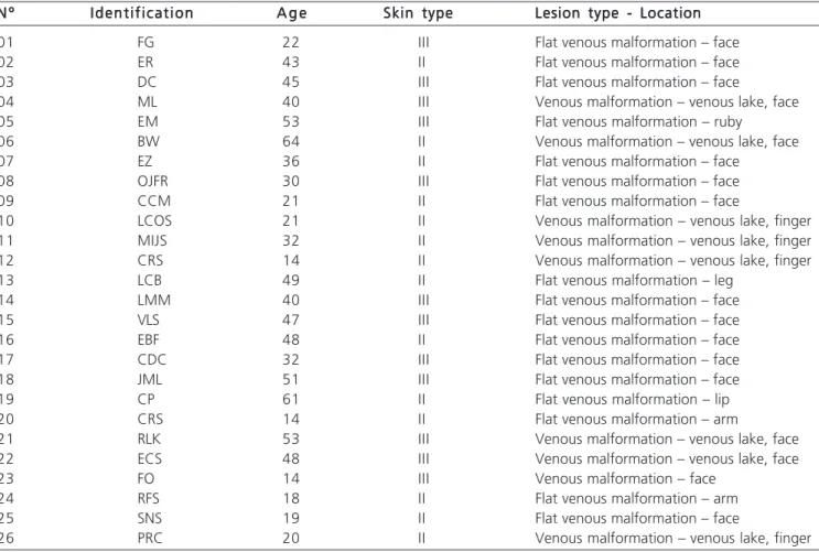
The treatment of vascular malformations needed on average 6.43 sessions, with intervals of six to eight weeks. Local anesthesia with Xylocaine® 1% without epinephrine or ice pack was applied to reduce pain. The data relating to the identification of patients, skin type and type of lesion are found in Figure 2.
Figure 3 shows the questionnaire for data collection. The series consisted mostly of flat venous malformations located on the face - 15 cases - and on the limbs (five).
Figure 1 – Figure 1 –Figure 1 – Figure 1 –
Figure 1 – Photoderm® and Vasculight® apparatus, used in the research (ESC Medical System Ltd./Sharplan, Yorkndam Industrial Park).
N º N º N º N º
N º I d e n t i f i c a t i o nI d e n t i f i c a t i o nI d e n t i f i c a t i o nI d e n t i f i c a t i o nI d e n t i f i c a t i o n A g eA g eA g eA g eA g e Skin typeSkin typeSkin typeSkin typeSkin type Lesion type - LocationLesion type - LocationLesion type - LocationLesion type - LocationLesion type - Location
01 FG 22 III Flat venous malformation – face
02 ER 43 II Flat venous malformation – face
03 DC 45 III Flat venous malformation – face
04 ML 40 III Venous malformation – venous lake, face
05 EM 53 III Flat venous malformation – ruby
06 BW 64 II Venous malformation – venous lake, face
07 EZ 36 II Flat venous malformation – face
08 OJFR 30 III Flat venous malformation – face
09 CCM 21 II Flat venous malformation – face
10 LCOS 21 II Venous malformation – venous lake, finger
11 MIJS 32 II Venous malformation – venous lake, finger
12 CRS 14 II Venous malformation – venous lake, finger
13 LCB 49 II Flat venous malformation – leg
14 LMM 40 III Flat venous malformation – face
15 VLS 47 III Flat venous malformation – face
16 EBF 48 II Flat venous malformation – face
17 CDC 32 III Flat venous malformation – face
18 JML 51 III Flat venous malformation – face
19 CP 61 II Flat venous malformation – lip
20 CRS 14 II Flat venous malformation – arm
21 RLK 53 III Venous malformation – venous lake, face
22 ECS 48 III Venous malformation – venous lake, face
23 FO 14 III Venous malformation – face
24 RFS 18 II Flat venous malformation – arm
25 SNS 19 II Flat venous malformation – face
26 PRC 20 II Venous malformation – venous lake, finger
Table 1 – Table 1 – Table 1 – Table 1 –
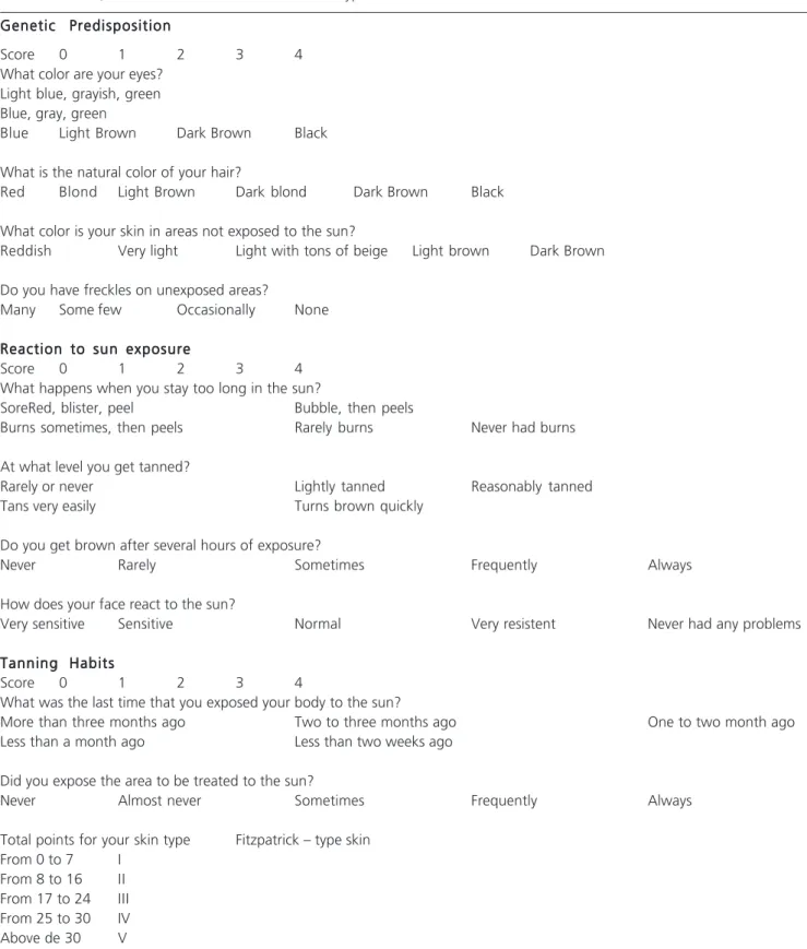
The classification of skin type was based on the proposal made by Fitzpatrick3. By filling out the questionnaire
and assigning points to the answers a value corresponding to the type of skin resulted (Figure 4).
Patients with skin type IV, V and VI were not included in the treatment because of the risk of burns and pigment loss resulting from destruction of melanocytes.
Intense Pulsed Light – Photoderm® “ exerts its effect by reaching different plans because of its wavelength. For this reason the treatment sessions were multiple. It ranged from two to 17 sessions, depending on the type, extension and depth of the malformation. The average was 6.42 sessions per patient.
In analyzing the results, the personal assessment ranks from the patients, considering the degree of satisfaction with outcomes, were satisfied, partially satisfied and unsatisfied.
The assessment by the medical professional was ranked in the disappearance of lesions, lightening and no result.
RESULTS
RESULTS
RESULTS
RESULTS
RESULTS
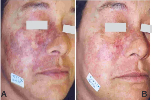
Of the 26 patients treated, 25 (96.16%) reported being satisfied, for they had observed a decrease or disappearance of the malformation. One (3.84%) reported the degree of satisfaction as partially satisfied and none reported dissatisfaction with the treatment. (Figures 5 and 6)
The observation of the physician as to the result showed disappearance of the lesions in 21 cases (80.76%), and lightening in five (19.24%).
DISCUSSION
DISCUSSION
DISCUSSION
DISCUSSION
DISCUSSION
Vascular malformations were not treated until 1667, due to the lack of sclerosing solutions. That year Tournay, cited by Macedo4, described the first attempt to
carry out vein thromboses by chemical methods made by Elshotz, which used a chicken bone like a needle tied in a pig’s bladder. This was the first idea of the syringe and
Name: Address:
Birth Date: ___/___/____ Age:
Town: State:
Phone:
FITZPATRICK’S SKIN TYPE CLASSIFICATION: FITZPATRICK’S SKIN TYPE CLASSIFICATION: FITZPATRICK’S SKIN TYPE CLASSIFICATION: FITZPATRICK’S SKIN TYPE CLASSIFICATION: FITZPATRICK’S SKIN TYPE CLASSIFICATION:
TYPE I ( ) TYPE II ( ) TYPE III ( ) TYPE IV ( )
TYPE OF LESION TYPE OF LESION TYPE OF LESION TYPE OF LESION TYPE OF LESION:
Flat venous malformation – face (port wine) ( ) Flat venous malformation – arm (port wine) ( )
Flat venous malformation – lip ( )
Venous malformation – ruby ( )
Venous malformation – venous lake – face ( ) Venous malformation – venous lake – finger ( )
I ) STHETIC RESULT
Disappeared YES ( ) NO ( ) ( ) %
Lightenning YES ( ) NO ( ) ( ) %
No Result YES ( ) NO ( ) ( ) %
II ) PERSONAL ASSESSMENT:
Satisfied YES ( ) NO ( )
Partially Satisfied YES ( ) NO ( )
Unsatisfied YES ( ) NO ( )
DATE: _______/______/_______ SESSIONS: ( )
Photo records: YES ( ) NO ( ) Table 2
Table 2 Table 2 Table 2
needle. By using the medicinal plant Plantago Major, L it caused phlebitis, sclerosis of the vein responsible for maintenance of the varicose ulcer leading ultimately to its healing.
Later local treatments were used - ethanol injection, hypertonic glucose infusion, Ethamolin® injection
associated with glucose, sodium morrhuate injection aethoxysclerol injection5,6, systemic treatments - the use of
corticosteroids, interferon5,6, and/or surgical excision,
cryotherapy, arterial embolization4 and betatherapy.
Some of these methods are still used by lack of knowledge or new technology or due to its high cost.
Genetic Predisposition Genetic Predisposition Genetic Predisposition Genetic Predisposition Genetic Predisposition
Score 0 1 2 3 4
What color are your eyes? Light blue, grayish, green Blue, gray, green
Blue Light Brown Dark Brown Black
What is the natural color of your hair?
Red Blond Light Brown Dark blond Dark Brown Black
What color is your skin in areas not exposed to the sun?
Reddish Very light Light with tons of beige Light brown Dark Brown
Do you have freckles on unexposed areas?
Many Some few Occasionally None
Reaction to sun exposure Reaction to sun exposure Reaction to sun exposure Reaction to sun exposure Reaction to sun exposure
Score 0 1 2 3 4
What happens when you stay too long in the sun?
SoreRed, blister, peel Bubble, then peels
Burns sometimes, then peels Rarely burns Never had burns
At what level you get tanned?
Rarely or never Lightly tanned Reasonably tanned
Tans very easily Turns brown quickly
Do you get brown after several hours of exposure?
Never Rarely Sometimes Frequently Always
How does your face react to the sun?
Very sensitive Sensitive Normal Very resistent Never had any problems
Tanning Habits Tanning Habits Tanning Habits Tanning Habits Tanning Habits
Score 0 1 2 3 4
What was the last time that you exposed your body to the sun?
More than three months ago Two to three months ago One to two month ago
Less than a month ago Less than two weeks ago
Did you expose the area to be treated to the sun?
Never Almost never Sometimes Frequently Always
Total points for your skin type Fitzpatrick – type skin From 0 to 7 I
From 8 to 16 II From 17 to 24 III From 25 to 30 IV Above de 30 V Table 3
Table 3 Table 3 Table 3
Figura 2 Figura 2 Figura 2 Figura 2
Figura 2 – Venous malformation in the face: pre-treatment (A) and post-treatment (B).
Figura 3 Figura 3 Figura 3 Figura 3
Figura 3 – Venous malformation in the forearm: pre-treatment (A) and post- treatment (B).
Laser devices are available in the Brazilian market since 1997. Skin lesions and complications arising from their use are dependent on training, skill, the parameters used and on the manipulation technique.
Several authors have shown good results in treating vascular malformations7-9 and Raulin et al.8 classify
PhotoDerm® as the gold standard.
Goldman et al.10 reported in 1998 the
occurrence of malformations on the face in 40% of ca-ses, and 35% in the limbs. Rather, this study found that the location on the face occurred in 57.70% of the time, and 19.23% in the extremities. The authors attributed the increased exposure of the face associated with the appreciation of aesthetics as a justification for increased demand of facial procedures.
Raulin et al.9 describe the use of a filter whose
wavelength is 550 nm and fluence of energy ranging from 25 to 40 j/cm2. In another study, the same author mentions
the use of 590 nm filter and fluence from 40 to 70 j/cm2 7.
Al Buainian et al.5 referred the use of 585 nm filter. Patients
treated by us were submitted to the parameters recommended by the PhotoDerm® apparatus program and the filters 515, 550 and 570 nm were used; the energy used ranged from 28 to 60 J/cm2.
Raulin et al.7,8 attributed different results in the
treatment with PhotoDerm® to the size of the filters with an area of 2.8 cm2, for it would cover a larger area per
shot. In that study the lower number of shots brought more comfort to the patient. In another publication, the author
A
B
A
reports that it took four sessions to get to the result. In this study the mean of sessions was 6.42, higher than those cited.
Based on the evolution of technology, routine use makes believe that the laser will be better indicated and better results will emerge. Importantly, professional
training in the technology minimizes consequences inherent to the method.
The treatment of venous vascular malformations with Photo-Derm® is safe and effective, having provided a high degree of patient satisfaction and good results in the disappearance of the lesions.
R E S U M O R E S U M O R E S U M O R E S U M O R E S U M O
Objetivo: Objetivo: Objetivo: Objetivo:
Objetivo: Avaliar se o tratamento das malformações vasculares venosas congênitas realizado com o emprego da tecnologia laser oferece clareamento ou desaparecimento das lesões com elevado grau de satisfação informado pelos pacientes e observa-do pelo médico. Métoobserva-dos:Métodos:Métodos:Métodos: O estudo retrospectivo reuniu 26 pacientes, portadores de malformação vascular venosa, dos quaisMétodos: 73,07% eram do sexo feminino e que foram tratados com o aparelho PhotoDerm®. O tratamento das malformações vasculares necessitou uma média de 6,43 sessões, com intervalos de seis a oito semanas. Os pacientes incluídos neste estudo apresentavam pele tipo II (57,40%) e tipo III (42,30%), conforme classificação de Fitzpatric. A idade média variou de 14 a 61 anos, com média de 36,70 anos. Os dados foram obtidos através do preenchimento de protocolo informado pelo paciente. Resultados:Resultados:Resultados:Resultados:Resultados: Foi informado elevado grau de satisfação (96,16%) e apenas um caso (3,84%) parcialmente satisfeito, considerando o clareamento ou desaparecimento das lesões. Houve desaparecimento das lesões avaliado pelo profissional médico em 80,76% dos casos e em 19,24% apenas clareamento. Conclusão:Conclusão:Conclusão:Conclusão:Conclusão: O tratamento das malformações vasculares venosas com o Photo-Derm® é seguro e eficiente tendo proporcionado elevado grau de satisfação dos pacientes assim como bons resultados em relação ao desapareci-mento das lesões.
Descritores: Descritores: Descritores: Descritores:
Descritores: Malformações vasculares. Terapia a laser.
REFERENCES
REFERENCES
REFERENCES
REFERENCES
REFERENCES
1. Maffei FHA. Doenças vasculares periféricas. Rio de Janeiro: MEDSI, 1988. cap. 48, p. 791-827.
2. Maiman, TH. Stimulated optical radiation in ruby. Nature. 1960;187:493.
3. Fitzpatrick TB. Soleil et peau. J. Méd. Esthétique. 1975;2:33. 4. Macedo JF. Alterações morfológicas vasculares e cutâneas
provocadas por solução de glicose hipertônica a 75% ou laser Nd:Yag de 1064 nm em coelhos. [Tese]. Curitiba: Universidade Federal do Paraná, Programa de Pós-Graduação em Clínica Cirúr-gica do Setor de Ciências da Saúde; 2001.
5. Buainian H, Verhaeghe E, Dierckxsens L, Naeyaert JM. Early treatment of hemangiomas with Lasers. Dermatology. 2003; 206:370-73.
6. Garzon MC, Huang JT, Enjolras O, Frieden IJ, Continuing medical Education: vascular malformation. J Am Acad. Dermatol. 2007;56:353-70.
7. Raulin C, Raulin SJ, Hellwig S, Schönermark MP. Treatment of benign venous with an intense light source (PhotoDerm®VL), European Journal of Dermatology. 1997 June;7(4):279-82. 8. Raulin C, Hellwig S, Schönermark MP. Treatment of nonresponding
port-wine stain with a new pulsed light source (PhotoDerm®VL). Lasers in Surgery and Medicine. 1997;21:203-08.
9. Raulin C, Goldman M.P, Weiss M. Treatment of adult port-wine stains using intense pulsed light therapy (PhotoDerm VL): brief clinical report. American Society for Dermatologic Surgery. 1997; 23(7):594-97.
10. Goldman MP. Escleroterapia: tratamento das veias varicosas e teleangiectasias dos membros inferiores. Rio de Janeiro: Interlivros, 1994. cap. 12, p. 334–65.
Received in 14/07/2009
Accepted for publication in 12/09/2009 Conflict of interest: none
Source of funding: none
How to cite this article: How to cite this article:How to cite this article: How to cite this article: How to cite this article:
Carvalho NTG, Ribas-Filho JM, Macedo JF, Malafaia O, Michaelis W, Macedo RAC. Laser treatment of venous malformations . Rev Col Bras Cir. [periódico na Internet] 2010; 37(5). Disponível em URL: http:// www.scielo.br/rcbc