Sao Paulo Med J. 2014; 132(4):239-42 239
SHORT COMMUNICATION
DOI: 10.1590/1516-3180.2014.1324703Systemic lupus erythematosus activity and beta two
microglobulin levels
Atividade do lúpus eritematoso sistêmico e níveis de beta dois microglobulina
Thelma Larocca Skare
I, Kellen Ferri
II, Marcela Aimone Santos
IIRheumatology Unit, Hospital Universitário Evangélico de Curitiba, Paraná, Brazil
ABSTRACT
CONTEXT AND OBJECTIVE: Systemic lupus erythematosus (SLE) is an autoimmune disease with a cyclical clinical course. Evaluation of the clinical activity of this disease is important for choosing the correct treat-ment. The objective of this study was to analyze the value of beta-2 microglobulin (β2M) serum levels in determining SLE clinical activity.
DESIGN AND SETTING: Cross-sectional analytical study conducted at the rheumatology outpatient clinic of a private university hospital.
METHODS: 129 SLE patients were studied regarding disease activity using SLEDAI (SLE Disease Activity Index) and cumulative damage using SLICC ACR (SLE International Collaborating Clinics/American Col-lege of Rheumatology Damage Index for SLE). At the same time, the β2M serum level, ESR (erythrocyte sedimentation rate), anti-dsDNA (anti-double-stranded DNA) and C3 and C4 complement fractions were determined.
RESULTS: β2M levels correlated positively with SLEDAI (P = 0.02) and ESR (P = 0.0009) and negatively with C3 (P = 0.007). Patients who were positive for anti-dsDNA had higher β2M serum levels (P = 0.009). CONCLUSION: β2M levels are elevated in SLE patients with active disease.
RESUMO
CONTEXTO E OBJETIVO: O lúpus eritematoso sistêmico (LES) é uma doença autoimune com curso clí-nico lutuante. Determinar a atividade clínica da doença é importante na escolha do tratamento correto. O objetivo deste estudo foi o de analisar se os níveis séricos de beta-2 microglobulina (β2M) têm valor na determinação da atividade clínica do LES.
TIPO DE ESTUDO E LOCAL: Estudo transversal, analítico, realizado no ambulatório de reumatologia de um hospital universitário particular.
MÉTODOS: 129 pacientes com LES foram estudados para atividade da doença pelo SLEDAI (SLE Disease Activity Index) e para dano cumulativo pelo SLICC (SLE International Collaborating Clinics/American Col-lege of Rheumatology Damage Index for SLE). Simultaneamente foram determinados os valores séricos de β2M, das frações C3 e C4 do complemento, VHS (velocidade de hemossedimentação) e anti dsDNA (DNA de dupla ita).
RESULTADO: Encontrou-se correlação positiva entre níveis de β2M com o SLEDAI (P = 0,02), com VHS (P = 0,0009) e correlação negativa com níveis de C3 (P = 0,007). Pacientes com presença de anti dsDNA tinham níveis mais altos de β2M (P = 0,009).
CONCLUSÃO: Níveis de β2M estão elevados em pacientes de LES com doença ativa.
IMD, PhD. Associate Professor, Discipline of
Rheumatology, Faculdade Evangélica do Paraná, and Head of Rheumatology Unit, Hospital Universitário Evangélico de Curitiba, Curitiba, Paraná, Brazil.
IIMedical Student. Faculdade Evangélica do
Paraná, Curitiba, Paraná, Brazil.
KEY WORDS:
Lupus erythematosus, systemic. beta 2-microglobulin. Inlammation. Blood sedimentation. Acute-phase reaction.
PALAVRAS-CHAVE: Lúpus eritematoso sistêmico. Microglobulina-2 beta. Inlamação.
SHORT COMMUNICATION | Skare TL, Ferri K, Santos MA
240 Sao Paulo Med J. 2014; 132(4):239-42 INTRODUCTION
Systemic lupus erythematosus (SLE) is an autoimmune disease with periods of lares and remissions.1 he treatment for SLE accompanies the degree of disease activity and, thus, determin-ing this activity level is very important, even though diicult. Commonly-used inlammatory markers such as sedimentation rate (ESR) and C-reactive protein are nonspeciic and unreli-able.1 On the other hand, composite measurements such as the SLE Disease Activity Index (SLEDAI) and the British Isles Lupus Assessment Group index (BILAG),2 which combine laboratory and clinical indings, are time-consuming and not easily appli-cable in daily practice.
Beta-2 microglobulin (β2M) is a light-chain subunit of class I human leukocyte antigen (HLA) that exists on the cell membrane of all nucleated cells.3 It sheds from the membrane and is measur-able in the bloodstream, where its levels are elevated in diseases with high lymphoproliferative activity.3 Some authors have noted increased plasma and/or urine β2M levels in Sjögren’s syndrome, rheumatoid arthritis and SLE,4 and have proposed that measur-ing its levels might be useful as an activity marker.
OBJECTIVE
he present analysis was undertaken to further examine the rela-tionship between β2M and lupus activity, along with the cumula-tive damage in a sample of Brazilian patients.
METHODS
his was a cross-sectional analytical study that was approved by the local Research Ethics Committee. All subjects signed an informed consent statement.
We included 129 patients from a single tertiary center, who were all over 18 years of age and met at least four of the classiica-tion criteria of the American College of Rheumatology for SLE.5 Patients with overlapping features, creatinine above 1.1 mg/dl, pre-vious history of lymphoproliferative disorders, pregnancy or asso-ciated infections were excluded. Demographic and clinical data were obtained through chart review.
All individuals underwent measurement of SLEDAI2 and SLICC/ACR6 (Systemic Lupus Erythematosus International Collaborating Clinics/American College of Rheumatology Damage Index for SLE) along with measurements of β2M, C3, C4, ESR (erythrocyte sedimentation rate), CRP (C-reactive pro-tein), hemoglobin (Hb) levels and anti-dsDNA (anti-double-stranded DNA).
β2M measurements were made in serum by means of chemi-luminescence (Immulite 2000, Diagnostic Products Corporation, USA), with normal values ranging from 604 to 2786 ng/ml; ESR was measured using the Westergreen method (normal value < 8 mm); CRP was measured by means of immunoturbidimetry
(normal values < 0.50 mg/dl); and Hb was measured using an automated method (normal values of 12.2-18.1 g/dl). C3 and C4 were evaluated in fasting serum by means of immuno-turbidimetry (normal values for C3 = 82-170 mg/dl; and for C4 = 12-36 mg/dl). Anti-dsDNA was analyzed by means of indi-rect immunoluorescence using Crithidia lucilae as the substrate (Immunoconcept, Alka, São Paulo, Brazil).
he data were compiled in frequency tables. he central trend was expressed as the mean and standard deviation for paramet-ric data and as the median and interquartile range (IQR) for non-parametric data. Correlation analyses were conducted using the Spearman and Pearson tests, according to sample distribution. An association analysis on β2M levels in the presence of anti-dsDNA was done using the Mann-Whitney test. he GraphPad Prism sotware, version 4.0, was used for calculations. he signif-icance level was taken to be 5%.
RESULTS
In this sample, 3.1% were male and 96.9% were female, with a mean age of 40.1 ± 11.3 years and median disease duration of 8.0 years. Regarding the cumulative clinical proile: 67.1% patients had photosensitivity; 60.1% arthritis; 43.3% oral ulcers; 39.8% butterly rash; 36.3% glomerulonephritis; 32.1% leucopenia; 9.0% pericar-ditis; 8.3% hemolytic anemia; 8.3% seizures; 6.9% discoid lesions; 5.5% pleuritis; 6.2% thrombocytopenia; and 3.4% psychosis.
In this population, 54.4% were using glucocorticoids; 71.2% antimalarials; 17.8% azathioprine; 15.8% methotrexate; 5.9% mycophenolate mofetil; 1.9% cyclophosphamide; 0.9% cyclospo-rine and 0.9% thalidomide.
SLEDAI ranged from 0 to 14 (median 0.0; IQR = 0.0-2.0); and SLICC ranged from 0 to 9 (median = 1.0; IQR = 0.0-2.0). he median ESR was 25.0 mm (IQR = 9.0-44.5 mm); the median C3 level was 111.0 mg/dl (IQR = 92.8-131.1 mg/dl) and median C4, 19.0 mg/dl (IQR = 13.8-23.7 mg/dl). he median Hb level was 13.0 g/dl (IQR = 12.3-14.0) and median CRP was 2.6 mg/dl.
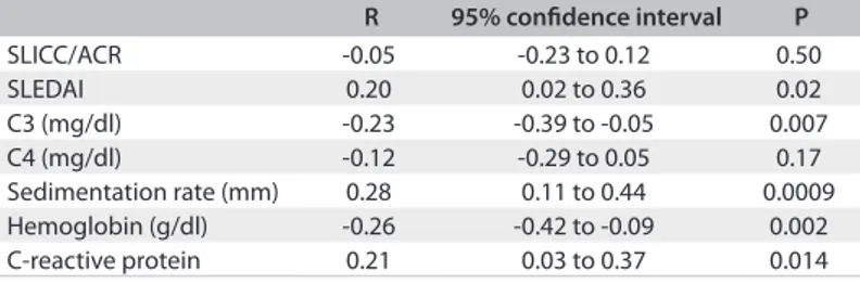
In 28.6% of the patients, anti-dsDNA was positive; β2M ranged from 1120 to 4943 ng/ml (median of 2045 ng/ml; IQR from 1679-2591 ng/ml). he correlations of β2M with SLEDAI, SLICC, complement, hemoglobin, C-reactive protein and ESR can be seen in Table 1. he median value for β2M in patients with positive anti-dsDNA was 2167 ng/ml (1829-3003 ng/ml), while in those without anti-dsDNA the median was 1950 ng/ml (1600-2307 ng/ml; P = 0.009).
Systemic lupus erythematosus activity and beta two microglobulin levels | SHORT COMMUNICATION
Sao Paulo Med J. 2014; 132(4):239-42 241 DISCUSSION
β2M is a small protein that has an amino acid sequence related to the constant parts of heavy and light chains of immunoglobu-lins.7 It locates on the surface of all nucleated cells and its best-characterized function is to interact with and stabilize the ter-tiary structure of the MHC class 1 chain.7 β2M is normally found in serum, urine and other body luids and is almost exclusively catabolized within the kidney; 95% to 100% of circulating β2M is eliminated through glomerular iltration.7
The reason why it has been found to be elevated in SLE patients is unknown. Some authors4 believed that this might result from the increased turnover of lymphocytes seen in this disease. Autoantibodies directed against β2M that pres-ent lymphocytotoxic activity have also been reported in SLE.8 Since immune complexes formed by β2M and anti-β2M have a larger size, they cannot be filtered by the kidney, thus rais-ing the serum levels of β2M and givrais-ing another explanation for this elevation.
Walters et al.9 detected higher levels of serum β2M in rheu-matoid arthritis patients and an even higher concentration in synovial luids, which suggested that there is intra-articular pro-duction of this protein in this disease. Interestingly, in studying the possible associations between β2M and speciic lupus mani-festations, Kim et al.4 found that the serum levels were higher in patients with serositis, oral ulcers and glomerulonephritis but not in those with arthritis. Experimental studies10 on β2M-deicient lupus mice showed that a lack of β2M caused dissociation in the clinical expression of the disease, with aggravation of skin dis-ease and amelioration of nephritis. his divergence of disdis-ease in the skin and kidneys of β2M-deicient mice suggests that target organs may respond in diferent ways to the autoimmune process in this context.
Another study on 26 lupus patients, by Hermansen et al.,11 found that serum β2M showed correlations with cytokines such as interleukin (IL)-6, IL-8, IL-10 and IL-18, and with serum interferon-α. hese authors believed that the increased β2M lev-els in active SLE relected the overall immunological activity.
In the present study, we found that ESR, CRP, C3 and SLEDAI were associated with β2M, thus showing that its level was elevated in cases of active disease. his result is similar to the indings of Evrin and Ström8 in 23 Swedish lupus patients, in whom they found a posi-tive correlation with ESR and a negaposi-tive association with hemoglo-bin levels. Moreover, Kim et al.4 studying Korean lupus population found a negative correlation between β2M and C3 and a positive cor-relation between β2M and SLEDAI, as we did. In a disease such as SLE, with great inluence of genetic background, our results showed that the levels of β2M in lupus activity in a sample of Brazilian pop-ulation follow the same pattern as others. Furthermore, we did not ind any association of β2M with SLE cumulative damage measured by SLICC/ACR, which had not been studied previously.
We also showed that, in this sample, SLEDAI correlated with ESR, C3 and anti-dsDNA, which raises the question of the additional value in measuring β2M. It needs to be noted that in this study we excluded patients with associated infections, which are a cause of great confusion in ESR interpretation. Also, C3 consumption and pres-ence of anti-dsDNA are associated with speciic lupus manifestations. Membranous glomerulonephritis (or class 5 glomerulonephritis) is a typical example in which the lupus manifestation may appear with a normal complement and without anti-dsDNA.12,13 In these situations, assaying forβ2M may help to determine disease activity.
CONCLUSIONS
Our indings provide conirmation that β2M levels are elevated in cases of active lupus, which thus may help in determining the degree of disease activity. Further studies are needed in order to understand the role of this protein in the pathophysiological process of lupus.
REFERENCES
1. Suh CH, Jeing YS, Park HC, et al. Risk factors for infection and
role of C-reactive protein in Korean patients with systemic lupus
erythematosus. Clin Exp Rheumatol. 2001;19(2):191-4.
2. Liang MH, Socher SA, Larson MG, Schur PH. Reliability and validity
of six systems for clinical assessment of disease activity on systemic
lupus erythematosus. Arthritis Rheum. 1989;32(9):1107-18.
3. McPhee CG, Sproule TJ, Shin DM, et al. MHC class I family proteins
retard systemic lupus erythematosus autoimmunity and B cell
lymphomagenesis. J Immunol. 2011;187(9):4695-704.
4. Kim HA, Jeon JY, Yoon JM, Suh CH. Beta 2-microglobulin can be a
disease activity marker in systemic lupus erythematosus. Am J Med
Sci. 2010;339(4):337-40.
5. Tan EM, Cohen AS, Fries JF, et al. The 1982 revised criteria for the
classiication of systemic lupus erythematosus. Arthritis Rheum.
1982;25(11):1271-7.
6. Stoll T, Seifert B, Isenberg DA. SLICC/ACR Damage Index is valid, and
renal and pulmonary organ scores are predictors of severe outcome
in patients with systemic lupus erythematosus. Br J Rheumatol.
1996;35(3):248-54.
Table 1. Result from correlating β2 microglobulin levels with the Systemic Lupus International Collaborating Clinics/American College of Rheumatology (SLICC/ACR) index, systemic lupus erythematosus disease activity index (SLEDAI), complement, hemoglobin, C-reactive protein and sedimentation rate
R 95% conidence interval P
SLICC/ACR -0.05 -0.23 to 0.12 0.50
SLEDAI 0.20 0.02 to 0.36 0.02
C3 (mg/dl) -0.23 -0.39 to -0.05 0.007
C4 (mg/dl) -0.12 -0.29 to 0.05 0.17
SHORT COMMUNICATION | Skare TL, Ferri K, Santos MA
242 Sao Paulo Med J. 2014; 132(4):239-42
7. Xie J, Wang Y, Freeman ME 3rd, Barlogie B, Yi Q. Beta 2-microglobulin
as a negative regulator of the immune system: high concentrations
of the protein inhibit in vitro generation of functional dendritic cells.
Blood. 2003;101(10):4005-12.
8. Evrin PE, Ström T. Beta 2-microglobulin and its binding activity in
serum from patients with SLE. Ann Rheum Dis.1984;43(2):267-74.
9. Walters MT, Stevenson FK, Goswami R, Smith JL, Cawley MI.
Comparison of serum and synovial luid concentrations of beta
2-microglobulin and C reactive protein in relation to clinical disease
activity and synovial inlammation in rheumatoid arthritis. Ann
Rheum Dis. 1989;48(11):905-11.
10. Chan OT, Paliwal V, McNif JM, et al. Deiciency in beta(2)-microglobulin,
but not CD1, accelerates spontaneous lupus skin disease while
inhibiting nephritis in MRL-Fas(lpr) mice: an example of disease
regulation at the organ level. J Immunol.2001;167(5):2985-90.
11. Hermansen ML, Hummelshøj L, Lundsgaard D, et al. Increased serum
β2-microglobulin is associated with clinical and immunological markers of disease activity in systemic lupus erythematosus patients.
Lupus. 2012;21(10):1098-104.
12. Falk RJ, Schur PH, Appel GB. Clinical features and therapy of
membranous lupus nephritis. Uptodate.com. Available from: http://
www.uptodate.com/contents/clinical-features-and-therapy-of-membranous-lupus-nephritis. Accessed in 2013 (Sep 4).
13. Sun HO, Hu WX, Xie HL, et al. Long-term outcome of Chinese patients
with membranous lupus nephropathy. Lupus. 2008;17(1):56-61.
Marcela Aimone Santos is a grantee of Programa Institucional de
Bolsas de Iniciação Cientíica – Conselho Nacional de Desenvolvimento
Cientíico e Tecnológico (PIBIC-CNPq), Brazil
Sources of funding: None Conlict of interest: None
Date of irst submission: April 17, 2013 Last received: September 11, 2013 Accepted: September 17, 2013
Address for correspondence:
Thelma Larocca Skare
Rua João Alencar Guimaraes, 796
Santa Quitéria — Curitiba (PR) — Brasil
CEP 80310-420
Tel. (+55 41) 3274-1659