4 4 6
Castier et al
Cardiac tamponade in systemic lupus erythematosus
Arq Bras Cardiol
volume 75, (nº 5), 2000
Report of Four Cases Rio de Janeiro, RJ - Brazil Hospital Universitário Pedro Ernesto - UERJ
Mailing address: Márcia Bueno Castier - Av. Epitácio Pessoa 4254/201 - 22471-001 - Rio de Janeiro, RJ, Brazil
English Version by Stela Maris C. Gandour
Objective – To report and assess the incidence of
cardiac tamponade in systemic lupus erythematosus as a
cardiac manifestation of the disease.
Methods – We reviewed the medical records of 325
patients diagnosed with systemic lupus erythematosus
ac-cording to the American Rheumatism Association and
their complementary laboratory tests compatible with
cardiac tamponade.
Results – In the 325 medical recors reviewed, we
found 108 patients with pericardial effusions
correspon-ding to 33.2% of the total and 54% of the patients studied
in the active phase of the disease. Clinical assessment and
transthoracic echocardiogram allowed the clinical
diagnosis of cardiac tamponade in only 4 (1.23%)
patients, 3 of whom were females, white, with ages ranging
from 25 to 44 years. The pericardial fluid was hemorrhagic
or serosanguineous with high levels of FAN and positivity
for LE cells. In the treatment, we successfully used
pericar-dicentesis associated with high doses of corticosteroids. In
clinical and laboratory follow-up performed for a period
of 3 years, neither recrudescence of the pericardial
effu-sion nor evolution to constriction occurred.
Conclusion – Even though rare (1.23%), cardiac
tamponade in patients with systemic lupus erythematosus
has a benign evolution when properly treated, according
to our experience.
Key words:
cardiac tamponade, systemic lupus
erythe-matosus, pericardial effusion
Arq Bras Cardiol, volume 75 (nº 5), 446-448, 2000
Márcia Bueno Castier, Elisa M. N eves Albuquerque, Maria Eduarda F. Costa Castro Menezes,
Evandro Klumb, Francisco Manes Albanesi Fº
Rio de Janeiro, RJ - Brazil
Cardiac Tamponade in Systemic Lupus Erythematosus.
Report of Four Cases
Brief Communication
Systemic lupus erythematous is a collagen disease
that often involves the heart, the pericardium being the most
commonly affected site, resulting in clinical
1-9and
anatomo-pathological
10-14manifestations. This involvement is
usually pericardial effusion, most of the time of mild
in-tensity
6,7,15-17. In a few patients, pericardial involvement may
be the initial form of presentation of systemic lupus
erythe-matosus
5-7. Cardiac tamponade is a rare clinical
manifes-tation
15, and very few cases are reported as the first
mani-festation of the disease
18-20.
Our study aims to assess the frequency of cardiac
tamponade in patients with systemic lupus erythematosus
being followed up in a specialized sector of a tertiary hospital.
Methods
We reviewed 325 medical records of patients with
sys-temic lupus erythematosus, who were being followed up in
the Sector of Collagen Diseases of the Rheumatology
Ser-vice of HUPE – UERJ, during the period from March ’91 to
October ’98. The diagnosis of systemic lupus
erythema-tosus met the criteria adopted by the American Rheumatism
Association (ARA)
21. To characterize disease activity, we
used the criteria of SLEDAI (Systemic Lupus
Erythema-tosus Disease Activity Index)
22. All patients studied had at
least one transthoracic echocardiography that was
perfor-med independently from the presence of cardiovascular
signals or symptoms, or both, because of the development
of several study protocols.
The echocardiographies were performed with Toshiba
SSH-65
Aand Apogee CX 200 devices, which provided
single-dimensional and two-dimensional recordings and
flow study with Doppler and color mapping. The
echocar-diographic measurements were obtained according to the
guidelines of the American Society of Echocardiography
23.
Once the presence of pericardial effusion was detected, it
was classified as mild, moderate, or severe
24. We assessed
Arq Bras Cardiol
volume 75, (nº 5), 2000
Castier et al
Cardiac tamponade in systemic lupus erythematosus
4 4 7
right ventricular dimensions
25, 26, collapse of the right atrial
wall during diastole
27, and diastolic invagination of the right
ventricular anterior wall
28. On Doppler echocardiography,
we assessed a drop in mitral and aortic flows during
inspiration
29.
For the diagnosis of cardiac tamponade, we used data
from clinical examinations, such as paradoxical pulse
as-sociated or not with reduction in intensity of the cardiac
sounds, pathological jugular venous distension, and
arteri-al hypotension. These data supported by
echocardiogra-phic findings of pericardial effusion associated with signals
of increase in the intrapericardial pressure established the
definitive diagnosis of cardiac tamponade.
Results
We reviewed 325 medical records and found 108
pa-tients with pericardial effusion, corresponding to 33.2% of
the total number of patients and to 54% of the patients
stu-died in the active phase of the disease. Only 4 (1.23%)
patients met the criteria for the diagnosis of cardiac
tampo-nade. Three of these 4 patients were females, white, with
ages ranging from 25 to 44 years (mean = 32.25 years), had a
previous diagnosis of systemic lupus erythematosus, and
underwent pericardicentesis.
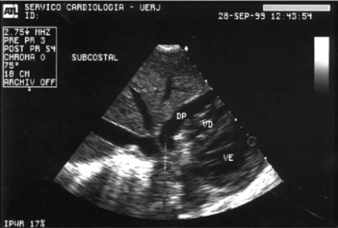
We were able to perform echocardiography during
and immediately after the procedure in 3 patients (fig. 1).
The analysis of the pericardial fluid revealed a hemorrhagic
aspect in 2 patients and a serosanguineous aspect in the
other 2. In all patients LE cells were present, the search for
ANF was positive (levels ranging from 1/32 to 1/128), and
cultures for microorganisms were negative. Later, other
causes of pericardial effusion, such as hypothyroidism,
uremia, and tuberculosis were discarded through laboratory
tests. Disease activity was confirmed in all patients.
In addition to pericardicentesis, the treatment included
high doses of corticosteroids (2-3 mg/kg of prednisone or
equivalent) with control of disease activity. The patients
were followed up with serial echocardiographic studies
during a period from 9 to 60 months (mean = 27 months). We
detected neither recrudesce of pericardial effusion nor
evolution to the constrictive form.
Discussion
Even though pericarditis in systemic lupus
erythemato-sus has already been well described in regard to its clinical
1-9,
anatomicopathological
10-14and echocardiographic
charac-teristics
30-36, occurrence of cardiac tamponade is rare, with
12 cases reported until 1986
37. To establish the differential
diagnosis with other etiologies, it is important to know the
characteristics of the fluid, which may have a hemorrhagic
aspect, low levels of complement, high levels of ANF, and
the presence of LE cells
38, 39.
Several isolated cases have been reported
40-44, and in
some of these cardiac tamponade was the initial
manifesta-tion of the rheumatic disease. All our patients had a
pre-vious diagnosis of systemic lupus erythematosus, which
certainly made the clinical diagnosis easier. An isolated case
of cardiac tamponade with associated polyarteritis has been
reported, in which the patient died because of the clinical
severity of both diseases
45. In our experience, even though
the initial findings indicated great instability, we had a very
good response to high doses of corticosteroids initiated
af-ter pericardial effusion withdrawal. These patients were
fol-lowed up during a period from 9 to 60 months, and neither
new pericardial effusion nor evolution to constriction was
detected with echocardiography.
Fig. 1 – Echocardiogram performed immediately after pericardicentesis with immediate relief of symptoms showing residual pericardial effusion
References
1. Libman E, Sacks B. A hithertho undescribed form of valvular and mural endocarditis. Arch Intern Med 1924; 2433: 701-37.
2. Dubois EL, Tuffanelli DL. Clinical manifestations of systemic lupus erythema-tosus. Computer analysis of 520 cases. JAMA 1964; 190: 104-11.
3. Jessar RA, Lamont-Havers RW, Ragan C. Natural history of lupus erythe ma-tosus disseminatus. Ann Intern Med 1953, 38: 717-31.
4. Harvey AM, Shulman LA, Tumulty PA. Systemic lupus erythematosus. Review of the literature and clinical analysis in 138 cases. Medicine 1959; 58: 291-437. 5. Shearn MA. The heart in systemic lupus erythematosus. Am Heart J 1959; 58:
452-66.
6. Mandell BF. Cardiovascular involvement in systemic lupus erythematosus. Semin Arthritis Rheum 1987; 17: 126-41.
7. Ansari A, Larson PH, Bates HD. Cardiovascular manifestations of systemic lupus erythematosus; current perspective. Prog Cardiovasc Dis 1985; 27; 421-34. 8. Carette S. Cardiopulmonary manifestations in systemic lupus erythematosus.
Rheum Dis Clin North Am 1988; 14; 135-47.
9. Bridgen W, Baywater GL, Lessof MH. The heart in systemic lupus erythe matosus. Br Heart J 1960; 22: 1-16.
4 4 8
Castier et al
Cardiac tamponade in systemic lupus erythematosus
Arq Bras Cardiol
volume 75, (nº 5), 2000
11. Griffith GC, Vural IL. Acute and subacute disseminated lupus erythematosus. A correlation of clinical and postmortem findings in eighteen cases. Circulation 1951; 3: 492-500.
12. Humpheys EM. The cardiac lesion of acute disseminated lupus. Ann Intern Med 1948; 28:12-4.
13. Kong TQ, Kellum RE, Haserick JR. Clinical diagnosis of cardiac involve ment in systemic lupus erythematosus. A correlation of clinical and authopsy findings in thirty patients. Circulation 1962; 26: 7-11.
14. Copeland GD, Van Capeller D, Stern TN. Systemic lupus erythema tosus: Clinical report of forty seven cases with pathological findings in eighteen. Am J Med Sci 1958; 236: 318-26.
15. Hetjmancik MR, Wright JC, Quint R, Jenning FL. The cardiovascular manifesta-tions of systemic lupus erythematosus. Am Heart J 1964; 68: 119-30. 16. Estes D, Chistian CL. The natural history of systemic lupus erythematosus.
Medicine 1971; 50: 85-95.
17. Doherty NE, Siegel RJ. Cardiovascular manisfestations of systemic lupus erythematosus. Am Heart J 1985; 110: 1257-65.
18. Reiner JS, Furie RA. Cardiac tamponade as an initial manifestation of systemic lupus erythematosus. J Rheumatol 1989; 16: 1127-9.
19. Lee IH, Yang SC, Kim TH, et al. Cardiac tamponade as an initial manifestation of systemic lupus erythematosus—single case report. J Korean Med Sci 1997; 12: 75-7. 20. Nour-Eddine M, Bennis A, Soulami S, Chraibi N. Cardiac tamponade
disclo-sing systemic lupus erythematosus. Ann Cardiol Angeiol 1996; 45: 71-3. 21. Tan EM, Cohen AS, Fries JF, et al. The 1982 revised criteria for the classification of
systemic lupus erythematosus. Arthritis Rheum 1982; 25: 1271-7. 22. Bombardier C, Gladman DD, Urowitz MB, Caoron D, Chang C. Derivation of the
SLEDAI. A disease activity index for lupus patients. Arthritis Rheum 1992; 35: 630-40.
23. Sahn DJ, De Maria A, Kisslo J, Weyman AE. Recommendations regarding quantitation in M-mode echocardiography: Results of a survey of echocar-diographic measurements. Circulation 1978; 58: 1072-83.
24. Horowitz MS, Schultz CS, Stirson EB, Harrison DC, Popp RL. Sensitivity and specificity of echocardiographic diagnosis of pericardial effusion. Circulation 1974; 50: 239-47.
25. D’Cruz IA, Cohen HC, Prabhu R. Diagnosis of cardiac tamponade by echocar-diography. Changes in mitral valve motion and ventricular dimensions with special reference to paradoxical pulse. Circulation 1975; 52: 460-5. 26. Schiller NB, Botvinick EH. Right ventricular compression as a sign of cardiac
tamponade. An analysis of echocardiographic ventricular dimensions and theur clinical implication. Circulation 1977; 56: 774-9.
27. Gillam LD, Guyer D, Gibson TC, King ME, Marshall JE, Weyman AE. Hydro-dynamic compression of the right atrial free wall, a knew highly sensitive echocardiographic sign of cardiac tamponade. Circulation 1983; 68: 294-301. 28. Armstrong WF, Schildt B, Helper DJ. Diastolic collapse of the right ventricle
with cardiac tamponade: an echocardiographic study. Circulation 1982; 65: 1491-6.
29. Appleton CP, Hatle LK, Popp RL. Cardiac tamponade and pericardial effusion: respiratory variation in transvalvular flow velocities studied by Doppler echocardiography. J Am Coll Cardiol 1988; 11: 575-9.
30. Li EK, Crozier IG, Milne MJ, Nocholls MG. Cardiac involvement in systemic lupus erythematosus. Arthritis Rheum 1989; 32: 116-6.
31. Chia BL, Mah EP, Feng PH. Cardiovascular abnormalities in systemic lupus erythematosus. J Clin Ultrasound 1981; 9: 237-43.
32. Maniscalco BS, Flener JM, McCans JL, Chiapella JA. Echocardiographic abnormalities in systemic lupus erythematosus. Circulation 1975; 52(supl.2): 211.
33. Ito M, Kugyama Y, Omura I. Cardiovascular manifestations of systemic lupus erythematosus. Jpn Circ J 1979; 43: 985-94.
34. Crozier IG, Li EK Milne MJ, Nicholls MG. Cardiac involvement in systemic lupus erythematosus detected by echocardiography. Am J Cardiol 1990; 65: 1145-8.
35. Doherty NE, Feldman G, Mauner G, Siegel RJ. Echocardiographic findings in systemic lupus erythematosus. Am J Cardiol 1988; 61: 1144.
36. Klinkoff AV, Thompson CR, Reid RD, Tomlinson CW. M-Mode and two-dimensional echocardiographic abnormalites in systemic lupus erythematosus, JAMA 1985; 252: 3273-7.
37. Rudra T, Evans PA, O’Brien EN. Systemic lupus erythematosus presenting with cardiac tamponade cardiac tamponade due to haemorragic effusion. Postgrad Med J 1987; 63: 567-8.
38. Hunder GC, Mullen BJ, McDuffie FC. Complement in pericardial fluid of lupus erythematosus. Ann Intern Med 1974; 80: 453-8.
39. Averbuch M, Bojko A, Levo Y. Cardiac tamponade in the early post partum period as the presenting and predominant manifestation of systemic lupus erythematosus. J Rheumatol 1986; 13: 444-5.
40. Zashin SJ, Lipsky PE. Pericardial tamponade complicating systemic lupus erythematosus. J Rheumatol 1989; 16: 374-7.
41. Carrol N, Barret JA. Systemic lupus erythematosus presenting with cardiac tamponade. Br Heart J 1984; 51: 542-3.
42. Ehrenfeld M, Asman A, Spilberg O, Samra Y. Cardiac tamponade as the presenting manifestation of systemic lupus erythematosus. Am J Med 1989; 86: 626-7. 43. Porcel JM, Selva A, Tornos MP, Galve E, Soler-Soler J. Resolution of cardiac
tamponade in systemic lupus erythematosus with indometacine. Chest 1989; 96: 1193-4.
44. Manresa JM, Gutierrez L, Viedma P, Alfani O. Cardiac tamponade as a clinical symptom of systemic lupus erythematosus. Rev Esp Cardiol 1997; 50: 600-2. 45. Costa AC, Mansur AJ, Assi VC, Grinberg M, Bellotti G, Pileggi F. Tamponamento