SOCIEDADE BRASILEIRA DE ORTOPEDIA E TRAUMATOLOGIA
w w w . r b o . o r g . b r
Original
Article
Radiographic
anatomy
of
the
proximal
femur:
femoral
neck
fracture
vs.
transtrochanteric
fracture
夽
Ana
Lecia
Carneiro
Leão
de
Araújo
Lima,
Saul
Caldas
Miranda,
Hudson
Felipe
Oliveira
de
Vasconcelos
∗HospitalOtáviodeFreitas,Recife,PE,Brazil
a
r
t
i
c
l
e
i
n
f
o
Articlehistory:
Received13June2016 Accepted4October2016 Availableonline18October2017
Keywords:
Hipfractures Femurneck Radiography
a
b
s
t
r
a
c
t
Objective:To evaluate thecorrelationbetween radiographicparameters oftheproximal femurwithfemoralneckfracturesortranstrochantericfractures.
Methods:Cervicodiaphysealangle(CDA),femoralneckwidth(FNW),hipaxislength(HAL), andacetabularteardropdistance(ATD)wereanalyzedin30pelvisanteroposteriorview X-raysofpatientswithfemoralneckfractures(n=15)andtranstrochantericfractures(n=15). The analysiswasperformedbycomparingtheresultsofthe X-rayswithfemoralneck fracturesandwithtranstrochantericfractures.
Results:Nostatisticallysignificantdifferencesbetweensampleswereobserved.
Conclusion: Therewasnocorrelationbetweenradiographicparametersevaluatedand spe-cificoccurrenceoffemoralneckfracturesortranstrochantericfractures.
©2017PublishedbyElsevierEditoraLtda.onbehalfofSociedadeBrasileiradeOrtopedia eTraumatologia.ThisisanopenaccessarticleundertheCCBY-NC-NDlicense(http:// creativecommons.org/licenses/by-nc-nd/4.0/).
Anatomia
radiográfica
do
fêmur
proximal:
fratura
de
colo
vs
.
fratura
transtrocantérica
Palavras-chave:
Fraturasdoquadril Colodofêmur Radiografia
r
e
s
u
m
o
Objetivo:Correlacionarparâmetrosradiográficosdofêmurproximalcomaocorrênciade fraturasdocolodofêmuroufraturastranstrocantéricasdofêmur.
Métodos:Foramavaliadosoângulocevicodiafisário(ACD),alarguradocolofemoral(LCF),o comprimentodoeixodoquadril(CEQ)eadistânciaentreaslágrimasacetabulares(DL)de radiografiasdebaciaemincidênciaanteroposteriorde30pacientescomfraturadecolo de fêmur(n=15)efraturatranstrocantéricadefêmur(n=15).Aavaliac¸ãofoi feitacom acomparac¸ãodospacientescomfraturadecolodefêmurcomospacientescomfratura transtrocantérica.
夽
StudyconductedatHospitalOtáviodeFreitas,Recife,PE,Brazil.
∗ Correspondingauthor.
E-mail:[email protected](H.F.Vasconcelos). http://dx.doi.org/10.1016/j.rboe.2017.10.007
Resultados: Não foram observadas diferenc¸as estatisticamente significantes entre as amostrasobtidasentreosdoisgruposcomparados.
Conclusão: Nãohouvecorrelac¸ãoentreosparâmetrosradiográficosavaliadoseocorrência específicadefraturasdecolodefêmuroufraturastranstrocantéricasdefêmur.
©2017PublicadoporElsevierEditoraLtda.emnomedeSociedadeBrasileirade OrtopediaeTraumatologia.Este ´eumartigoOpenAccesssobumalicenc¸aCCBY-NC-ND (http://creativecommons.org/licenses/by-nc-nd/4.0/).
Introduction
Advancesinmedicineandpharmacologyhaveledtoa sig-nificantincreaseingloballifeexpectancy,reflectedpositively inthegrowingnumberofelderlypeople.However,thereisa realconcernabout thequalityoflifeoftheseagingadults, andespecially,regardinghowtoadequatelypreventandtreat thecomplicationsinherenttothis agegroup.Amongthese complicationsare low-energyfractures,or thosethat are a consequenceofassociatedpathologicalcomplications.1–3
Hipfractureshaveseriousimpactonelderlypatients, espe-ciallytheveryelderly(over80years).3Thisissueisrelevant due to the high morbidity and mortality, high postopera-tivedisabilityindex,andincreasingcoststosocietywithless beneficialresultsrelatedtotreatment.4 These fracturesare consideredoneofthelargestpublichealthproblemsinthe world.4AccordingtoAmericanstatistics,over250,000hip frac-turesoccureachyear;itisexpectedthatoverthenext30years, therewillbeanincreaseof100%inthenumberofcases/year. InBrazil,in2010,theincidencewas100,000fracturesperyear, andthemeanmortalityoneyearafterthefracturewas30%. Femoralfractures,especiallyproximalfractures,are among themostrelevant.4
Adequatesurgicaltreatmentisparamountforgood prog-nosis; the method chosen is directly related to the type of hip fracture, specifically the types of femoral fractures (distal orproximal). Proximalfractures can bedivided into two types: intracapsular and extracapsular. The first type includesfracturesofthefemoralneck,andthesecondtype, transtrochantericfractures.Bothhavelow-energytraumaas themainetiology,andbothhavegreatinfluenceinassociated pathologies,suchasosteoporosis.5–7
Osteoporosis, undoubtedly the most common of bone diseases,hasbecomeaburdenofconsiderableeconomic sig-nificance.Factorssuchasethnicity,gender,physicalactivity, andnutritioninfluencethemaximumbonequalityachieved byeachindividual,butarenottheonlydeterminingfactors forfractures.Thespecializedliteratureemphasizesthatbone mineraldensity(BMD),anage-relatedpredictoroffracture,is notalwaysconsistent:individualswithverylowfemoralneck BMDmaynotpresentfracture,whilethosewithnormalBMD might.8Theremaybeotherrelevantvariablesthatdetermine fracturesandespeciallytheirtypes,suchasboneanatomy.8,9 Bonegeometryoftheproximalfemurhasbeenstudied10 asapotentialriskfactor,andhasbeenpositivelyassociated inthepredictionoffracturerisk.However,mosthipfracture studiesdonotdistinguishthepredispositionbetweenthetwo maintypesoffracture(femoralneckandtranstrochanteric), whichinclinical practicewouldbefundamental,sincethe
surgicalapproachofchoicecanbedifferentduetothehigh rateofhiparthroplastyindicationinfemoralneckfractures, whichinturnhasfinancialrepercussionsandaffectspatient recoveryinthepostoperativeperiod.
Thus, this study isaimed atanalyzing the influenceof proximalfemoralbonegeometryinthetypeoffemurfracture presented,bymeasuringstandardpelvicradiographs.
Material
and
methods
Thiswas aprospective,cross-sectional study performedin anorthopedicandtraumaserviceinBrazilbetweenAugust 10,2015andSeptember8,2015.Thestudyincluded30 radio-graphsofpatientswithhip fractures,randomlyselectedas caseswereadmitted.ThestudyfollowedtheDeclarationof HelsinkiandwasapprovedbytheinternalEthicsCommittee (No.1.221.094).
Radiographsweretakenintheanteroposteriorview,with the X-ray generator located one meter from the chassis. Patientswereplacedinahorizontaldorsalrecumbent posi-tion,withthelowerlimbsrotatedinternallyat15◦.
Theinclusioncriteriawerepanoramicradiographsofthe hip of patients aged over 60 years, of both genders, with femoralneckandtranstrochantericfractures.
Exclusioncriteriaincludedradiographsofskeletally imma-turepatients; bilateralhipfracture;and presenceoftumor, infectiouslesions,ormetabolicdiseasesthatcouldalterthe hipandproximalfemuranatomy.
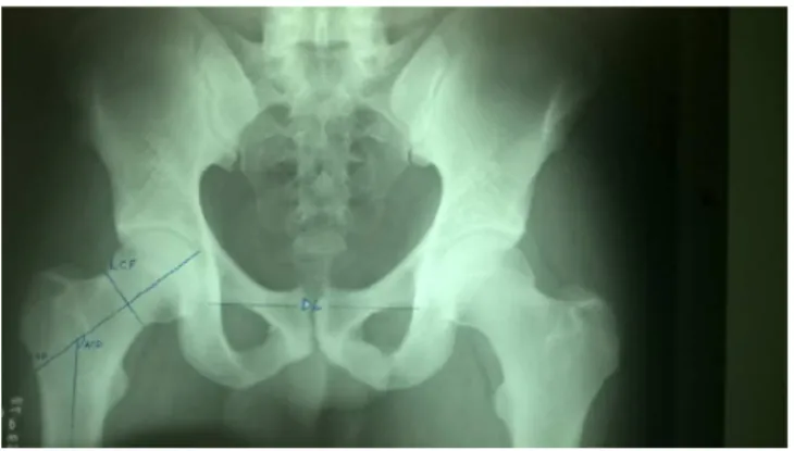
After classification and selection, the radiographs were anatomicallyevaluated,accordingtothefollowingmeasures:
• Cervicodiaphysealangle(CDA):anglebetweentheaxisof thefemoralneckandthediaphysis.
• Femoralneckwidth(FNW):distancebetweencorticallines, atthemidpointofthefemoralneck,perpendiculartoits axis.
• Hipaxislength(HAL):thedistanceinastraightlinebetween thebaseofthegreattrochantertotheendofthefemoral head,followingthelineoftheaxisofthefemoralneck. • Acetabular tear drop distance (ATD): the distance in a
straightlinebetweentheacetabularteardrops.
Thechoiceofthesemeasurementindexeswasbasedon previous studies that conductedmorphometricanalysesof theproximalfemur.11Allmeasurementsweremadebytwo blindedexaminersusingagoniometer(MSD,Europe BVBA-Belgium).
Fig.1–Representationofanglesmeasuredinan anteroposteriorpelvicradiograph.
usecomputerprogramsformeasuring,astheprocessof scan-ningtheradiographscouldleadtounevenmagnificationofthe imagesandthusgeneratecalibrationbias,sincethesystem availableatthismedicalcenterisnotdigital(Fig.1).
The Kolmogorov–Smirnov test was used to assess the intrinsicparametersofthesampleregardingitsnormalityand distribution.Datawereexpressedasmean,standard devia-tion,andpercentage(SPSSStatisticalSoftware).
The variables were analyzed descriptively through the mean,standarddeviation,minimumandmaximumvalues, and 95% confidenceintervals. Student’st-testwas used to comparethedifferencebetweenthemeansoftwovariables, andPearson’scorrelationcoefficientwasusedtoassessthe correlationindex.Thelevelofsignificancewassetat5% (ROS-NER,B.FundamentalsofBiostatistics.Boston,PWSPublishers, 2nded.)
Results
Thestudy included 30 patients, male (n=6;mean age=76, SD=3.48)andfemale(n=24;meanage=77.37,SD=8.53),that were divided into two large groups offractures with their respectiveanatomicevaluations,asshowninTable1.
Parametricevaluationofthecollecteddata
Inordertoestablishreliableindexesinthecomparisons,the normalityofthesampleswasfirstdeterminedaccordingtothe
Table1–Characterizationofthegroupsaccordingtothe evaluatedangles.
Angles Fractures
Transtrochanteric Neck
CDA FNW HAL ATD CDA FNW HAL ATD
Max 139 42 126 135 139 39 132 134
Min 125 30 99 110 120 29 90 114
M 131.7 34.7 110.2 125.1 131.8 33.2 112.6 122.1 SD 1.2 0.98 2.22 1.96 1.33 0.65 3.08 1.36
n 15 15 15 15 15 15 15 15
Max,maximum;Min,minimum;M,mean;SD,standarddeviation; n,numberofcases.
Table2–Pearson’scorrelationtesttode-characterizethe correlationbetweenage,gender,andmeasurements.
Measurements CDA FNW HAL ATD
Age Pearson’scorrelation 0.124 0.049 0.159 0.094 p-value 0.392 0.735 0.116 0.318 Gender Pearson’scorrelation −0.094−0.064−0.225−0.144 p-value 0.387 0.516 0.657 0.318
Total n 50 50 50 50
Table3–Numericalrepresentationofthecomparison data(unpairedt-test)betweentypesoffracturesforeach pairofanglesstudied.
Pairs n MD 95%CI R p
CDATrans×Neck 30 0.13±1.7 −3.5to3.8 0.0001 0.69 FNWTrans×Neck 30 −1.46±1.18 −3.89to0.95 0.05 0.14 HALTrans×Neck 30 2.4±3.8 −5.39to10.19 0.01 0.53 ATDTrans×Neck 30−3±2.3 −7.9to1.9 0.05 0.22
MD,meandeviation;n,numberofpatients;p,statisticalvaluefor significanceofcorrelation;r,Pearsoncorrelationindex.
150
140
130
120
110
CDA trans
Degree
CDA neck
Fig.2–Representationofthestatisticalrelationshipofthe pairedt-testbetweenCDAintranstrochantericfractures
andCDAinfemoralneckfractures.
Kolmogorov–Smirnovtest,i.e.,itwasdeterminedwhethertwo underlyingprobabilitydistributionswoulddifferinrelationto thenormalityhypothesis,inanyoneofthecases.The normal-ityhypothesiswasnotrejectedforthevariablesinvestigated withp>5%.
Then,tocharacterizepossibleinterferencebiasesbetween theanglesmeasuredbytheobserversofthestudyregarding genderandage,Pearson’scorrelationtestwasused.No posi-tiveassociationwasobservedbetweenthevariables,asshown inTable2.
Afterdeterminingthenormalityofthesampledistribution and ruling out interferencebias, the measurements made by the observers were compared to assess the difference betweenthetypesoffractures,representedbythemeasured angles,andwhetherthesevalueswerecorrelated.Although there were differences between the mean angles (around 3◦–7◦),Student’st-testindicatedthattheywerenotsignificant
45
40
35
30
25
FNW trans
Degree
FNW neck
Fig.3–Representationofthestatisticalrelationshipof pairedt-testbetweenFNWintranstrochantericfractures andFNWinfemoralneckfractures.
140
100 120
80
HAL trans
Degree
HAL neck
Fig.4–Representationofthestatisticalrelationshipofthe pairedt-testbetweenHALintranstrochantericfractures andHALinfemoralneckfractures.
150
140
130
120
110
100
ATD trans
Degree
ATD neck
Fig.5–Representationofthestatisticalrelationshipofthe pairedt-testbetweenDLintranstrochantericfracturesand DLinfemoralneckfractures.
Inordertoestablishacorrelation betweenthevariables measured according to the type of fracture, Pearson’s cor-relationtestwasapplied,whichshowednegativityandlow correlationindexes,allofwhichwerenon-significant(Table4, Figs.6–9).
Table4–Numericaldescriptionofthevaluesassigned tothePearsoncorrelationpairsbetweenfemoralneck andtranstrochantericfractures.
Pairs n r 95%CI R p
CDATrans×Neck 30 0.38 −0.17to0.6 0.15 0.15 FNWTrans×Neck 30 0.394 −0.16to0.74 0.145 0.14 HALTrans×Neck 30 0.04 −0.47to0.54 0.002 0.43 ATDTrans×Neck 30 −0.06 −0.55to0.46 0.003 0.82
n,numberofpatients;p,statisticalvalueforsignificanceof corre-lation;r,Pearsoncorrelationindex.
150
150 140
140 130
130
CDA trans
CDA trans
CDA neck
CDA neck
120
120 110
110
Fig.6–Representationofthenegativecorrelationbetween CDAintranstrochantericfracturesandCDAinfemoralneck fractures.
45
45 40
40 35
35
FNW trans
FNW trans FNW neck
FNW neck
30
30 25
25
Fig.7–Graphicrepresentationofthenegativecorrelation
betweenFNWintranstrochantericandFNWfracturesin
femoralneckfractures.
Studylimitations
Ahigherandmorerepresentativesamplingofthepopulation affectedbyhipfractures,astudyindifferentgroupswith asso-ciatedpathologies,andtheadditionofahealthycontrolgroup wouldbenecessary.
Discussion
Inthepresentstudy,itwasdemonstratedthat,although
radio-graphy is a good method to evaluate bone structures and
140
140 120
130 120
HAL trans
HAL trans HAL neck
HAL neck
100
110 100 80
90
Fig.8–Graphicrepresentationofthenegativecorrelation betweenHALintranstrochantericfracturesandHALin femoralneckfractures.
150
150 140
140 130
130
ATD trans
ATD trans
ATD neck
ATD neck 120
120 110
110 100
100
Fig.9–Graphicalrepresentationofthenegativecorrelation betweenATDintranstrochantericandATDfracturesin femoralneckfractures.
fractureswhencomparedwithahealthy controlgroup.No significant geometricdifference was observed betweenthe groupsstudied.
Theincreasedriskofbonefracturesduetolossofbone massduringadiseaseoragingprocessisamajorclinical prob-lem,whichleadstoanestimateofhealthcostsofaroundUS$ 17billionintheUnitedStatesalone.12,13Inadditiontothe eco-nomicburden,non-vertebralfractures,especiallythoseofthe hip,areanimportantcauseofmorbidityandmortalityinthe agingpopulation.14,15Over4%ofpatientswithpelvicfracture dieduringhospitalization,and24%diewithinayear.16Thus, concentratedeffortsareneededtoidentifytreatment strate-giesthatmaintainskeletalhealthaspatientsage.However,it isofparamountimportancetoimproveaccuracyinidentifying thoseatriskforbonefractures.
BMD measurements are widely used to assess bone mineralstatus, especially in women; theycan account for upto70%ofbonestrength.Althoughstudies have demon-stratedthecorrelationbetweenBMD(commonlydetermined through dual-emission X-ray absorptiometry [DXA]) and fracture risk, predictive models based on DXA alone often presentlowsensitivityinidentifyingindividualssusceptible
tofractures,particularlyinwomenofmenopauseageandin olderpopulations.2,17
Thestructuralintegrityofthis tissueinany mechanical loadingenvironmentisdependentonthespatialdistribution ofBMD, size, and shape,as wellas the propertiesofbone material.18,19
Inliterature,severalstudieshavedemonstratedtheclinical potentialofbonetextureanalysisthroughpelvicradiographs in predicting the risk of femoral neck fractures. In a ret-rospective study, Thevenot et al.10 observed a high intra-and interobserver reproducibility and reliability, and con-cluded that the structural analysis of pelvic radiographs allowstheidentificationofpatientswithriskoffemoralneck fractures. This finding corroborates with studies described in theliterature,which suggest that thetrabecular texture parameters,especiallytheentropicparameter,allowthe sep-aration of individuals at risk from the control individuals; however, no parameter suggests theability todifferentiate among the types of injury, as was studied in the present study.20–22
Oneofthegreatfoesinboneinjuryisosteoporosis, the mostcommonbonedisease.Ithasbecomeaburdenof con-siderable economic significance. Factors such as ethnicity, gender, physicalactivity,and nutritioninfluencethe maxi-mumbonemassqualityachievedbyeachindividual.However, bonemassalone isnotadeterminingfactor.3,4 A studyby Cummengs et al.22 foundthat Japanese womenhad lower BMDthantheirCaucasianpeers;however,theformersuffered fewerfractures.Likewise,age and BMImaynot bedirectly relatedtolossinbonemass.23
Wheeleretal.,8intheirstudyofthecross-sectional geom-etry oflong bonediaphysesthat correlatedBMD, BMI, and age,demonstratedthatbonestrengthissignificantlyhigher inobeseindividualswhencomparedwiththosewithnormal BMI. However,thejoint dimensionsdonotdiffer apprecia-bly; older individuals with a higher BMIare less likely to develop a fracture than younger individuals with normal BMI.
In attempting to establish a risk assessment based on DXA,amultifactortoolwasdevelopedtodeterminehip frac-turepropensity,amethodrecommendedbytheWorldHealth Organization. Thistooltakes into accountdifferent factors (anthropometricvariables,medicalhistory,anddruguse)to evaluatetheten-yearriskoffracture,usingclinicalrisk fac-torswithorwithoutBMDvalues.24Nonetheless,thismethod still has low sensitivity for fracture prediction, since it is improvedinagenericway,andcannotreflectthecomplexity ofthepersonalizedevaluationofindividualsand/orspecific populations.25,26
Differentimagingmethodssuch,asperipheral quantita-tivecomputedtomographyandmagneticresonanceimaging (MRI), can be used to obtain three-dimensional geometry and bonearchitecture in vivo. These methodsmay provide some relevant information in assessing bone quality.27 However, the limited availability and high cost of these methodshasledtothedevelopmentofothertypesof low-cost analyses that may be clinically applicable, such as radiographs.
evaluation ofthe geometry, the structure, and, eventually, the risk of bone fracture. Nonetheless, new prospective studies with geometric measurements are still needed to confirmthe clinical capability ofthebone texture analysis throughthistool,aswellasthepossibilityofpredictingand defining risk groups for specific types of hip fractures, especially transtrochanteric and those of the femoral head.
Conclusion
Inthepresentstudy,itwasdemonstratedthatalthough radio-graphy is a good method to evaluatebone structures and predicthipfractures,itwasnotsensitiveenoughtocapture differencesbetweenfemoralneckandtranstrochanteric frac-tureswhencomparedwithahealthycontrolgroup.Further prospectivestudiesareneededtoestablishparameters capa-bleofmeasuringsuchdifferences.
Conflicts
of
interest
Theauthorsdeclarenoconflictsofinterest.
r
e
f
e
r
e
n
c
e
s
1.JohnellO,KanisJA,OdénA,SernboI,Redlund-Johnell
I,PettersonC,etal.Mortalityafterosteoporotic fractures.OsteoporosInt.2004;15(1):38–42.
2.KanisJA.Diagnosisofosteoporosisandassessment
offracturerisk.Lancet.2002;359(9321):1929–36. 3.SchuitSC,vanderKliftM,WeelAE,deLaetCE,Burger
H,SeemanE,etal.Fractureincidenceandassociation
withbonemineraldensityinelderlymenand
women:theRotterdamStudy.Bone.
2004;34(1):195–202.
4.LouresFB,ChaoubahA,OliveiraVM,AlmeidaAM,
CamposEM,PaivaEP.Economicanalysisofsurgical
treatmentofhipfractureinolderadults.RevSaúde
Pública.2015;49:12.
5.DaniachiD,SantosNettoA,OnoNK,GuimarãesRP,
PoleselloGC,HondaEK.Epidemiologyoffracturesof
theproximalthirdofthefemurinelderlypatients. RevBrasOrtop.2015;50(4):371–7.
6.FormosaMM,Xuereb-AnastasiA.Biochemical
predictorsoflowbonemineraldensityandfracture
susceptibilityinmaltesepostmenopausalwomen.
CalcifTissueInt.2016;98(1):28–41.
7.PalmH,TeixidorJ.Proximalfemoralfractures:canwe
improvefurthersurgicaltreatmentpathways?Injury.
2015;46Suppl.5:S47–51.
8.WheelerRL,HamptonAD,LangleyNR.Theeffectsof
bodymassindexandageoncross-sectional
propertiesofthefemoralneck.ClinAnat.
2015;28(8):1048–57.
9.FritzJ,CösterME,NilssonJÅ,RosengrenBE,Dencker
M,KarlssonMK.Theassociationsofphysicalactivity
withfracturerisk-a7-yearprospectivecontrolled
interventionstudyin3534children.OsteoporosInt.
2016;27(3):915–22.
10.ThevenotJ,HirvasniemiJ,PulkkinenP,MäättäM,
KorpelainenR,SaarakkalaS,etal.Assessmentofrisk
offemoralneckfracturewithradiographictexture
parameters:aretrospectivestudy.Radiology.
2014;272(1):184–91.
11.PiresRE,PrataEF,GibramAV,SantosLE,Lourenc¸oPR,
BellotiJC.Radiographicanatomyoftheproximal
femur:correlationwiththeoccurrenceoffractures.
ActaOrtopBras.2012;20(2):79–83.
12.BurgeR,Dawson-HughesB,SolomonDH,WongJB,
KingA,TostesonA.Incidenceandeconomicburden
ofosteoporosis-relatedfracturesintheUnitedStates,
2005–2025.JBoneMinerRes.2007;22(3):465–75.
13.KanisJA,JohnellO,OdenA,SemboI,Redlund-Johnell
I,DawsonA,etal.Long-termriskofosteoporotic
fractureinMalmö.OsteoporosInt.2000;11(8):669–74.
14.ZhouZ,RedaelliA,JohnellO,WillkeRJ,MassiminiG. Aretrospectiveanalysisofhealthcarecostsforbone
fracturesinwomenwithearly-stagebreast
carcinoma.Cancer.2004;100(3):507–17.
15.KayanK,KanisJ,McCloskeyE.Osteoporosis
managementbygeriatriciansintheUK.AgeAgeing.
2003;32(5):553.
16.KhoslaS,MeltonLJ3rd,DekutoskiMB,AchenbachSJ,
ObergAL,RiggsBL.Incidenceofchildhooddistal
forearmfracturesover30years:apopulation-based
study.JAMA.2003;290(11):1479–85.
17.KanisJA,BlackD,CooperC,DargentP,
Dawson-HughesB,DeLaetC,etal.Anewapproachto
thedevelopmentofassessmentguidelinesfor
osteoporosis.OsteoporosInt.2002;13(7):527–36.
18.JepsenKJ,HuB,TommasiniSM,CourtlandHW,Price
C,TerranovaCJ,etal.Geneticrandomizationreveals
functionalrelationshipsamongmorphologicand
tissue-qualitytraitsthatcontributetobonestrength
andfragility.MammGenome.2007;18(6–7):492–507.
19.TommasiniSM,NasserP,HuB,JepsenKJ.Biological
co-adaptationofmorphologicalandcomposition
traitscontributestomechanicalfunctionalityand
skeletalfragility.JBoneMinerRes.2008;23(2):236–46.
20.ChappardD,BasléMF,LegrandE,AudranM.
Trabecularbonemicroarchitecture:areview.
Morphologie.2008;92(299):162–70.
21.PulkkinenP,SaarakkalaS,NieminenMT,JämsäT.
Standardradiography:untappedpotentialinthe
assessmentofosteoporoticfracturerisk.EurRadiol. 2013;23(5):1375–82.
22.CummingsSR,CauleyJA,PalermoL,RossPD,
WasnichRD,BlackD,etal.Racialdifferencesinhip axislengthsmightexplainracialdifferencesinrates ofhipfracture.StudyofOsteoporoticFractures
ResearchGroup.OsteoporosInt.1994;4(4):226–9.
23.KanisJA,JohnellO,OdenA,JohanssonH,McCloskey
E.FRAXandtheassessmentoffractureprobabilityin
menandwomenfromtheUK.OsteoporosInt.
2008;19(4):385–97.
24.TrémollieresFA,PouillèsJM,DrewniakN,LaparraJ,
RibotCA,Dargent-MolinaP.Fractureriskprediction
usingBMDandclinicalriskfactorsinearly
postmenopausalwomen:sensitivityoftheWHO
FRAXtool.JBoneMinerRes.2010;25(5):1002–9.
25.KorthoewerD,ChandranM,EndocrineandMetabolic
SocietyofSingapore.Osteoporosismanagementand
careprofessionalsoftheAsia-Pacific.Arch
Osteoporos.2012;7:193–200.
26.ChappardD,GuggenbuhlP,LegrandE,BasléMF,
AudranM.TextureanalysisofX-rayradiographsis
correlatedwithbonehistomorphometry.JBoneMiner
Metab.2005;23(1):24–9.
27.HansD,GoertzenAL,KriegMA,LeslieWD.Bone
microarchitectureassessedbyTBSpredicts
osteoporoticfracturesindependentofbonedensity:
theManitobastudy.JBoneMinerRes.