SHORT COMMUNICATION
Analyses of the 16S-23S intergenic region of the phytoplasma
causing the sugarcane white leaf disease in Yunnan Province,
China
Xiao-Yan Wang1, Wen-Feng Li1, Ying-Kun Huang1*, Rong-Yue Zhang1, Zhi-Ming Luo1, Hong-Li Shan1, Jiong Yin1, Ke Shen1, Ying-Ming Jia2 & Zhi-Gang Bai2
1Sugarcane Research Institute, Yunnan Academy of Agricultural Science, Yunnan Key Laboratory of Sugarcane Genetic Improvement, Kaiyuan 661699, P. R. China; 2Baoshan Sugarcane Research Institute, Tongren Street No. 114, Longyang District, Baoshan 678000, P. R. China
Author for correspondence: Ying-Kun Huang, e-mail: [email protected]
ABSTRACT
Sugarcane white leaf (SCWL) is an important disease caused by a phytoplasma. In Baoshan, Yunnan, China, SCWL was firstly observed in 2012, and has extended its area of occurrence to 600 hm2 . Up to 52% of the plants may become diseased in a field and even
complete loss of cane yield may result in the heavily infected fields, posing a serious threat to Yunnan sugar industry. To ascertain the causal agent of suspected SCWL disease in Yunnan, nested PCR using two sets of phytoplasma primer pairs (MOLX/MLOY and P1/ P2) was used to successfully amplify a genomic region of the 16S ribosomal DNA (16S rDNA) from 36 suspected SCWL samples. On the basis of sequencing, phylogenetic analysis and nucleotide alignments of 17 nested PCR products randomly selected from positive samples, identical fragments of 210bp in length were obtained that could be clustered into the 6Sr group XI (Rice Yellow Dwarf group) and shared 100% identity with the 16S-23S intergenic spacer region (ISR) of a member of this group, the SCWL phytoplasma (GenBank: HQ917068), and 99.52% with Sugarcane grassy shoot phytoplasma, of the same group. These results indicate that the SCWL disease in Baoshan is caused by a phytoplasma of the 6Sr group XI.
Key words:Saccharum, disease detection, etiology, nested PCR.
Sugarcane white leaf (SCWL) disease, caused by a phytoplasma, is one of the most important diseases of sugarcane (Nakashima et al., 1994; Wongkaew et al., 1997; Hanboonsong et al., 2002; Wongkaew & Fletcher, 2004; Hanboonsong et al., 2006). SCWL was first observed in Lumpang province in the northern part of Thailand in 1954 (Marcone, 2002), and was reported in Taiwan in 1958 (Ling, 1962). At present, SCWL is known from India, Sri Lanka, Laos, and Myanmar (Kumarasinghe & Jones, 2001; Rao et al., 2005; Thein et al., 2012). SCWL causes severe losses, reducing the number of millable canes in the planted crop, and the disease severity can increase in the ratoon crop. In Thailand, a 5%-35% disease incidence for SCWL has been recorded, resulting in losses to sugarcane farmers amounting to US$20 million every year (Hanboonsong et al., 2006). With 100% losses in the cultivar ‘Ragna’ in New Guinea, SCWL was responsible for considerable economic losses for the local sugar industry (Rao et al., 2005).
Phytoplasmas have been reported to be associated with two major sugarcane diseases, SCWL and SCGS. SCWL is characterised by leaf chlorosis and white striations. Diseased leaves are smaller and narrower than uninfected leaves, and have a soft texture. Plants with SCGS are dwarfed and develop witches’ broom-type symptoms by
producing a large number of tillers (Chona et al. 1960; Sarosh, 1986; Rishi & Chen, 1989). SCWL phytoplasma is naturally transmitted by the leafhopper Matsumuratettix hiroglyphicus (Matsumura) and Yamatotettix flavovittatus
(Hanboonsong et al., 2002; Hanboonsong et al., 2006), but the insect vector responsible for the natural spread of SCGS has not yet been identified
sensitivity of the PCR assay, researchers often use nested PCR (Andersen et al., 1998; Lee et al., 1994). A nested polymerase chain reaction (PCR)-based method was hence used for the confirmation of the etiology of this sugarcane disease.
In October of 2012, 36 sugarcane leaf samples from varieties PY3120, Yunzhe86-61, Yunzhe03-194, Yuetang86-368, and Yuetang93-159 with suspected SCWL symptoms were collected from Shidian and Longyang of Baoshan City, Yunnan, China.
Total DNA was extracted from 0.2 g of each leaf sample using the improved CTAB extraction procedure described by Harrison et al. (1994). A DNA extracted from an infected sugarcane individual (cultivar UT3) was used as a positive control (provided by the Yunnan Key Laboratory of Sugarcane Genetic Improvement, Kaiyuan, China). The nested-PCR product of this positive control was sequenced The result of a BLAST search showed that it shared 100% sequence identity with the sequence from the Thailand SCWL phytoplasma (GenBank accession HQ917068) and over 99% identity with isolates from India and Hawaii (GenBank accession DQ380345, JN223448). DNA extracted from phytoplasma-free healthy sugarcane plants was used as a negative control, and double distilled water was used as a blank control.
Nested PCR assays were carried out using primer pairs with sequences complementary to the 16S rDNA of the phytoplasmas (Hanboonsong et al., 2002). The first set consisted of primers MLOX (5’-GTTAGGTTAAGTCCTAAAACGAGC-3’) and MLOY (5’-GTGCCAAGGCATCCACTGTATGCC-3’), that amplify a 700 bp DNA fragment. The second set of primers P1 GTCGTAACAAGGTATCCCTACCGG-3’)/P2 (5’-GGTGGGCCTAAATGGACTTGAACC-3’) amplify a 210 bp fragment. The first PCR amplification was performed in a 20 μL reaction mixture containing 1 μL total DNA template, 2.5 μL 10× PCR buffer, 1 μL each primer (20 μmol/L), 0.5 μL MgCl2 (25 mmol/L), 1 μL dNTPs (10 mmol/L), 0.2 μL
Taq DNA polymerase (5 U/μL) and 12.8 μL ddH2O. The
reaction was heated to 94ºC for 5 min, followed by 25 cycles of denaturation for 1 min at 94ºC, annealing for 1 min at 55ºC, and extension for 1 min at 72ºC, with a final extension for 10 min at 72ºC. For nested PCR, 1 µL of the first PCR product (diluted 1:100 in sterile deionized water) was used as template in the same reaction mixture as in the first PCR except for the primers; the amplification conditions were 5 min at 94ºC followed by 35 cycles, each consisting of denaturation at 94ºC for 1 min, annealing at 62ºC for 1 min, and extension at 72ºC for 1 min, followed by extension for 10 min at 72ºC. The positive control, negative control and blank control were included. PCR products were analysed by electrophoresis through a 2% agarose gel in Tris-acetate-EDTA (TAE) buffer, stained with ethidium bromide and visualized with a UV transilluminator.
Seventeen nested PCR products were randomly selected from positive samples, excised from the gel and
purified using the TIANgel Midi Purification kit (TIANGEN, Beijing, China). DNA fragments were ligated into the plasmid vector pMD18-T (Takara, Dalian, China), and the recombinant plasmids were introduced into Escherichia coli
strain DH5ɑ (Sangon, Shanghai, China) by transformation. The six positive clones from each sample were selected and the inserts were sequenced at BGI Sequencing (Beijing) Co. Ltd. Next a BLAST search against the NCBI (http://www. ncbi.nlm.nih.gov) sequence databases, homology analysis was performed with DNAMAN, version 6.0. A phylogenetic tree was constructed using the neighbor-joining program as implemented in the genetic analysis software MEGA, version 4.0 (Tamura et al., 2007) with Acholeplasma brassicae (GenBank: AY974060) as the outgroup.
The suspected SCWL symptoms observed on sugarcane in Baoshan were similar to those described previously in sugarcane (Marcone, 2002; Wongkaew & Fletcher, 2004). Disease started with leaves showing pale streaks and mottling and progresssed towards the entire leaves turning white. Younger leaves became infected first, and diseased plants showed white leaves in the spindle area, with the older leaves remaining green. The infected plants showed stunting, proliferating tillers, shortened internodes, and thin stalks (Figure 1).
Nested PCR products of approximately 200 bp were amplified from DNA extracted from all 36 symptomatic SCWL tissue samples with primers MLOX/MLOY followed by P1/P2, but no amplification product was obtained from the negative control DNA isolated from a healthy sugarcane plant or from double distilled water control (Figure 2).
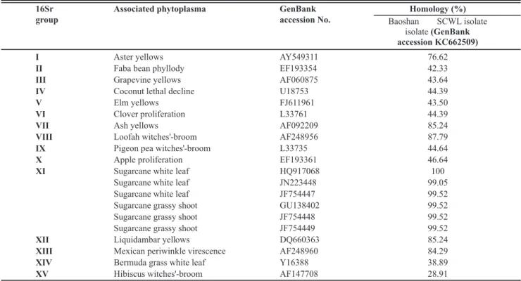
Seventeen nested PCR products were randomly selected from positive samples, cloned and six clones from each sample were sequenced. The results showed that the sequences were all 210-bp in length and were all identical. One of these was deposited in GenBank under accession number KC662509. A BLAST search indicated that the sequence obtained in the present study (GenBank accession KC662509) shared 100% nucleotide sequence identity with the 16S-23S intergenic spacer region (ISR) sequence from the SCWL phytoplasma from Thailand (GenBank accession HQ917068) and 99.52% with those of Sugarcane grassy shoot phytoplasma (Table 1).
A phylogenetic tree was constructed with 19 16S rDNA sequences from typical phytoplasmas of the 16Sr group. The resulting tree showed that the sequence identified in this study (GenBank accession KC662509) is closely related to that from the SCWL phytoplasma and also the sugarcane grassy shoot (SCGS) phytoplasma, which are members of the 16Sr XI group (Figure 3).
FIGURE 1 - Sugarcane infected
with Sugarcane white leaf disease (SCWL) in Banshan, Yunnan, China. A. Infected
leaves on plants at shooting stage. B. Infected plants at
mature stage.
FIGURE 2 - Nested PCR
amplification of phytoplasma 16S rDNA from plants suspected to be infected with SCWL collected from Baoshan,
Yunnan, China. M, DNA
Marker. 1-13, symptomatic
samples collected from the
SCWL-infected areas. PC,
positive control. NC, negative
control. CK, blank control.
observed sporadically in fields of Longyang in Baoshan, Yunnan, China in 2012. Confirmation of the etiology of diseases is critical for making effective management decisions to prevent the spread of SCWL and ensure the sustainable development of the sugar industry. In this study all 36 suspected SCWL samples were positive for the presence of the phytoplasm. Sequence comparison showed that the DNA sequence obtained in this study (GenBank
16Sr
group
Associated phytoplasma GenBank
accession No.
Homology (%)
Baoshan SCWL isolate
isolate(GenBank
accession KC662509)
I Aster yellows AY549311
II Faba bean phyllody EF193354
III Grapevine yellows AF060875
IV Coconut lethal decline U18753
V Elm yellows FJ611961
VI Clover proliferation L33761
VII Ash yellows AF092209
VIII Loofah witches'-broom AF248956
IX Pigeon pea witches'-broom L33735
X Apple proliferation EF193361
XI Sugarcane white leaf HQ917068
Sugarcane white leaf JN223448
Sugarcane white leaf JF754447
Sugarcane grassy shoot GU138402
Sugarcane grassy shoot JF754448
Sugarcane grassy shoot JF754449
XII Liquidambar yellows DQ660363
XIII Mexican periwinkle virescence AF248960
XIV Bermuda grass white leaf Y16388
XV Hibiscus witches'-broom AF147708
76.62
42.33
43.64
44.39
43.50
44.39
85.24
87.79
44.64
46.64
100
99.05
99.52
99.52
99.52
99.52
85.24
84.29
38.89
28.91
TABLE 1 - Comparison of 16S rDNA nucleotide sequences from Baoshan SCWL isolate with other phytoplasmas of the 16Sr groups.
FIGURE 3 - Phylogenetic distance tree constructed by the neighbor-joining method, comparing the 16S rDNA sequence of the present
study (sugarcane white leaf from Baoshan, Yunnan, China) with those of other representative phytoplasmas from GenBank. Acholeplasma brassicae was used as an outgroup species. Accession numbers are shown in parentheses. Numbers at branch nodes are bootstrap
The sequence comparison analysis performed here revealed that the 16S-23S ISR sequences from the phytoplasma identified in this study (GenBank accession KC662509) and the SCGS phytoplasma (GenBank: GU138402, JF754448, JF754449), which is also a member of the 16Sr XI group, shared 99.52% nucleotide identity, and there was no significant difference between SCWL phytoplasma and SCGS phytoplasma. The unequivocal identification at the species level this phytoplasma needs further in-depth studies.
Only part of the cane-growing areas in Baoshan City, Yunnan Province were monitored for SCWL in this study. This survey needs to be expanded. Also, in order to genereate a theoretical basis and technical support for the effective control of SCWL, further studies are needed to complete the experimental work for the detection of insect species which might be responsible for the transmission of SCWL in China, and a systematic and comprehensive study of the occurrence and field epidemic dynamics of SCWL in all cane-growing regions of China is also necessary.
ACKNOWLEDGEMENTS
This work was supported by grants from the Earmarked Fund for China Agriculture Research System (CARS-20-2-2) and the Earmarked Fund for Yunnan Province Agriculture Research System for project support.
REFERENCES
Andersen MT, Beever RE, Gilman AC, Liefting LW, Balmori E, Beck DL, Sutherland PW, Bryan GT, Gardner RC, Forster RLS (1998) Detection of Phormium yellow leaf phytoplasma in New Zealand flax (Phormium tenax) using nested PCRs. Plant
Pathology47:188-196.
Chona BL, Capoo SP, Varma PM, Seth ML (1960) Grassy shoot disease of sugarcane. Indian Phytopathology 13:37-47.
Hanboonsong Y, Choosai C, Panyim S, Damak S (2002) Transovarial transmission of sugarcane white leaf phytoplasma in the insect vector Matsumuratettix hiroglyphicus (Matsumura).
Insect Molecular Biology 11:97-103.
Hanboonsong, Y., Ritthison, W., Choosai, C., & Sirithorn, P. (2006). Transmission of sugarcane white leaf phytoplasma by Yamatotettix flavovittatus, a new leafhopper vector. Journal of Economic Entomology 99:1531-1537.
Harrison NA, Richardson PA, Jones P, Tymon AM, Eden-Green SJ., Mpunami AA (1994) Comparative investigation of MLOs associated with Caribbean and African Coconut lethal decline
diseases by DNA hybridization and PCR assays. Plant Disease 78:507-511.
Kumarasinghe NC, Jones P (2001) Identification of white leaf disease of sugarcane in Sri Lanka. Sugar Tech 3:55-58.
Lee I-M, Gundersen DE, Hammond RW, Davis RE (1994) Use of mycoplasmalike organism (MLO) group-specific oligonucleotide primers for nested-PCR assays to detect mixed-infections in a single host plant. Phytopathology84:559-566.
Ling KC (1962) White leaf disease of sugarcane. Taiwan Sugar 9:1-5.
Marcone C (2002) Phytoplasma disease of sugarcane. Sugar Tech 4:79-85.
Nakashima K, Chaleeprom W, Womgkaew P, Sirithorn P (1994) Detection of mycoplasma-like organisms associated with white leaf disease of sugarcane in Thailand using DNA probes. Japan International Research Center for Agricultural Sciences 1:57-67. Rao GP, Singh A, Singh HB, Sharma SR (2005) Phytoplasma diseases of sugarcane: characterization, diagnosis and management. Indian Journal of Plant Pathology 23:1-21.
Rishi N, Chen CT (1989) Grassy shoot and white leaf disease. In: Ricaus BC, Egan BT (Eds.) Diseases of sugarcane. Amsterdam, The Netherlands. Elsevier Science. pp. 289-300.
Sarosh, Misra SR, Kishan-Singh, Singh K (1986) Anatomical studies on sugarcane affected with grassy shoot disease. Indian Phytopathology 39:499-502.
Tamura K, Dudley J, Nei M, Kumar S (2007) MEGA4: Molecular Evolutionary Genetics Analysis (MEGA) software version 4.0. Molecular Biology and Evolution 24:1596-1599.
Thein MM, Jamjanya T, Kobori Y, Hanboonsong Y (2012) Dispersal of leafhoppers Matsumuratettix hiroglyphicus and
Yamatotettix flavovittatus (Homoptera: Cicadellidae), vectors of sugarcane white leaf disease. Applied Entomology and Zoology 47:255-262.
Viswanathan R (1997) Detection of phytoplasmas associated with grassy shoot disease of sugarcane by ELISA techniques. Journal of Plant Diseases and Protection 104:9-16.
Wongkaew P, Hanboonsong Y, Sirithorn P, Choosai C, Boonkrong S, Tinnangwattana T, Kitchareonpanya R, Damak S (1997) Differentitation of phytoplasmas associated with sugarcane and gramineous weed white leaf disease and sugarcane grassy shoot disease by RFLP and sequencing. Theoretical and Applied Genetics 95:660-663.
Wongkaew P, Fletcher J (2004) Sugarcane white leaf phytoplasma in tissue culture: long-term maintenance, transmission, and oxytetracycline remission. Plant Cell Reports 23:426-434. Zhou ZJ, Lin QY, Xie LH (1987) Sugarcane white leaf disease occurrence and observation of its pathogens by electron microscopic. Journal of Fujian Agricultural College 16:165-168.