Hypo tro phy o f co nduit arte ry walls o f
the o ffspring o f nitric o xide -de fe ctive
rats
Institute of Normal and Pathological Physiology, Slovak Academy of Sciences, Bratislava, Slovakia F. Kristek
and M. Gerová
Abstract
The objective of the present study was to investigate the structure of the arterial walls of the offspring stemming from nitric oxide (NO)-defective hypertensive parents. The parents were treated with NG -nitro-L-arginine methyl ester (40 mg kg-1 day-1) for 5 weeks. Blood pressure was measured noninvasively in six 30-day-old rats and nine age-matched controls. The cardiovascular system was perfused with glutaraldehyde at 120 mmHg. The thoracic aorta and carotid artery were processed for electron microscopy, and geometry was deter-mined by light microscopy. Endothelial cells, smooth muscle cells (SMC) and extracellular matrix (ECM) were determined by the point counting method in electron micrographs of the carotid artery. The blood pressure of experimental offspring was 150.0 ± 2.3 vs 104.6 ± 2.1 mmHg (P < 0.01) for the controls and their heart/body weight ratio of 3.9 ± 0.1 vs 4.4 ± 0.2 (P < 0.05) for the controls indicated cardiac hypotrophy. The wall thickness (tunica intima and media) of the thoracic aorta and carotid artery of experimental offspring was de-creased to 78.9% (P < 0.01) and 83.8% (P < 0.01), respectively, compared to controls, as confirmed by a respective cross-sectional area of 85.3% (P < 0.01) and 84.1% (P < 0.01). The wall thickness/ inner diameter ratio was reduced to 75% (P < 0.01) in the thoracic artery and to 81.5% (P < 0.01) in the carotid artery. No change in endothelial cell volume density or ECM was observed in the tunica intima of the carotid artery, and SMC volume density was lower in the tunica media (37.6 ± 0.9 vs 44.7 ± 1.1% for controls, P < 0.01), indicating compromised SMC development. Interference with argi-nine metabolism, a decrease in NO, and other factors are possible mechanisms underlying the structural alterations of the cardiovascular system of offspring from NO-defective hypertensive rats.
Co rre spo nde nce
F. Kristek
Institute of Normal and Pathological Physiology Slovak Academy of Sciences Sienkiewiczova 1 813 71 Bratislava Slovakia
Fax: + 421-2-5296-8516 E-mail: frantisek.kristek@ savba.sk
Research supported by VEGA (Nos. 2/7240/21, 2/7241/20) and Slovakofarma Joint Stock Company.
Received February 4, 2003 Accepted January 5, 2004
Ke y words
•Artery
•Nitric oxide
•O ffspring
•Hypertension
•Vascular smooth muscle
•Extracellular matrix
•Morphometry
Intro ductio n
Very early after the discovery of the en-dothelium-derived relaxing factor a series of arginine analogues were found to compete for the key enzyme nitric oxide (NO)
pres-sure (2-4). Adult animals were chosen for this novel experimental model of hyperten-sion and extensive alterations in the struc-ture of the cardiovascular system were found, i.e., cardiac hypertrophy, and increased wall thickness of conduit and resistance arteries (5-8).
In studies started in 1996 and continuing over the subsequent years, we addressed the ontogenetic aspect of the activity of NO synthase and the controlling role of NO in the vascular system (9). In experiments with isolated conduit vessels of canine fetuses and newborns, we detected a distinct re-sponse to NO synthase activators (acetyl-choline, bradykinin) which was even more marked compared with that of vessels from adult animals (10). Experiments with con-duit vessels of canine offspring who received an NO synthase inhibitor for 5 weeks after birth demonstrated only moderate alterations of smooth muscle relaxation, indicating that NO synthase was already fully operative during the early ontogenetic period (9).
We next addressed the characteristics of the cardiovascular system of offspring from parents with sustained NO-defective hyper-tension (11). The first experiments provided information on high blood pressure in these offspring. Unexpected findings, particularly with respect to high blood pressure, were a low heart weight and heart/body weight ra-tio. The next natural issue was to determine the structural characteristics of the arterial tree of these newborns, as justified by the fact that no relevant data were available.
Mate rial and Me thods
Adult Wistar-Kyoto rats and their male newborns were used for the study. All proce-dures followed the guidelines of the Guide for the Use of Laboratory Animals issued by the Ethics Committee for Experimental Work, Slovak Academy of Sciences, 1995. The animals (parents) were housed in individual cages on a 12-h dark-light cycle, with
con-stant temperature (22-24ºC) and free access to pellet food and water.
Pare nts
Experimental group. The parents
con-sisted of 5 females and 5 males aged ten weeks receiving the NO synthase inhibitor NG-nitro-L-arginine methyl ester (L-NAME)
at the dose of 40 mg kg-1 day-1 in drinking
water for a period of 5 weeks. Fertilization occurred during the fifth week of L-NAME administration. In females, L-NAME admin-istration continued during pregnancy and suckling up to the fourth week of offspring age.
Control group. The control group
con-sisted of 3 females and 3 males age matched to the experimental group and housed under the same conditions.
Blood pressure was measured noninva-sively in the tail artery weekly in both groups of parents, using the plethysmographic method.
O ffspring
Experimental group. This group consisted
of 6 newborns from 5 hypertensive NO-deficient parents fed by dams receiving L-NAME continuously.
Control group. The control group
con-sisted of 9 newborns from 3 control parents fed by control mothers.
The blood pressure of the offspring was measured noninvasively in the tail artery twice on the 23rd and 29th day using a miniaturized cuff.
On the 30th day, the newborn pups were sacrificed by an overdose of anesthetic (thio-pental, 100 mg/kg body weight, ip). The
ca-rotid artery and the middle part of the tho-racic aorta were excised, cleaned, divided into segments of about 1 mm in length and fixed in the same fixative overnight at 4º to 8ºC. After fixation, the segments were post-fixed with 40 mM OsO4 in 100 mM sodium
phosphate buffer, washed in 100 mM so-dium phosphate buffer, and stained en bloc
with uranyl acetate. The blocks were dehy-drated with increasing concentrations of al-cohol, washed in propylene oxide and em-bedded in Durcupan ACM.
Two randomly selected blocks of each artery from each offspring were cut perpen-dicularly to the longitudinal axis. The inner circumference and arterial wall thickness (tunica intima + tunica media) of individual vessels were measured on semithin sections under light microscopy. Arterial wall thick-ness was measured at about 45º intervals around the vessel circumference. The cross-sectional area (tunica intima + tunica media) of the arterial wall, the inner diameter, and wall thickness/inner diameter ratio of the vessels were then calculated from these data. The volume densities of cellular and ex-tracellular components of the tunica media and tunica intima of the carotid artery wall were estimated using the point counting method of Weibel et al. (12). Briefly, the grid was randomly placed on the respective sec-tion and 5000 points were counted. Secsec-tions from three randomly selected blocks from vessels of control and experimental animals were similarly processed.
Data are reported as means ± SEM. ANOVA and the Bonferroni test for un-paired variables were used to assess statisti-cal significance. Results were considered to be significant when P < 0.05.
Re sults
The blood pressure of the dams (102.8 ± 1.1 mmHg, N = 5) increased to 129.9 ± 4.3 mmHg (P < 0.001) after the first week of L-NAME administration and remained elevated
throughout the pregnancy and suckling pe-riod (146.8 ± 7.9 mmHg, P < 0.001 at the end of experiment). The blood pressure of males (107.4 ± 2.8 mmHg, N = 5) increased to 137.0 ± 3.2 mmHg (P < 0.001) after the first week of L-NAME administration and re-mained at this significantly increased value throughout the first week of pregnancy(154.8 ± 5.2 mmHg, P < 0.001).
All control newborns and 75.5% of the experimental newborns survived until the end of experiment. The blood pressure of offspring measured at the age of 3 and 4 weeks was 140.9 ± 4.6 (P < 0.001) and 150.0 ± 2.3 mmHg, N = 6 (P < 0.001), respectively, significantly higher than the blood pressure of age-matched controls, 94.6 ± 4.5 and 104.6 ± 2.1 mmHg, N = 9.
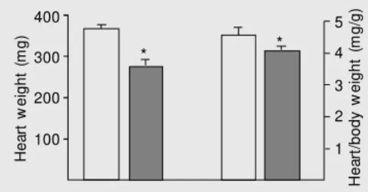
The body weight of the hypertensive off-spring, 68.7 ± 3.2 g, was significantly lower (P < 0.01) than the body weight of control offspring, 83.6 ± 2.7 g. The heart weight of hypertensive offspring, 276.8 ± 15.4 mg, was significantly lower (P < 0.05) than that of control offspring, 367.7 ± 11.4 mg. The heart/body weight ratio (mg/g) of the exper-imental offspring, 3.9 ± 0.1, was significant-ly lower (P < 0.05) than that of control offspring (4.4 ± 0.2; Figure 1).
The general parameters of the thoracic aorta and carotid artery are given in Table 1 and Figure 2. As shown in the figure, NO-defective offspring showed a decrease in wall thickness (tunica intima + tunica me-dia) of both thoracic aorta and carotid artery to 78.9 (P < 0.01) and 83.8% (P < 0.01), respectively, compared to controls. This was also confirmed by calculating the cross-sec-tional area of the arterial wall, which was
H
e
a
rt
w
e
ig
h
t
(m
g
)
400
300
200
100
H
e
a
rt
/b
o
d
y
w
e
ig
h
t
(m
g
/g
)
5
4
3
2
1
* *
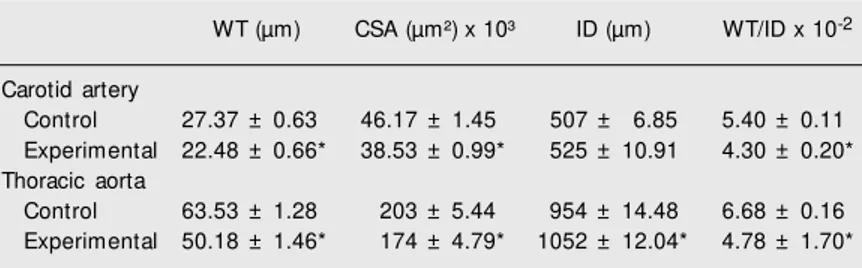
only 85.3% (P < 0.01) of the control thoracic aorta and 84.1% (P < 0.01) of the control carotid artery. The internal diameters were wider in the experimental animals, being 110.2% of control (P < 0.01) in the thoracic aorta and 102.6% (nonsignificant) in the carotid artery. The values of the wall thick-ness/inner diameter ratio were 75.0% (P < 0.01) in the thoracic aorta and 81.5% (P < 0.01) compared to the respective controls (Table 1).
Abnormalities of individual cellular and non-cellular components of the wall were determined for the carotid artery of NO-defective offspring and of control offspring (Figures 3 and 4). No alterations were found in volume density of the intima components, i.e., endothelial cells and respective extra-cellular matrix (Figure 3). Remarkable changes were detected in the tunica media, especially a significant decrease in smooth muscle cell volume density. A respective relative increase in extracellular matrix vol-ume density was found. In agreement, the calculated smooth muscle cell cross-sectional areas of the tunica media confirmed the im-paired development of the cellular compo-nent of the tunica media. No change in extra-cellular matrix cross-sectional area was de-tected (Figure 4).
D iscussio n
The blood pressure of offspring from NO-deficient hypertensive parents at the age of 3-4 weeks was significantly higher than in age-matched offspring from normotensive control rats. In contrast to the high blood pressure, the heart weight and heart-body weight ratio were inappropriately lower in hypertensive newborns than in controls. The main goal of this study was to determine the geometry and structure of the main conduit arteries.
The wall thickness (tunica media and tunica intima) of the aorta and carotid artery was lower in NO-defective offspring com-Table 1. Geometry of the carotid artery and thoracic aorta of offspring of nitric
oxide-defective rats.
WT (µm) CSA (µm²) x 10³ ID (µm) WT/ID x 10-2
Carotid artery
Control 27.37 ± 0.63 46.17 ± 1.45 507 ± 6.85 5.40 ± 0.11 Experimental 22.48 ± 0.66* 38.53 ± 0.99* 525 ± 10.91 4.30 ± 0.20* Thoracic aorta
Control 63.53 ± 1.28 203 ± 5.44 954 ± 14.48 6.68 ± 0.16 Experimental 50.18 ± 1.46* 174 ± 4.79* 1052 ± 12.04* 4.78 ± 1.70*
CSA = cross-sectional area (tunica intima plus tunica media); ID = inner diameter; WT = w all thickness (tunica intima plus tunica media).
* P < 0.01 compared to control (ANOVA and Bonferroni test).
W a ll th ic k n e s s ( µ m ) 80 60 40 20 0
Thoracic aorta Carotid artery
* *
Figure 2. Wall thickness of tho-racic aorta and carotid artery (tu-nica intima and tu(tu-nica media) of control (open columns) and NO-defective offspring (closed col-umns). * P < 0.01 compared to control (ANOVA and Bonferroni test). V o lu m e d e n s it y ( % ) 60 50 40 30 20 10 0
Tunica intima Tunica media
EC ECM 1 SM C ECM 2
* *
Figure 3. Volume density as per-cent of individual w all compo-nents of the tunica media and tunica intima of the carotid ar-tery. Tunica intima: endothelial cells (EC) and extracellular ma-t rix (ECM 1); ma-t unica m edia: smooth muscle cells (SM C) and ext racellular m at rix (ECM 2). Cont rol of f spring (open col-umns), NO-defective offspring (closed colum ns). * P < 0.01 compared to control (ANOVA and Bonferroni test).
C ro s s -s e c ti o n a l a re a ( µ m 2) x 1 0 3 24 20 16 12 8 4 0
Tunica intima Tunica media
EC ECM 1 SM C ECM 2
*
pared to controls. Since the cross-sectional area measured and calculated in both vessels was also significantly lower, the low wall thickness value was not due to high intravas-cular pressure, as one would expect from the increase in the inner diameter, and thus was not a passive consequence of the increase in blood pressure. It seems reasonable to sug-gest that an inhibition of growth processes in conduit vessel walls was a consequence of compromised arginine metabolism. With re-spect to the individual layers of the vessel wall, no change was found in volume density of endothelial cells or in the extracellular matrix of the tunica intima of the carotid artery. On the other hand, a significantly lower volume density of smooth muscle cells was found in the tunica media of the carotid artery from NO-defective offspring, support-ing the above suggestion. Since the reduced arterial wall thickness was demonstrated to be due to the low volume density of smooth muscle cells, it would be warranted to also consider the role of apoptotic processes in these cells. These considerations are justi-fied by the fact that in adult NO-defective rats apoptotic processes were found even in hypertrophic myocardium and in hyper-trophic resistance vessel walls (6,8,13,14). No data on this topic are available for new-born rats from NO-defective hypertensive parents and therefore there is a strong need for experimental proof of the above mechan-isms.
However, these findings contradict the respective wall thickness values repeatedly found in the conduit vessels of adult NO-defective hypertensive rats (5,7,15,16). In adult hypertensive NO-defective rats the wall thickness of conduit arteries increased sig-nificantly in terms of both extracellular and cellular components. Increased wall thick-ness in the arteries of adult animals was also repeatedly found in other experimental mod-els of hypertension, i.e., renal or spontane-ously hypertensive rats and others (17-20). To understand the unexpected finding of
hypotrophy of the wall of conduit arteries in NO-defective hypertensive newborns, and hypotrophy of the heart as well, the follow-ing considerations might be of help. Indeed, arginine, which is considered to be a semi-essential amino acid in adults, was demon-strated to be essential in newborns (21). Any interference with the balance of arginine metabolism might have consequences for the structure of the vessel walls and of the heart as well. Furthermore, NO was shown to be essential in cell differentiation (22). Thus, intervention in arginine metabolism by inhibitors of NO synthase causing a lower NO production might be the reason for hy-potrophy of the cardiovascular system.
When analyzing the factors involved in the decline of the wall thickness of the aorta and carotid artery it is necessary to consider two further points: since the mothers had long-term sustained hypertension due to NO deficiency, the blood supply for the fetus might be assumed to have been compro-mised. This would have contributed to low body weight, low heart weight, and low me-dia thickness of the aorta and carotid artery. Nevertheless, the low heart/body weight in-dex indicates a specific inhibition of growth processes of the cardiovascular system.
hardly be explained by the above analogy. Four-week-old offspring from L-NAME-treated NO-defective hypertensive parents had high blood pressure and decreased wall thickness of the thoracic aorta and carotid artery, together with low heart weight and heart/body weight ratio. They also had a significantly decreased wall thickness/inner diameter ratio in both conduit arteries stud-ied. Analysis of the rate of involvement of individual components of the vessel wall revealed hypotrophy of vascular smooth muscle in the tunica media with a relative increase in the extracellular matrix in the carotid artery. Several factors, namely the
interaction between arginine metabolism and compromised NO levels, suggest that a com-promised blood supply to the fetus, as well as other factors might be responsible for the hypotrophy of the cardiovascular system in the offspring studied.
Ackno wle dgm e nts
The authors are grateful to Marek Danay for valuable technical assistance, to Anna Buzalková for assistance with the statistical analysis, to Katarina Šoltésová for help with the preparation of the manuscript, and to I. Haná…ková for help with animal care.
Re fe re nce s
1. Rees DD, Palmer RM J, Schulz R, Hodson HF & M oncada S (1990). Characterization of three inhibitors of endothelial nitric oxide syn-thase in vitro and in vivo. British Journal of Pharmacology, 101: 746-752.
2. Baylis C, M itruka B & Deng A (1992). Chronic blockade of nitric oxide synthesis in the rat produces systemic hypertension and glomerular damage. Journal of Clinical Investigation, 90: 278-281. 3. Ribeiro M O, Antunes E, Nucci G, Lovisolo SM & Zatz R (1992).
Chronic inhibition of nitric oxide synthesis: a new model of arterial hypertension. Hypertension, 20: 298-303.
4. Dananberg J, Sider RS & Grekin RJ (1993). Sustained hypertension induced by orally administered nitro-L-arginine. Hypertension, 21: 359-363.
5. Kristek F & Gerová M (1996). Long-term NO synthase inhibition affects heart w eight and geometry of coronary and carotid artery. Physiological Research, 45: 361-367.
6. Pessanha M G & M andarim-de-Lacerda CA (2000). Influence of the chronic nitric oxide synthesis inhibition on cardiomyocyte number. Virchow s Archiv, 437: 667-674.
7. Delacretaz E, Hayoz D, Osterheld M C, Genton CY, Brunner HR & Waeber B (1994). Long-term nitric oxide synthase inhibition and distensibility of carotid artery in intact rats. Hypertension, 23: 967-970.
8. Sharifi AM & Schiffrin EL (1998). Apoptosis in vasculature of sponta-neously hypertensive rats. American Journal of Hypertension, 11: 1108-1116.
9. Török J & Gerová M (1996). Vascular responses after long term inhibition of nitric oxide synthesis in new born dogs. Physiological Research,45: 323-328.
10. Török J & Gerová M (1997). Developmental dynamics of endothelial and neurogenic control of canine thoracic aorta. M echanisms of Ageing and Development, 95: 143-152.
11. Gerová M , Bernátová I, Török J & Juráni M (2002). Cardiovascular system of offspring of hypertensive rats w ith defective nitric oxide production. Physiological Research, 51: 465-474.
12. Weibel ER, Kistler GS & Scherle WF (1966). Practical stereological methods for morphometric cytology. Journal of Cell Biology, 30: 23-38.
13. DeBlois D, Orlov SN & M hamet P (2001). Apoptosis in cardiovascu-lar remodeling - effect of medication. Cardiovascular Drugs and Therapy, 15: 539-545.
14. Chillon JM , Ghoneim S & Baumbach G (1997). Effects of chronic nitric oxide synthase inhibition on cerebral arterioles in rats. Hyper-tension, 30: 1097-1104.
15. Kristek F, Gerová M , Devát L & Varga I (1996). Remodelling of septal branch of coronary artery and carotid artery in L-NAM E treated rats. Physiological Research, 45: 329-333.
16. Kristek F (2000). Pentaerythrityl tetranitrate attenuates structural changes in conduit arteries evoked by long-term NO-synthase inhi-bition. British Journal of Pharmacology, 130: 450-456.
17. Cox RH & Bagshaw RJ (1988). Effects of hypertension and its reversal on canine arterial w all properties. Hypertension, 12: 301-309.
18. Dzau VJ & Gibbons GH (1991). Endothelium and grow th factors in vascular remodeling of hypertension. Hypertension, 18: 15-21. 19. London GM , Guerin AP, Pannier B, M archais SJ & Safar M E (1998).
Large artery structure and function in hypertension and end-stage renal disease. Journal of Hypertension, 16: 1931-1938.
20. Zícha J & KuneÓ J (1999). Ontogenetic aspects of hypertension development analysis in the rat. Physiological Review s, 79: 1227-1281.
21. Reyes AA, Karl IE & Klahr S (1994). Role of arginine in health and in renal disease. American Journal of Physiology, 267: F331-F346. 22. Peunova N & Enikolopov G (1995). Nitric oxide triggers a sw itch to
grow th arrest during differentiation of neuronal cells. Nature, 375: 68-73.