Uppe r e so phage al sphincte r pre ssure
in patie nts with Chagas’ dise ase and
prim ary achalasia
Departamento de Clínica Médica, Faculdade de Medicina de Ribeirão Preto, Universidade de São Paulo, Ribeirão Preto, SP, Brasil
R.O . Dantas
Abstract
The most important component of the upper esophageal sphincter (UES) is the cricopharyngeal muscle. During the measurement of sphincter pressure the catheter passed through the sphincter affects the pressure value. In Chagas disease and primary achalasia there is an esophageal myenteric plexus denervation which may affect UES pressure. We measured the UES pressure of 115 patients with Chagas disease, 28 patients with primary achalasia and 40 healthy volunteers. We used a round manometric catheter with continuous perfusion and the rapid pull-through method, performed in triplicate during apnea. Pressures were measured in four directions, and the direction with the highest pressure (anterior/posterior) and the average of the four direc-tions were measured. The highest UES pressure in Chagas disease patients without abnormalities upon radiologic esophageal examina-tion (N = 63) was higher than in normal volunteers (142.8 ± 47.4 mmHg vs 113.0 ± 46.0 mmHg, mean ± SD, P<0.05). There was no difference in UES pressure between patients with primary achalasia and patients with Chagas disease and similar esophageal involvement and normal volunteers (P>0.05). There was no difference between patients with or without esophageal dilation. In the group of subjects less than 50 years of age the UES pressure of primary achalasia (N = 21) was lower than that of Chagas disease patients with normal radiologic esophageal examination (N = 41), measured at the site with the highest pressure (109.3 ± 31.5 mmHg vs 149.6 ± 45.3 mmHg, P<0.01) and as the average of the four directions (64.2 ± 17.1 mmHg vs 83.5 ± 28.6 mmHg, P<0.05). We conclude that there is no differ-ence in UES pressure between patients with Chagas disease, primary achalasia and normal volunteers, except for patients with minor involvement by Chagas disease, for whom the UES pressure at the site with the highest pressure was higher than the pressure of normal volunteers and patients with primary achalasia.
Co rre spo nde nce
R.O . Dantas
Departamento de Clínica Médica Faculdade de Medicina de Ribeirão Preto, USP 14049-900 Ribeirão Preto, SP Brasil
Fax: + 55-16-633-6695 Research supported by FINEP and Pronex (No. 42/97). Publication supported by FAPESP.
Received O ctober 21, 1999 Accepted February 8, 2000
Ke y wo rds
·Upper esophageal sphincter ·Megaesophagus
·Achalasia ·Chagas’ disease
Intro ductio n
The most important component of the upper esophageal sphincter (UES) is the cricopharyngeal muscle (1-3) with the adja-cent portions of the cervical esophagus
ageal balloon distention (7,8), intraesoph-ageal air infusion (9), anesthesia (10), respi-ration (5), and the movement of a catheter inside the sphincter (7,10).
UES pressure is controlled by the central nervous system through the vagus nerve (6,11) in response to sensory input from the oropharynx and esophagus and also directly by the central nervous system (11).
Primary achalasia, of unknown etiology (12,13), and Chagas disease, caused by the
flagellate protozoan Trypanosoma cruzi
(14,15), cause a loss of esophageal myen-teric plexus (13,14) with similar clinical, radiological and manometric manifestations, although some differences between the two conditions may be seen (16). It is suggested that esophageal sensitivity decreases in these diseases (17,18), a fact that may change the UES response to different stimuli.
As the presence and movement of a mano-metric catheter in the UES reflexively in-creases sphincter pressure, our hypothesis is that in diseases with impairment of esoph-ageal innervation the pressure response to the catheter movement is not the same as in normal subjects. Also, it is possible that the UES pressure of patients with esophageal dilation differs from that of patients who do not have esophageal dilation. The aim of this investigation was to compare the UES pres-sure meapres-sured by the rapid pull-through
tech-nique in normal volunteers, patients with Chagas disease and patients with primary achalasia.
Mate rial and Me tho ds
We studied 115 patients with a positive serological test for Chagas disease, 28 pa-tients with a clinical, radiological and mano-metric diagnosis of primary achalasia (12), and 40 healthy volunteers. The patients with primary achalasia had a negative serological
test for Trypanosoma cruzi, no heart or
co-lon diseases and did not live in places where Chagas disease was endemic.
The results of radiological esophageal examination in Chagas disease patients showed normal transit in 63 and abnormal transit in 52 patients. Of the latter patients, 10 had dilation and 42 did not (Table 1). All patients with primary achalasia (N = 28) showed esophageal retention of barium sul-fate in the esophagus during radiological examination, with dilation in 14 and no dila-tion in 14. Normal volunteers ranged in age from 21 to 70 years, Chagas disease patients from 19 to 70 years and patients with achala-sia from 19 to 69 years. Dysphagia was a complaint in all patients with achalasia, in 49% of patients with Chagas disease, and was absent in all volunteers (Table 1). In-formed written consent was obtained from each volunteer and patient. The study was approved by the Human Research Commit-tee of the Hospital das Clínicas of Ribeirão Preto.
For measurement of UES pressure we used a manometric method previously de-scribed (19,20) with a round eight-lumen polyvinyl catheter measuring 4.5 mm in ex-ternal diameter and 0.8 mm in inex-ternal diam-eter (Arndorfer Specialties Inc., Greendale, WI, USA). The four distal openings were at
the same level, at 90o angles, and were used
to measure UES pressure. The four proximal
openings were spaced 5 cm apart, also at 90o
angles. The lumens of the catheter were
Table 1 - Characteristics of the population studied.
Number Gender Age Dysphagia
(mean ± SD)
M F Yes No
Normal volunteers 40 20 20 37.5 ± 14.3 0 40
Chagas’ disease
Normal radiology 63 47 16 43.3 ± 12.7 15 48
Abnormal radiology 52 30 22 51.3 ± 10.8 41 11
No dilation 42 24 18 51.5 ± 10.8 31 11
Dilation 10 6 4 50.7 ± 11.3 10 0
Primary achalasia
Abnormal radiology 28 8 20 39.5 ± 14.4 28 0
No dilation 14 4 10 44.5 ± 14.0 14 0
connected to an external pressure transducer (Model RP 1500, Narco Bio Systems, Narco Scientific, Houston, TX, USA) connected in turn to a four-channel physiograph (Model MK IV, Narco Bio Systems). The lumens were perfused with distilled water at a rate of 0.5 ml/min by low-compliance continuous perfusion.
All volunteers and patients were studied in the supine position. The catheter was in-serted through the nose into the stomach after an overnight fast. After the manometric examination of the lower esophageal sphinc-ter and esophageal body (19,20) the UES pressure was recorded using the four distal openings and the rapid pull-through tech-nique. The subjects were instructed to stop breathing during the movement of the cath-eter, which was pulled by hand at the ve-locity of 1 cm/s by a trained technician. UES pressures were recorded in triplicate, with the intraesophageal pressure used as refer-ence. The results were the mean of the three pressures measured at the site where they were highest, and the mean of the twelve values recorded.
The results are reported as mean ± SD. Analysis of variance and the Tukey-Kramer test for multiple comparisons were used for data analysis. Differences were considered to be significant when P<0.05.
Re sults
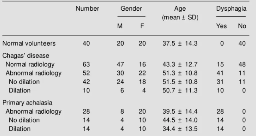
There was a considerable overlap of UES pressures between patients and volunteers (Figure 1).
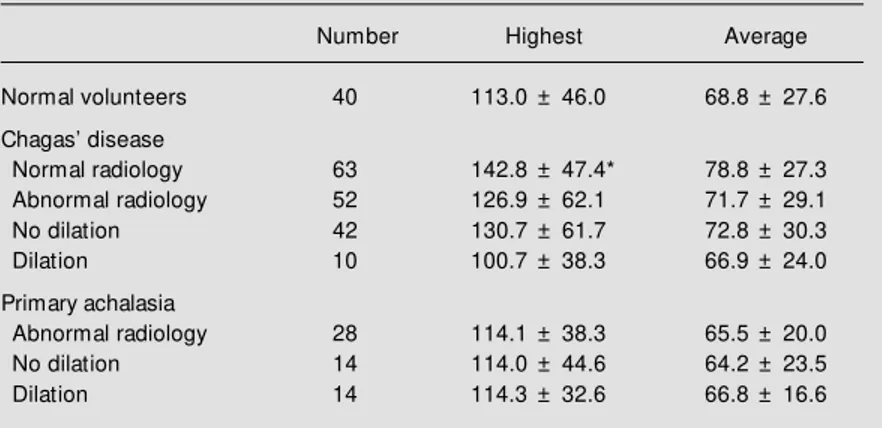
The highest UES pressure in Chagas disease patients without abnormalities upon radiologic esophageal examination was higher than in normal volunteers (P<0.05, Table 2). There was no difference between the UES pressure of patients with primary achalasia and normal volunteers, or between patients with primary achalasia and patients with Chagas disease (P>0.05). There was no difference between groups when UES
Figure 1 - Upper esophageal sphinct er (UES) pressure of healthy volunteers (controls), pa-tients w ith Chagas’ disease and normal esophageal transit by ra-diologic esophageal examination (A) or abnormal esophageal tran-sit (B), and patients w ith primary achalasia, measured at the site w ith the highest pressure (top) or at the four sites of the sphinc-ter (bottom). * P<0.05 vs con-trols.
*
U
E
S
p
re
s
s
u
re
(
m
m
H
g
) 250
200
150
100
50
0 Controls A B Achalasia
Chagasic patients
U
E
S
p
re
s
s
u
re
(
m
m
H
g
) 150
120
90
60
30
0 Controls A B Achalasia
Chagasic patients
Table 2 - Upper esophageal sphincter pressures (mmHg) of normal volunteers, pa-tients w ith Chagas’ disease and papa-tients w ith primary achalasia, measured at the site w ith the highest pressure and as the average of the four sites w here the pressures w ere measured.
Data are reported as mean ± SD; * P<0.05 compared to volunteers (Tukey-Kramer test).
Number Highest Average
Normal volunteers 40 113.0 ± 46.0 68.8 ± 27.6
Chagas’ disease
Normal radiology 63 142.8 ± 47.4* 78.8 ± 27.3 Abnormal radiology 52 126.9 ± 62.1 71.7 ± 29.1
No dilation 42 130.7 ± 61.7 72.8 ± 30.3
Dilation 10 100.7 ± 38.3 66.9 ± 24.0
Primary achalasia
Abnormal radiology 28 114.1 ± 38.3 65.5 ± 20.0
No dilation 14 114.0 ± 44.6 64.2 ± 23.5
Dilation 14 114.3 ± 32.6 66.8 ± 16.6
pressure was calculated as the average of the four directions, or when patients with or without esophageal dilation were compared (P>0.05).
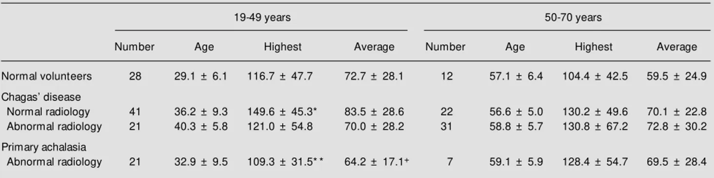
sta-tistical significance (P>0.05, Table 3). In the group of subjects aged 19-49 years the UES pressure measured at the site with the high-est pressure was higher in patients with Chagas disease and normal radiologic ex-amination than in normal volunteers or pa-tients with primary achalasia (P<0.05, Table 3). The average pressure of patients with primary achalasia was also lower than that of patients with Chagas disease and normal radiologic esophageal examination (P<0.05, Table 3). There were no differences in the group of subjects aged 50-70 years. Patients with Chagas disease without dysphagia (N = 59) had higher UES pressure (highest: 149.3 ± 57.2 mmHg, average: 81.7 ± 30.4 mmHg) than patients with Chagas disease and dysphagia (N = 56, highest: 121.0 ± 50.3 mmHg, average: 69.3 ± 25.1 mmHg) and patients with primary achalasia (N = 28), all with dysphagia (highest: 114.1 ± 38.3, aver-age: 65.5 ± 20.0 mmHg) (P<0.01).
D iscussio n
There were no differences in UES pres-sure between patients with similar esoph-ageal involvement by Chagas disease and achalasia. These pressures also did not differ from those of normal volunteers. The pa-tients with Chagas disease and no
radiologi-cal esophageal abnormalities showed higher pressure than normal volunteers at the site with the highest UES pressure. In the 19-49 year group UES pressure was also higher than in patients with achalasia.
The UES is a tonically contracted seg-ment with the resting pressure within the sphincter exibiting marked radial asymme-try (7,21,22). The highest UES pressures are in the anteroposterior direction of the crico-pharyngeal muscle (7,21,22). With the cath-eter movement the muscle increases its to-nus (7,10). The resting pressure shows con-siderable variation with environmental stimuli. The continuous rapid pull-through technique itself or water perfusion into the UES zone may stimulate the UES to contract (7). The cricopharyngeal response to the stimulus may be different in patients with Chagas disease and minor esophageal in-volvement.
The cricopharyngeus muscle receives its motor nerve supply from the pharyngeal branch of the vagus (23). The afferent sen-sory pathways from the pharynx and larynx that control swallowing travel via the glos-sopharyngeal, trigeminus, and vagus nerves to the nucleus tractus solitarius. Spinal affer-ents from the proximal esophagus, although sparse, arise from cervical and dorsal root ganglia (11). Some sensory structures are
Table 3 - Effect of age on upper esophageal sphincter pressures (mmHg) of tw o groups of normal volunteers, patients w ith Chagas’ disease and patients w ith primary achalasia, measured at the site w ith the highest pressure and as the average of the four sites w here the pressures w ere measured.
Data are reported as mean ± SD. * P<0.05 vs volunteers; * * P<0.01 vs Chagas w ith normal radiology; +P<0.05 vs Chagas w ith normal radiology (Tukey-Kramer test).
19-49 years 50-70 years
Number Age Highest Average Number Age Highest Average
Normal volunteers 28 29.1 ± 6.1 116.7 ± 47.7 72.7 ± 28.1 12 57.1 ± 6.4 104.4 ± 42.5 59.5 ± 24.9
Chagas’ disease
Normal radiology 41 36.2 ± 9.3 149.6 ± 45.3* 83.5 ± 28.6 22 56.6 ± 5.0 130.2 ± 49.6 70.1 ± 22.8 Abnormal radiology 21 40.3 ± 5.8 121.0 ± 54.8 70.0 ± 28.2 31 58.8 ± 5.7 130.8 ± 67.2 72.8 ± 30.2
Primary achalasia
located in the ganglia of the myenteric plexus (11).
The pressures recorded in the UES arise from two sources: contraction of the sphinc-ter muscle and passive elastic forces gener-ated in the tissues in and around the sphinc-ter (10,11). Because the UES is composed of striated muscle, its level of tonic contraction depends upon the activity of the somatic nerves supplying it (3,11). The greater the neural input, the more intense the spike ac-tivity recorded on the electromyogram (EMG) and the greater the tone (3,11).
The response of the UES to intraesoph-ageal air infusion in patients with achalasia differs from the response in normal subjects (24). UES pressure decreases after intrae-sophageal air infusion in normal subjects whereas it increases in patients with achala-sia, trapping the air inside the esophagus (24). In patients with megaesophagus conse-quent to Chagas disease intraesophageal water infusion increases intraesophageal and UES pressure (25). During swallowing, in-creased residual pressure in the UES and reduction in the duration of UES relaxation occur in patients with achalasia (26,27). Our personal observations suggest that the same abnormalities are present in patients with megaesophagus caused by Chagas disease, but we do not have enough reliable data to reach any conclusion. Some patients with achalasia may have a serious problem with UES relaxation (28,29).
The most striking abnormal microscopic finding in the esophageal body of cases of primary achalasia and Chagas disease is a significantly lower number or total absence of myenteric ganglion cells in the proximal and distal esophagus compared with normal controls (13,14). We do not know of any studies about the innervation of the UES in achalasia or Chagas disease. Some publica-tions have suggested that esophageal sensi-tivity may be partly impaired by the diseases (17,18), a fact that could change the UES response to the catheter movement. We did
not find differences in UES pressure in pa-tients with esophageal dilation, who have more intense denervation (14), compared with patients without dilation and normal volunteers, an indication that the sensitivity controlled by the myenteric plexus should have no influence on the UES pressure of these patients. Studies that made reference to UES pressure in achalasia did not find differences in UES pressure from normal subjects (24,26,27). In Chagas disease one study described a low UES pressure meas-ured by the rapid pull-through technique in patients with more marked esophagopathy (30).
We do not have an explanation for the higher pressure found in patients with Chagas disease and minor esophageal in-volvement. One hypothesis is related to the premise under which these patients were studied. The patients with known esoph-ageal disease already knew there was some-thing wrong with their esophagus. Findings on manometry may therefore cause less anxi-ety. However, Chagas disease patients with-out dysphagia and with no radiologic abnor-mality might have been more anxious, a situation which can increase UES pressure (4). Since UES pressure is dependent on the electrical activity of the cricopharyngeus muscle (3) we speculate that these patients may have a more marked increase in EMG activity during UES pressure measurement than volunteers. These patients show changes in esophageal motility, at times correspond-ing to complete aperistalsis (31) or simulta-neous contractions (32), showing that they have some degree of esophageal denerva-tion.
the difference did not achieve statistical sig-nificance.
We did not find differences in UES pres-sure between normal volunteers, patients with Chagas disease and patients with primary achalasia, as determined by the rapid
pull-through technique, except for patients with minor manifestations of esophageal involve-ment by Chagas disease, when UES pres-sure at the site with the highest prespres-sure was higher than the pressure of normal volun-teers and patients with primary achalasia.
Re fe re nce s
1. Kahrilas PJ, Dodds WJ, Dent J, Logemann JA & Shaker R (1988). Upper esophageal sphincter function during deglutition. Gas-troenterology, 95: 52-62.
2. Cook IJ, Dodds WJ, Dantas RO, M assey B, Kern M K, Lang IM , Brasseur JG & Hogan WJ (1989). Opening mechanisms of the human upper esophageal sphinc-ter. American Journal of Physiology, 257: G748-G759.
3. Lang IM , Dantas RO, Cook IJ & Dodds WJ (1991). Videoradiographic, manomet-ric and electromyographic analysis of ca-nine upper esophageal sphincter. Ameri-can Journal of Physiology, 260: G911-G919.
4. Cook IJ, Dent J & Collins SM (1987). M easurem ent of upper esophageal sphincter pressure: Effect of acute emo-tional stress. Gastroenterology, 93: 526-532.
5. Kahrilas PJ, Dodds WJ, Dent J, Haeberle B, Hogan WJ & Arndorfer RC (1987). Ef-fect of sleep, spontaneous gastroageal reflux, and a meal on upper esoph-ageal sphincter pressure in normal hu-man volunteers. Gastroenterology, 92: 466-471.
6. Shaker R, Ren J, Xie P, Lang IM , Bardan E & Sui Z (1997). Characterization of the pharyngo-UES contractile reflex in hu-mans. American Journal of Physiology, 273: G854-G858.
7. Kahrilas PJ, Dent J, Dodds WJ, Hogan WJ & Arndorfer RC (1987). A method for con-tinuous monitoring of upper esophageal sphincter pressure. Digestive Diseases and Sciences, 32: 121-128.
8. Andreollo NA, Thompson DG, Kendall GPN & Earlam RJ (1988). Functional rela-t ionships berela-t w een cricopharyngeal sphincter and oesophageal body in re-sponse to graded intraluminal distension.
Gut, 29: 161-166.
9. Kahrilas PJ, Dodds WJ, Dent J, Wyman JB, Hogan WJ & Arndorfer RC (1986). Upper esophageal sphincter function dur-ing belchdur-ing. Gastroenterology, 91: 133-140.
10. Jacob P, Kahrilas PJ, Herzon G & M cLaughlin B (1990). Determinants of up-per esophageal sphincter pressure in dogs. American Journal of Physiology, 259: G245-G251.
11. Conklin JL & Christensen J (1994). M otor functions of the pharynx and esophagus. In: Johnson LR (Editor), Physiology of the Gastrointestinal Tract. 3rd edn. Raven Press, New York, 903-928.
12. Feldman M (1988). Esophageal achalasia syndromes. American Journal of M edical Sciences, 295: 60-81.
13. Goldblum JR, Whyte RI, Orringer M B & Appelman HD (1994). Achalasia - A mor-phologic study of 42 resected specimens.
American Journal of Surgical Pathology, 18: 327-337.
14. Köberle F (1968). Chagas’ disease and Chagas’ syndrome: the pathology of A-merican trypanosomiasis. Advances in Parasitology, 6: 63-116.
15. Oliveira RB, Troncon LEA, Dantas RO & M eneghelli UG (1998). Gastrointestinal manifestations of Chagas’ disease. Ameri-can Journal of Gastroenterology, 93: 884-889.
16. Dant as RO, Godoy RA, Oliveira RB, M eneghelli UG & Troncon LEA (1990). Low er esophageal sphincter pressure in Chagas’ disease. Digestive Diseases and Sciences, 35: 508-512.
17. Paterson WG (1997). Esophageal and low er esophageal sphincter response to balloon distention in patients w ith achala-sia. Digestive Diseases and Sciences, 42: 106-112.
18. Ejima FH, Dantas RO, Simões M V, M arin Neto JA & M eneghelli UG (1998). Intrae-sophageal balloon distension test in Cha-gas’ disease patients w ith noncardiac chest pain. Digestive Diseases and Sci-ences, 43: 2567-2571.
19. Ferriolli E, Oliveira RB, M atsuda NM , Braga FJHN & Dantas RO (1998). Aging, esophageal motility, and gastroesophage-al reflux. Journal of the American Geriat-rics Society, 46: 1534-1537.
20. Dantas RO, Ferriolli E & Souza M AN
(1998). Gender effects on esophageal motility. Brazilian Journal of M edical and Biological Research, 31: 539-544. 21. Winans CS (1972). The
pharyngoesoph-ageal closure mechanism: a manometric study. Gastroenterology, 63: 768-777. 22. Weihrauch TR, Brummer A, Biew ener H
& Ew e K (1980). Assessment of various factors influencing esophageal pressure measurement. I. Significance of methodi-cal factors in intraluminal manometry.
Klinische Wochenschrift, 58: 279-285. 23. Lund WS & Ardran GM (1964). The motor
nerve supply of t he cricopharyngeal sphincter. Annals of Otology, Rhinology and Laryngology, 73: 599-617.
24. M assey BT, Hogan WJ, Dodds WJ & Dantas RO (1992). Alteration of the upper esophageal sphincter belch reflex in pa-tients w ith achalasia. Gastroenterology, 103: 1574-1579.
25. Godoy RA (1972). Estudo da esofagopatia chagásica crônica por meio do método eletromanométrico e da prova de meta-colina em pacientes com e sem dilatação de esôfago. Revista Goiana de M edicina, 18: 1-73.
26. Dudnick RS, Castell JA & Castell DO (1992). Abnormal upper esophageal sphinc-ter function in achalasia. American Jour-nal ofGastroenterology, 87: 1712-1715. 27. Yoneyama F, M iyachi M & Nimura Y
(1998). M anometric findings of the upper esophageal sphincter in esophageal acha-lasia. World Journal of Surgery, 22: 1043-1047.
28. Ali GN, Hunt DR, Jorgensen JO, DeCarle DJ & Cook IJ (1995). Esophageal achala-sia and coexistent upper esophageal sphincter relaxation disorder presenting w ith airw ay obstruction. Gastroenterol-ogy, 109: 1328-1332.
29. Souza M AN & Dantas RO (1998). Crico-pharyngeal dysfunction in a patient w ith achalasia. Journal of Clinical Gastroenter-ology, 26: 216-218.
Acta Gastroenterologica Latinoamericana, 24: 105-111.
31. Oliveira RB, Rezende Filho J, Dantas RO & Iazigi N (1995). The spectrum of esoph-ageal motor disorders in Chagas’ disease.
American Journal of Gastroenterology, 90: 1119-1124.
32. Dantas RO, Deghaide NHS & Donadi EA (1999). Esophageal manometric and ra-diologic findings in asymptomatic sub-jects w ith Chagas’ disease. Journal of Clinical Gastroenterology, 28: 245-248. 33. Weihrauch TR, Vallerius P, Alpers H &
Ew e K (1980). Assessment of various