Braz. Arch. Biol. Technol. v.60: e17160243, Jan/Dec 2017
1
Vol.60: e17160243, January-December 2017 http://dx.doi.org/10.1590/1678-4324-2017160243
ISSN 1678-4324 Online Edition
BRAZILIAN ARCHIVES OF BIOLOGY AND TECHNOLOGY
A N I N T E R N A T I O N A L J O U R N A L
Expression analysis of three immune genes
Interferon-gamma, Mx and Interferon regulatory factor-1 of Japanese
flounder (
Paralichthys olivaceus
)
Feng Zhang
1, Xuemei Qiu
1*, Yang Liu
1, Juan Wang
1, Xipeng Li
1, Xiuli Wang
1.
1 Dalian Ocean University - College of Fisheries and Life Science, Dalian, China.
ABSTRACT
IFN-γ (Interferon-gamma), Mx and IRF-1 (Interferon regulatory factor-1) are main immune-related genes and they play important roles in the innate immune system of vertebrates. In this study, the expression level of the three immune-related genes in twelve tissues of normal adult Japanese flounder was detected using semi-quantitative RT-PCR. Thirteen time points (3h, 6h, 9h, 12h, 24h, 36h, 48h, 60h, 72h, 84h, 96h, 108h, 120h) were selected to analyze the expression of IFN-γ, Mx and IRF-1 in spleen, head kidney, liver of Japanese flounder infected with Lipopolysaccharide (LPS). The Japanese flounder IFN-γ, Mx and IRF-1 genes were differently expressed in these tissues and had high expression levels in classical fish immune organs like spleen and head kidney. It was found that the highest expression levels of the Japanese flounder IFN-γ were detected at 24h in spleen, 36h in head kidney and 48h in liver after challenge with LPS. Interestingly, the highest expression levels of Mx in spleen and head kidney were both at 36h and IRF-1 in spleen and liver were both at 24h. The highest expression level of Mx in liver was at 48h and IRF-1 in head kidney was at 12h. The study provides a basis for further research on immune mechanism of IFN-γ, Mx, IRF-1 and the production of recombinant IFN-γ, Mx or IRF-1 used in Japanese flounder cultivation in future.
Key words: Interferon-gamma, Mx, Interferon regulatory factor-1, Paralichthys olivaceus, tissue expression
*
Author for correspondence: [email protected]
Zhang, F et al. 2
Abbreviations: IFN-γ, Interferon-gamma; IRF-1, Interferon regulatory factor-1;
LPS, Lipopolysaccharide; IPNV, infectious pancreatic necrosis virus; CCV, channel catfish herpesvirus; ISKNV, infectious spleen and kidney necrosis virus; ISRE, Interferon-stimulated response element; ISGs, interferon stimulated genes; DBD, DNA-binding domain; MHC, major histocompatibility complex; PS, physiological saline; IOD, integrated optical density; CK, uninfected adult's tissues; IHNV, infectious hematopoietic necrosis virus; HRV, Hirame rhabdovirus; HINAE, Hirame natural embryonic;
INTRODUCTION
The immune system is commonly divided into the innate (non-specific) and the adaptive (specific) immune system. The innate response generally precedes the adaptive response and determines the nature of the adaptive response 1 and 2. The fish immune system is very different from mammals and the innate immune system of fish is the primarily important defense barrier since the adaptive immune system is inefficient due to its evolutionary status 3. Compared with the adaptive immune system, the innate immune response is instant and relatively temperature independent 4,5. The main parameters of the innate immune system of fish include lectins, lysozyme, complement proteins, antibacterial peptides, IgM, transferrin, interferon and Mx protein 5, 6, 7.
Interferon (IFN) which plays an important role in innate immune system is a protein secreted from cells in response to invasion by pathogens and virus. In mammals, IFN is classified into type-I IFN (α, β, ε, δ, τ and ω), type-II IFN (γ) and type-III IFN (λ) based on their differences in primary structure, serology and their most likely time of evolutionary divergence 8, 9, 10.
Interferon-gamma (IFN-γ) is a pleiotropic proinflammatory and antiviral cytokine, which was initially identified in PHA-activated mammalian lymphocyte supernatants
11
. It was demonstrated that IFN-γ is a central participant in host resistance to various infections by using IFN-γ knockout mice infected with Leishmania major 12, Listeria monocytogenes 13 and Mycobacteria 14.
Mx proteins present a 70-80kDa GTPases of dynamin super family among vertebrates. The Mx gene in fish was first detected in 1989 15 and then seven forms of Mx genes were found in zebrafish 16, three forms in rainbow trout 17, two forms in crucian carp (Carassius auratus) 18 and a single form in Japanese flounder (Paralichthys olivaceus) 19 and Pufferfish 20. However, the antiviral property of fish Mx proteins had been demonstrated only in Japanese flounder and Atlantic salmon against fish rhabdoviruses, infectious pancreatic necrosis virus, and viral haemorrhagic septicaemia virus 21, 22, 23.
Interferon regulatory factors (IRFs) were transcription factors which bound to the ISREs (Interferon-stimulated response element) and mainly regulated the expression of type-I IFN gene, interferon stimulated genes (ISGs) and other cytokines 24, 25. Up to now as many as ten members of IRFs had been reported in vertebrates 26 and they all showed significant homology within the N-terminal 120 amino acids which contained the DNA-binding domain (DBD) forming a helix-turn-helix structure 27. Over the last two decades, these IRFs have been the focus of many immunological as well as medical studies 26 and it has been confirmed that they play important roles in antiviral activity and immune regulation 28, 29.
Expression analysis of three immune genes
Braz. Arch. Biol. Technol. v.60: e17160243, Jan/Dec 2017
3
Japanese flounder is an important economic marine fish species in the world due to its rapid growth, good taste and high nutritive value. In recent years, the scale of Japanese flounder cultivation has been expanding continuously in China. In northern China, the seawater temperature is comparatively lower than in southern China, especially in winter. The lower temperature might affect the growth of Japanese flounder and its immune response. In this study, the expression regularity of the three immune genes (IFN-γ, Mx and IRF-1) of Japanese flounder at different time points and different tissues after infection with Lipopolysaccharide (LPS) at lower temperature was well revealed. This study might be helpful for further research on the antiviral mechanism of these three immune genes and for the production and application of relevant recombinant IFN-γ, Mx and IRF-1.
MATERIAL AND METHODS
Japanese flounder
Normal adult individuals weighing 700g on average were raised at Key Laboratory of Mariculture, Ministry of Agriculture, Dalian Ocean University. They were kept in two tanks (15m3) separately for one week before experimentation and the water temperature was 8 °C to 10 °C.
Challenge and sampling
Lipopolysaccharide (LPS) (sigma, L2880, 055:B5), the major constituent of the external layer of Gram-negative bacteria, was dissolved with physiological saline (PS) to a concentration of 1mg/ml. Thirty-nine Japanese flounders were injected intraperitoneally with LPS (400ug/kg) 34 as the experimental group and thirteen were injected with an equal does of PS as the control group. Then three LPS-infected fish and one PS-injected fish were sacrificed at each sampling time (3h, 6h, 9h, 12h, 24h, 36h, 48h, 60h, 72h, 84h, 96h, 108h and 120h) after infection respectively. Tissues of interest (spleen, head kidney and liver) were dissected, quickly frozen in liquid nitrogen and stored at -80 °C until RNA extraction. Meanwhile, two normal adult fish were sacrificed and tissues of heart, blood, spleen, head kidney, brain, intestine, kidney, liver, gill, skin, muscle and gonad were removed and kept at -80℃ until use
35
. All procedures involving the handling and treatment of the individuals during this study were approved by the Animal Care and Use committee of Key Laboratory of
Mariculture & Stock Enhancement in North China’s Sea at Dalian Ocean University.
Primer design
Zhang, F et al. 4
Table1 Primer information of IFN-γ, Mx, IRF-1 and GAPDH gene of Japanese flounder used for semi-quantitative RT-PCR
Primer ID Primer sequence Gene Accession
number
Fragement
length Tm(℃) Location*
IFN-F 5'-TGGTCTGTCTGTCCCTGTG-3'
IFN-γ gene AB435093 371bp 56 26-396
IFN-R 5'-CTTGAAGCGATTTGTCCTC-3'
Mx-F 5'-AAGAGGAAAAGGCATAGTG-3'
Mx gene AB110446 395bp 57 177-571
Mx-R 5'-CCTCTGTGGTTGCTATGTC-3'
IRF-F 5'-AGCAAAGGTCACAAAGCAG-3'
IRF-1 gene AB005883 303bp 59 366-668
IRF-R 5'-ACAATGTCGTCGGAGTAGC-3'
GAPDH-F 5'-ATGCTGGTGCCCACTATGT-3'
GAPDH gene AB029337 577bp 56 260-836
GAPDH-R 5'-ACCTGGTGCTCGGTGTATG-3'
Expression analysis of three immune genes
Braz. Arch. Biol. Technol. v.60: e17160243, Jan/Dec 2017
5
Isolation of RNA
Total RNA was extracted using TRIzol reagent (Invitrogen) according to the reagent instructions. Briefly, 50mg samples were homogenized with 1ml TRIzol reagent and after incubation at 20℃ for 5 min, 0.2ml of chloroform was added. The tubes were shaken vigorously by hand for 15 seconds and incubated them at 20℃ for 3min. Then the mixture was centrifuged at 12000rpm, 4℃ for 15 min. Subsequently, supernatant was collected and 0.5ml of isopropyl alcohol was added. After incubation at 20℃ for 10min, the sample was centrifuged at 12000rpm, 4℃ for 10 min. Finally, the precipitation was collected,washed with 1ml 75% ethanol once, then dried at room temperature and dissolved in RNase-free water. The dissolved RNA was kept at -80℃ for cDNA synthesis.
Semi-quantitative RT-PCR analysis of IFN-γ, Mx and IRF-1 gene expression
cDNA synthesis was carried out using M-MLV Transcriptase (TaKaRa) according to
the manufacture’s instructions. Total RNA from the same time points or tissues were
equally mixed for reverse transcription, separately.
Primers (IFN-F/R, Mx-F/R, IRF-F/R and GAPDH-F/R) were separately used for amplifying IFN-γ fragment of 371bp, Mx fragment of 395bp, IRF-1 fragment of 303bp and GAPDH fragment of 577bp. Polymerase chain reaction (PCR) were used for amplification with conditions as follows: 1 cycle of 5 min at 94℃; 20-25 cycles of 30s at 94℃, 30s at 56℃-59℃ (Details seen in Table 1), 45s at 72℃; 1 cycle of 7 min at 72℃. PCR products were analyzed in a 1.0% agarose gel stained with ethidium bromide and the amplified DNAs were recorded with the Kodak Image Station 440CF. The integrated optical density (IOD) ratios of IFN-γ, Mx and IRF-1 versus GAPDH were determined with Gel-Pro analyzer to analyze the relative mRNA expression 36 and 37. Each column represents the average value of triple determinations.
RESULTS
IFN-γ, Mx and IRF-1 expression in twelve tissues of normal adult Japanese
flounder
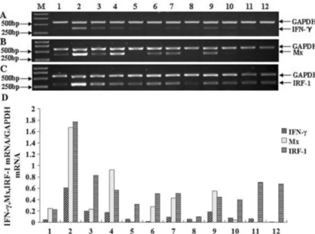
Semi-quantitative RT-PCR was conducted to analyze expressions of the IFN-γ, Mx and IRF-1 gene in normal Japanese flounders. The expression level of Japanese flounder IFN-γ was the highest in blood and high in spleen, head kidney, kidney and gill. The expression level was very faint in heart, brain, intestine, skin, liver and muscle. And IFN-γ was not detected in gonad (Fig.1 A).
Zhang, F et al. 6
Densitometric analysis of semi-quantitative RT-PCR was used to obtain the relative IOD of IFN-γ, Mx and IRF-1 versus GAPDH; M: molecular weight standard; 1-12: heart, blood, spleen, head kidney, brain, intestine, kidney, skin, Gill, liver, muscle, gonad. The Japanese flounder IFN-y specific RT-PCR products (371bp), Mx gene-specific RT-PCR products (395 bp) and IRF-1 gene-gene-specific RT-PCR products (303 bp) are indicated on the right margin, along with GAPDH RT-PCR products (577 bp) as na internal control.
The expression level of Japanese flounder Mx was the highest in blood and head kidney and was high in heart, spleen, intestine, kidney and gill. The expression of Mx was very faint in liver and not detected in brain, skin, muscle and gonad (Fig.1 B).
Seen from Figure.1 C, the expression of Japanese flounder IRF-1 was detected in all tissues and was very high in blood, spleen, intestine, kidney, gill, muscle and gonad.
IFN-γ expression in infected Japanese flounder tissues
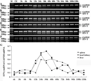
The expression of Japanese flounder IFN-γ was analyzed in the spleen, head kidney and liver after challenge with LPS. In the three tissues, the expression of IFN-γ was dramatically induced after infection. The highest expression levels of the IFN-γ were detected at 24h in spleen, 36h in head kidney and 48h in liver. The expression in spleen started at 12h, decreased gradually at 36h and returned to the level of background after 48h. The expression levels of IFN-γ in spleen at 24h, 36h and 48h were approximately 3.2-, 4.3- and 2.8-fold compared with the uninfected adult's tissues (CK), respectively. At the rest time points, no significant increases of the gene were observed (Fig.2 A, G). In head kidney, the expression levels kept high from 12h to 48h and then decreased gradually to background. The relative IOD value showed the expression levels from 12h to 48h were approximately 4.9-, 8.8-, 9- and 7.1-fold compared with CK (Fig.2 C, G). The high expression level in liver lasted from 24h to 84h and then become faint consistent with background. The highest expression level was at 48h and it was 4.5-fold compared with CK (Fig.2 E, G). We also injected the Japanese flounder with physiological saline (PS) as control and the results showed that the IFN-γ expression in spleen, head kidney and liver was consistent with CK, indicating the injection of PS was not harmful to Japanese flounder (Fig.2 B, D, F).
Expression analysis of three immune genes
Braz. Arch. Biol. Technol. v.60: e17160243, Jan/Dec 2017
7
Mx expression in infected Japanese flounder tissues
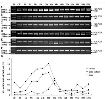
The expression of Mx in spleen, head kidney and liver was dramatically induced after challenge. The expression in spleen started at 9h and peaked at 36h and then decreased to background level at 60h. The expression levels of Mx in spleen from 9h to 48h were approximately 1.7-, 2-, 2.1-, 2.5- and 1.3-fold compared with CK (Fig.3 A, G). The highest expression level in head kidney was detected at 36h and went back to background level at 60h. The expression levels were approximately , 5.1-, 7.9-5.1-, 8.6- and 4.3-fold compared with CK from 9h to 48h (Fig.3 C5.1-, G). Seen from Figure.3 E, the expression of Mx in liver was high at 36h, peaked at 48h and decreased at 60h and the expression of Mx at other time points was very faint. Compared with CK, the expression level peaked as high as 15-fold in liver at 48h (Fig.3 G). The control group also showed that the injection of PS did not influence the expression of Mx (Fig.3 B, D, F).
Figure 3. Semi-quantitative RT-PCR analysis of Japanese flounder Mx in infected tissues (spleen, head kidney, liver). A, C, E: spleen, head kidney, liver from the Japanese flounder injected with LPS, respectively; B, D, F: spleen, head kidney, liver from the Japanese flounder injected with PS, respectively; G: the relative IOD of Mx versus GAPDH in infected spleen, head kidney and liver; M: molecular weight standard; CK: uninfected tissues as controls; Different time points after the challenge with LPS are indicated on the above margin. The Japanese flounder Mx gene-specific RT-PCR products (395 bp) and GAPDH RT-PCR products (577 bp) are indicated on the right margin.
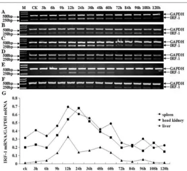
IRF-1 expression in infected Japanese flounder tissues
Zhang, F et al. 8
expression of IRF-1 in spleen, head kidney and liver injected with PS was consistent with the expression of IFN-γ and Mx injected with PS (Fig.4 B, D, F).
Figure 4. Semi-quantitative RT-PCR analysis of Japanese flounder IRF-1 in infected tissues (spleen, head kidney, liver). A, C, E: spleen, head kidney, liver from the Japanese flounder injected with LPS, respectively; B, D, F: spleen, head kidney, liver from the Japanese flounder injected with PS, respectively; G: the relative IOD of IRF-1 versus GAPDH in infected spleen, head kidney and liver; M: molecular weight standard; CK: uninfected tissues as controls; Different time points after the challenge with LPS are indicated on the above margin. The Japanese flounder IRF-1 gene-specific RT-PCR products (303 bp) and GAPDH RT-PCR products (577 bp) are indicated on the right margin.
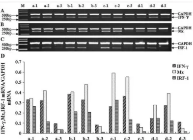
Expression of IFN-γ, Mx and IRF-1 in spleen, head kidney and liver at 12h, 24h,
36h and 48h
According to the results, the main expression time points of IFN-γ, Mx and IRF-1 were at 12h, 24h, 36h and 48h. So the four time points were selected to analyze the expression of the three genes in spleen, head kidney and liver.
Expression analysis of three immune genes
Braz. Arch. Biol. Technol. v.60: e17160243, Jan/Dec 2017
9
Figure 5. Semi-quantitative RT-PCR analysis of IFN-γ, Mx and IRF-1 in infected tissues of Japanese flounder at 12h, 24h, 36h, 48h. A, B, C: Expression of Japanese flounder IFN-γ, Mx, IRF-1 in infected tissues (spleen, head kidney, liver) at different time points (12h, 24h, 36h, 48h). M: molecular weight standard; a-d: 12h, 24h, 36h, 48h. 1-3: spleen, head kidney, liver. D: the relative IOD of IFN-γ, Mx and IRF-1 versus GAPDH; The Japanese flounder IFN-γ specific RT-PCR products (371bp), Mx specific RT-PCR products (395bp) and IRF-1 gene-specific RT-PCR products (303bp) are indicated on the right margin, along with GAPDH RT-PCR products (577bp) as an internal control.
DISCUSSION
The innate immune system is primarily important to combat infections in fish. It is commonly stated that the innate immune system are relatively temperature independent because of its evolutionary status and poikilothermic nature 3, 4, 5. Interferon, Mx and IRF play important roles in immune response in fish. It was considered that innate parameters were active at the lower temperature range of the fish. It was proved that in cod the spontaneous haemolytic and antiprotease activity was more active at 1℃ and 7℃ than at 14℃38. However, there were some examples of low temperature having adverse effects on innate parameters. Rainbow trout acclimatized at different temperatures showed lower phagocytic and complement activity at 5℃ than at 20℃39. Similarly, a drop in water temperature from 22℃ to 10℃ had adverse effects on the protective role of channel catfish mucus 40. In this study the Japanese flounders were kept for one week with lower water temperature at 8°C-10°C before the experiment was carried out. The results showed that the expression of IFN-γ, Mx and IRF-1 in spleen, head kidney and liver of Japanese flounders injected with LPS was different and regular at thirteen time points, which indicated the innate system of Japanese flounder could resist external infection well at lower temperature.Our results showed that the three immune-related genes were differently expressed in Japanese flounder tissues, which indicated the sites of function in the body. There were high expression levels of IFN-γ, Mx and IRF-1 in classical fish immune organs like spleen and head kidney (Fig. 1), which suggested their functional roles in the immune system. The expression of three genes was very intense in blood, which showed that abundant cytokines existed in blood that transcribes these genes. When organism was invaded by virus or bacteria, cytokines in blood could perform their function in time. Besides, the expression of IFN-γ and IRF-1 were also detected in muscle tissue of Japanese flounder but Mx was not detected. Interestingly, the expression of IFN-γ was also detected in goldfish muscle
41
Zhang, F et al. 10
existed not only in immune organs but also in the organs which did not take part in immune response.
IFNs are best known for their vital roles in innate immune system. Recently, studies focused on the immune-related functions of IFN-γ had been carried out in many teleosts, such as goldfish 42 and zebrafish 10. We have carried out the expression analysis of the Japanese flounder IFN-γ in tissues infected with LPS and revealed the expression regularity. The Japanese flounder IFN-γ was highly induced in spleen at 24h, in head kidney at 36h and in liver at 48h (Fig. 2). The results were very similar to the expression of type-I IFN in head kidney of Atlantic Salmon 43, which suggested the Japanese flounder IFN-γ played important roles in immune responses to external invasion.
Mx induced by type-I IFNs (IFN-α and IFN-β) is a kind of anti-viral protein which specially prevents growth, in vivo and in vitro, of certain classes of virus. Up to date, many experiments have been carried out to study the immune-related functions of Mx in many teleosts. It was reported that Rainbow trout Mx1, Mx2 and Mx3 showed no significant inhibition against a fish rhabdovirus, infectious hematopoietic necrosis virus (IHNV) 44. The expression analysis of Japanese flounder Mx in infected tissues was carried out in the study. The Mx was highly expressed in spleen, head kidney at 36h and in liver at 48h after challenge with LPS (Fig. 3). The expression of Mx after challenge was instant, steady and abundant; therefore it could serve as a molecular indicator to the expression of type-I IFNs in Japanese flounder.
IRFs are best known for their vital roles in regulating the expression of IFN-I, ISGs and other cytokines 24, 25. Recently, studies on IRFs in teleosts also focused on their immune-related functions. We carried out the expression analysis of the Japanese flounder IRF-1 in infected tissues. Interestingly, the Japanese flounder IRF-1 was highly expressed at 12h in spleen, liver and at 24h in head kidney (Fig. 4). The result was very similar to the expression of IRF-1 in spleen of large yellow croaker 45. However, it was very different from the results of the relative expression of Japanese flounder IRF-1 infected with HRV and Edwardsiella tarda in 1998 30, which might be caused by the difference of fish, seawater temperature and area. The expression of IRF-1 gene may reflect its importance in acute stresses in Japanese flounders.
Expression analysis of three immune genes
Braz. Arch. Biol. Technol. v.60: e17160243, Jan/Dec 2017
11
CONCLUSION
In conclusion, the highest expression level of the IFN-γ gene was detected at 24h in spleen, 36h in head kidney and 48h in liver after normal adult Japanese flounder challenged with lipopolysaccharide. In the same manner,the highest expression levels of Mx gene in spleen and head kidney were both at 36h and IRF-1 gene in spleen and liver were both at 24h. And the highest expression level of Mx gene in liver was at 48h and IRF-1gene in head kidney was at 12h. This study provides a basis for further research on immune mechanism of IFN-γ, Mx, IRF-1 and the production of recombinant IFN-γ, recombinant Mx or recombinant IRF-1 used in Japanese flounder cultivation in future.
ACKNOWLEDGMENTS
This study was supported by the grant of Dalian Ocean University (No. 2012HYDX07). We thank John Wheaton (College of International Exchange and English Education, Jishou University) for polishing the manuscript.
REFERENCES
1. Fearon D.T. & Locksley R.M. The instructive role of innate immunity in the acquired immune response. Science. 1996; 272: 50-53.
2. Fearon D.T. Seeking wisdom in innate immunity. Nature. 1997; 388: 323-324.
3. Magnadóttir B. Innate immunity of fish (overview). Fish Shellfish Immunol. 2006; 20: 137-151.
4. Du Pasquier L. Antibody diversity in lower vertebrates--why is it so restricted? Nature. 1982;296: 311-313.
5. Ellis A.E. Innate host defence mechanism of fish against viruses and bacteria. Dev. Comp. Immunol.2001;25: 827-839.
6. Rombout J.H, Taverne N, van de Kamp M, Taverne-Thiele A.J. Differences in mucus and serum immunoglobulin of carp (Cyprinus carpio L.).Dev. Comp. Immunol. 1993; 17: 309-317.
7. Stafford J.L, Belosevic M. Transferrin and the innate immune response of fish: identification of a novel mechanism of macrophage activation. Dev. Comp. Immunol. 2003; 27: 539-554.
8. Muller U, Steinhoff U, Reis L.F, Hemmi S, Pavlovic J, Zinkernagel R.M, et al. Functional role of type I and type II interferons in antiviral defense. Science. 1994; 264: 1918-1921. 9. Roberts R.M, Liu L, Guo Q, Leaman D, Bixby J. The evolution of the type I interferons. J.
Interferon Cytokine Res. 1998; 18: 805-816.
10. Sieger D, Stein C, Neifer D, van der Sar A.M, Leptin M. The role of gamma interferon in innate immunity in the zebrafish embryo. Dis. Model Mech. 2009; 2: 571-581.
11. Wheelock E.F. Interferon-like virus-inhibitor induced in human leukocytes by phytohemagglutinin. Science. 1965; 149: 310-311.
12. Wang Z.E, Reiner S.L, Zheng S, Dalton D.K, Locksley R.M. CD4+ effector cells default to the Th2 pathway in interferon gamma-deficient mice infected with Leishmania major. J. Exp. Med. 1994; 179: 1367-1371.
13. Huang S, Hendriks W, Althage A, Hemmi S, Bluethmann H, Kamijo R, et al. Immune response in mice that lack the interferon-gamma receptor. Science. 1993; 259: 1742-1745. 14. Cooper A.M, Dalton D.K, Stewart T.A, Griffin J.P, Russell D.G,Orme I.M. Disseminated
tuberculosis in interferon gamma gene-disrupted mice. J. Exp. Med. 1993; 178: 2243-2247. 15. Staeheli P, Yu Y.S, Grob R, Haller O. A double stranded-RNA inducible fish gene
Zhang, F et al. 12
16. Altmann S.M, Mellon M.T, Johnson M.C, Paw B.H, Trede N.S, Zon L.I, et al. Cloning and characterization of an Mx gene and its corresponding promoter from the zebrafish, Danio rerio. Dev. Comp. Immunol .2004;28: 295-306.
17. Trobridge G.D, Chiou P.P, Leong J.A. Cloning of the rainbow trout (Oncorhynchus mykiss) Mx2 and Mx3 cDNAs and characterization of trout Mx protein expression in salmon cells. J. Virol. 1997; 71: 5304-5311.
18. Zhang Y.B, Li Q, Gui J.F. Differential expression of two Carassius auratus Mx genes in cultured CAB cells induced by grass carp hemorrhage virus and interferon. Immunogenetics. 2004; 56: 68-75.
19. Lee J.Y, Hirono I, Aoki T. Cloning and analysis of expression of Mx cDNA in Japanese flounder, Paralichthys olivaceus. Dev. Comp. Immunol. 2000; 24: 407-415.
20. Yap W.H, Tay A, Brenner S, Venkatesh B. Molecular cloning of the pufferfish (Takifugu rubripes) Mx gene and functional characterization of its promoter. Immunogenetics. 2003; 54: 705-713.
21. Caipang C.M.A, Hirono I, Aoki T. In vitro inhibition of fish rhabdoviruses by Japanese flounder. Paralichthys olivaceus Mx, Virology. 2003; 317:373-382.
22. Larsen R, Rokenes T.P, Robertsen B. Inhibition of infectious pancreatic necrosis virus replication by atlantic salmon Mx1 protein. J. Virol. 2004; 78: 7938-7944.
23. Pakingking R. Jr, Okinaka Y, Mori K, Arimoto M, Muroga K, Nakai T. In vivo and in vitro analysis of the resistance against viral haemorrhagic septicaemia virus in Japanese flounder (Paralichthys olivaceus) precedingly infected with aquabirnavirus. Fish Shellfish Immunol. 2004; 17: 1-11.
24. Mamane Y, Heylbroeck C, Génin P, Algarté M, Servant M.J, LePage C, et al. Interferon regulatory factors: the next generation. Gene. 1999; 237: 1-14.
25. Taniguchi T, Ogasawara K, Takaoka A, Tanaka N. IRF family of transcription factors as regulators of host defense. Annu. Rev. Immunol. 2001; 19:23-55.
26. Tamura T, Yanai H, Savitsky D, Taniguchi T. The IRF family transcription factors in immunity and oncogenesis. Annu. Rev. Immunol. 2008; 26: 535-584.
27. Escalante C.R, Yie J, Thanos D, Aggarwal A.K. Structure of IRF-1 with bound DNA reveals determinants of interferon regulation. Nature. 1998; 391: 103-106.
28. Barnes B, Lubyova B, Pitha P.M. On the role of IRF in host defense. J. Interferon Cytokine Res. 2002; 22: 59-71.
29. Nguyen H, Hiscott J, Pitha P.M. The growing family of interferon regulatory factors. Cytokine Growth Factor Rev. 1997;8: 293-312.
30. Yabu T, Hirose H, Hirono I, Katagiri T, Aoki T, Yamamoto E. Molecular cloning of a novel interferon regulatory factor in Japanese flounder Paralichthys olivaceus. Mol. Mar. Biol. Biotechnol. 1998; 7: 138-144.
31. Collet B, Hovens G.C, Mazzoni D, Hirono I, Aoki T, Secombes C.J. Cloning and expression analysis of rainbow trout Oncorhynchus mykiss interferon regulatory factor 1 and 2 (IRF-1 and IRF-2). Dev. Comp. Immunol. 2003; 27: 111-126.
32. Ordás M.C, Abollo E, Costa M.M, Figueras A, Novoa B. Molecular cloning and expression analysis of interferon regulatory factor-1 (IRF-1) of turbot and sea bream. Mol. Immunol. 2006; 43: 882-890.
33. Sun B.J, Chang M.X, Song Y, Yao W.J, Nie P. Gene structure and transcription of IRF-1 and IRF-7 in the mandarin fish Siniperca chuatsi.Vet. Immunol. Immunopathol. 2007; 116: 26-36.
34. Lee E.Y, Park H. H, Kim Y. T, Choi T. J. Cloning and sequence analysis of the interleukin-8 gene from flounder (Paralichthys olivaceous). Gene. 2001; 274: 237-243. 35. Liu Y, Chen S.L, Meng L, Zhang Y.X. Cloning, characterization and expression analysis
of a novel CXC chemokine from turbot (Scophthalmus maximus). Fish Shellfish Immunol . 2007;23: 711-720.
36. Hayashi H, Nishimoto A, Oshima N, Iwamuro S. Expression of the estrogen receptor alpha gene in the anal fin of Japanese medaka, Oryzias latipes, by environmental concentrations of bisphenol A. J. Toxicol Sci. 2007; 32: 91-96.
Expression analysis of three immune genes
Braz. Arch. Biol. Technol. v.60: e17160243, Jan/Dec 2017
13
38. Magnadóttir B, Jónsdóttir H, Helgason S, Björnsson B, Jørgensen T.O, Pilström L. Humoral immune parameters in Atlantic cod (Gadus morhua L.) I: The effects of environmental temperature. Comp. Biochem Physiol B. 1999; 122: 173-180.
39. Nikoskelainen S, Bylund G, Lilius E.M. Effect of environmental temperature on rainbow trout (Oncorhynchus mykiss) innate immunity. Dev. Comp. Immunol. 2004; 28: 581-592. 40. Quiniou S.M.A, Bigler S, Clem L.W, Bly J.E. Effects of water temperature on mucous
cell distribution in channel catfish epidermis: a factor in winter saprolegniasis. Fish Shellfish Immunol. 1998; 8: 1-11.
41. Grayfer L. & Belosevic M. Molecular characterization, expression and functional analysis of goldfish (Carassius aurutus L.) interferon gamma. Dev. Comp. Immunol. 2009; 33: 235-246.
42. Grayfer L, Belosevic M. Molecular characterization, expression and functional analysis of goldfish (Carassius aurutus L.) interferon gamma. Dev. Comp. Immunol. 2009; 33: 235-246.
43. Robertsen B, Bergan V, Rokenes T, Larsen R, Albuquerque A. Atlantic salmon interferon genes: cloning, sequence analysis, expression, and biological activity. J. Interferon Cytokine Res. 2003; 23: 601-612.
44. Trobridge G.D, Chiou P.P, Leong J.A. Cloning of the rainbow trout (Oncorhynchus mykiss) Mx2 and Mx3 cDNAs and characterization of trout Mx protein expression in salmon cells. J. Virol. 1997; 71: 5304-5311.
45.Yao C.L, Kong P, Huang X.N, Wang Z.Y. Molecular cloning and expression of IRF1 in large yellow croaker, Pseudosciaena crocea. Fish Shellfish Immunol. 2010; 28: 654–660.