Comparison to a Promastigote Screen and Identification
of a Host Cell-Specific Hit
Geraldine De Muylder1*, Kenny K. H. Ang2, Steven Chen2, Michelle R. Arkin2, Juan C. Engel1, James H.
McKerrow1
1Department of Pathology, Sandler Center for Drug Discovery, University of California San Francisco, San Francisco, California, United States of America,2Small Molecule Discovery Center, University of California San Francisco, San Francisco, California, United States of America
Abstract
The ability to screen compounds in a high-throughput manner is essential in the process of small molecule drug discovery. Critical to the success of screening strategies is the proper design of the assay, often implying a compromise between ease/ speed and a biologically relevant setting. Leishmaniasis is a major neglected disease with limited therapeutic options. In order to streamline efforts for the design of productive drug screens againstLeishmania, we compared the efficiency of two screening methods, one targeting the free living and easily cultured promastigote (insect–infective) stage, the other targeting the clinically relevant but more difficult to culture intra-macrophage amastigote (mammal-infective) stage.
Screening of a 909-member library of bioactive compounds against Leishmania donovani revealed 59 hits in the
promastigote primary screen and 27 in the intracellular amastigote screen, with 26 hits shared by both screens. This suggested that screening against the promastigote stage, although more suitable for automation, fails to identify all active compounds and leads to numerous false positive hits. Of particular interest was the identification of one compound specific to the infective amastigote stage of the parasite. This compound affects intracellular but not axenic parasites, suggesting a host cell-dependent mechanism of action, opening new avenues for anti-leishmanial chemotherapy.
Citation:De Muylder G, Ang KKH, Chen S, Arkin MR, Engel JC, et al. (2011) A Screen againstLeishmaniaIntracellular Amastigotes: Comparison to a Promastigote Screen and Identification of a Host Cell-Specific Hit. PLoS Negl Trop Dis 5(7): e1253. doi:10.1371/journal.pntd.0001253
Editor:Ana Rodriguez, New York University School of Medicine, United States of America ReceivedMarch 14, 2011;AcceptedJune 10, 2011;PublishedJuly 19, 2011
Copyright:ß2011 De Muylder et al. This is an open-access article distributed under the terms of the Creative Commons Attribution License, which permits unrestricted use, distribution, and reproduction in any medium, provided the original author and source are credited.
Funding:This work was supported by the Sandler Foundation, NIH grant AI090605, and the Belgian American Education Foundation (BAEF). The funders had no role in study design, data collection and analysis, decision to publish, or preparation of the manuscript.
Competing Interests:The authors have declared that no competing interests exist.
* E-mail: [email protected]
Introduction
Leishmaniasis is caused by protozoan parasites of the genus
Leishmania. The disease is endemic in the tropics, subtropics and the Mediterranean basin. There are three main clinical syndromes caused by different species of Leishmania. Cutaneous and muccocutaneous leishmaniasis result in large, painful sores that can take many months to heal [1]. Visceral leishmaniasis results in fever, weight loss, and damage to internal organs such as the spleen and the liver and may be fatal if left untreated [2].
Leishmania parasites are transmitted to mammalian hosts through the bite of phlebotomine sandflies. The parasites that develop in the mid-gut of the flies, called promastigotes, are flagellated and extracellular. Upon injection in the bloodstream of a mammalian host, promastigotes are rapidly phagocytosed by macrophages where they differentiate into the amastigote form. Amastigotes multiply in the macrophage parasitophorous vacuole, leading to destruction of the host cell and release of free amastigotes into the bloodstream, where they are capable of infecting new phagocytic cells [3].
Current treatment for leishmaniasis relies on chemotherapy, as no efficient vaccine is available. Sodium stibogluconate and amphotericin B have been the first line treatment; however, they have significant side effects and unresponsiveness to sodium
stibogluconate has been reported for many years [4–6]. A few new anti-leishmanial drugs have been recently released (miltefosine, paromomycin), but they also have drawbacks including cost and toxicity [7]. In addition, it has been shownin vitro that in some cases resistance can be easily induced [8].
New therapeutics are therefore urgently needed. Recognition of this need in recent years has led to partnerships between a number of foundations, agencies and universities to support the discovery of anti-parasitic agents, including anti-leishmanials. Lead discov-ery, one of the bottlenecks in the pipeline for novel anti-leishmanial drugs, would be facilitated by improved high-throughput technology allowing for the ability to screen large number of candidates [9,10]. Several anti-leishmanial high-throughput screens have been reported [11–13]. Primary screens often target the parasite promastigote stage because of ease of culture and manipulation. Indeed, promastigotes from several
from macrophages have shown differences in several cellular processes, including metabolism, intracellular transport and response to oxidative stress [18]. These observations highlight the importance of the host macrophage in driving the parasite to specific adaptations. The axenic amastigote model therefore has limitations as it does not encompass many aspects of intracellular parasite development [19]. Compounds active against axenic forms might be unable to reach the intracellular amastigote because of their inability to cross host cell membranes or maintain stability under low pH. Other compounds may need to be metabolized by the macrophage to gain activity. Finally, the macrophage itself might be directly targeted, thereby leading to parasite growth inhibition [20].
We have developed a host cell-based screening assay using a human macrophage cell line infected withL. donovani, one of the agents of visceral leishmaniasis. This assay format enables screening of compounds directly against the intracellular stage of the parasite. This assay was used to screen a library of 909 bioactive compounds consisting largely of FDA approved small molecules. In order to compare the efficiency of this screening method with traditional high-throughput screening assays, the same compound library was screened against free living promastigotes. A compound leading to sixty percent parasite growth inhibition at 10mM was considered a hit in both assays. 59 hits were identified in the promastigote assay of which only 26 were also considered hits in the intracellular amastigote assay. Only one compound was specifically active against the intracel-lular amastigote stage. We conclude that the promastigote assay fails to identify all active compounds and leads to a rate of 56% false positives.
Materials and Methods
Cell culture
THP-1 cells (human acute monocytic leukemia cell line – ATCC TIB202) were grown in RPMI supplemented with 10% Foetal Bovine Serum (FBS) and 50mM 2-Mercaptoethanol at 37uC in 5% CO2.L. donovanipromastigotes [strain 1S, clone 2D (MHOM/SD/62/1S-cl2D)] were grown at 27uC in RPMI supplemented with 10% FBS and 10% Brain Heart Tryptose medium (BHT) [21]. Differentiation of promastigotes into axenic
amastigotes was achieved by dilution of 56105 promastigotes in 3 ml of low-pH axenic amastigote media (15 mM KCl; 136 mM KH2PO4; 10 mM K2HPO4?3H2O; 0.5 mM MgSO4?7H2O;
24 mM NaHCO3; 22 mM glucose; 1 mM glutamine, 16RPMI 1640 vitamin mix, 10mM folic acid, 100mM adenosine, 16 RPMI amino acid mix, 5mg/ml hemin, 50 U/ml of penicillin, 50mg/ml of streptomycin, 25 mM MES and 20% FBS. The pH was adjusted to pH 5.66 at 22uC, yielding a final pH of 5.5 at 37uC) [22]. Axenic amastigotes were grown in ventilated flasks at 37uC in 5% CO2.
Compounds
A library of 909 bioactive compounds was donated by Iconix Biosciences. These compounds were dissolved in DMSO at a stock concentration of 1 mM. Amphotericin B (Sigma) was used as a positive control.
Promastigote high-throughput assay
L.donovanipromastigotes from an exponentially growing culture were diluted to 106/ml in RPMI containing 10% FBS and 10% BHT. The diluted culture (99ml/well) was dispensed in sterile 96-well flat white opaque assay plates (Greiner Bio-One) using a WellMate multichannel dispenser (Matrix). 1ml of 1 mM test compound dissolved in DMSO was added to the plates for a final concentration of 10mM compound and 1% DMSO. Amphoter-icin B was added as a positive control (final concentration 2mM, 1% DMSO) and as a negative control, 1ml DMSO was added (1% final concentration). Compounds and controls were added to the assay plate with the robotic dispenser Biomek FXp liquid handler (Beckman Coulter). Promastigotes were incubated with the compounds for 72 h at 27uC. The parasites were then lysed by adding 50ml of CellTiter-Glo (Promega) and placed on an orbital shaker for 5 min at room temperature. After lysis, the resulting ATP-bioluminescence was measured using the Analyst HT plate reader (Molecular Devices). Percentage inhibition of parasite growth was calculated for each well as [1-(RLUx-RLU+)/(RLU-
-RLU+
)]*100 where RLUx, RLU+and RLU-are respectively the
Relative Light Units for each well, positive (amphotericin B) and negative (DMSO) controls. A screening window coefficient, denoted Z’ factor, was used to evaluate the performance of the assay. The Z’ factor, calculated as 1-(3sc++3sc2)/(mc+2mc2) where sc+, sc2, mc+ and mc2 are respectively the standard deviation and mean values of positive and negative controls, is reflective of the assay signal dynamic range and the data variation associated with signal measurement [23]. For GI50determinations
(half maximal inhibitory concentration), compounds were serially diluted 3-fold in DMSO, with final assay concentrations ranging from 50mM to 0.02mM (1% final concentration of DMSO). GI50
curve fitting was carried out using GraphPad Prism 4 Software (GraphPad Software Inc., San Diego, CA).
Intracellular amastigote high-throughput assay
Sterile, black, 96-well, clear bottom plates (Greiner Bio-One) were seeded with exponentially growing THP-1 (56105cells/ml). THP-1 were treated with 0.1mM phorbol myristate acetate (PMA, Sigma) at 37uC for 48 h to achieve differentiation into adherent, non-dividing macrophages. Maturation of THP-1 cells towards monocyte-macrophage like cells is essential to avoid parasitized cells being overgrown by replicating cells. After activation by PMA, cells were washed and incubated with complete RPMI medium containing stationary phaseL. donovanipromastigotes at a macrophage/promastigote ratio of 1/15. After 4 h incubation at 37uC, non-internalized promastigotes were removed by 2–3 successive washes with RPMI containing 5% FBS and 5% horse
Author Summary
serum. Test compounds (10mM), positive control (2mM ampho-tericin B) or negative control (1% DMSO) were then added to the cultures using a Biomek FXp liquid handler (Beckman Coulter). Cultures were incubated at 37uC for 72 h. Cells were then washed with phosphate-buffered saline (PBS), fixed for 30 minutes with 4% formaldehyde, rinsed again with PBS, stained for 2 h with 49,69-diamidino-2-phenylindole (DAPI 300 nM) and finally washed with PBS. For GI50 determination, compounds were
serially diluted 3-fold in DMSO, with final assay concentrations ranging from 50mM to 0.02mM (1% final concentration of DMSO). Images were acquired with an INCell Analyzer 1000 automated epi-fluorescent microscope (G.E. Healthcare). The excitation and emission filters used to detect DAPI were 350/ 50 nm and 460/40 nm respectively. Eight image fields were acquired per well with a 20X objective. The proprietary INCell Developer Toolbox 1.7 software was used for image analysis. Segmentation parameters were set to identify host nuclei with a minimum area of 250mm2 and parasite kinetoplast with an average area of 1mm2. The intensity of parasite nucleus was too low to be detected with a 20X objective. A border, representing the boundary of the cell, was drawn around the nucleus (total area between 700 and 2000mm2). Only parasites found within this area were included in the calculation to eliminate extracellular parasites. False positive parasite detection in the nucleus was also excluded from the calculation. Host cell nuclei and parasite kinetoplasts were counted and the ratio of parasites DNA to host nuclei was selected as the measurement output. Percentage inhibition of parasite growth was calculated as [1-(P/hcx-P/hc+)/
(P/hc2-P/hc+
)]*100 where P/hcx, P/hc+and P/hc2are parasite
per host cell ratio for every well, positive control (amphotericin B) and negative control (DMSO) respectively. Calculation of Z’ factor and GI50curve fitting were carried out as described above.
Dose response study against axenic amastigotes
L. donovani axenic amastigotes (56105 cells/ml in axenic amastigote media) were dispensed in sterile 96-well flat white opaque assay plates (Greiner Bio-One) using a WellMate multichannel dispenser (Matrix). Compounds were serially diluted 3-fold in DMSO, with final assay concentrations ranging from 50mM to 0.02mM (1% final concentration of DMSO). 1% DMSO and amphotericin B (2mM, 1% DMSO final concentra-tion) were added as negative and positive controls respectively. Axenic amastigotes were incubated with the compounds for 72 h at 37uC with 5% CO2. Parasite viability was then measured using CellTiter-Glo as described above. Calculation of Z’ factor, percentage of parasite growth inhibition and GI50 curve fitting
were carried out as described above.
Results
Development of an image-based high-throughput assay for drug screening against intracellularL. donovani
We developed a 96-well plate, cell-based assay simple to manipulate and reproducible, enabling screening of a high number of compounds against intra-macrophageL. donovani.
The human leukemia monocyte cell line THP-1 has been commonly used as a model forLeishmaniainfection and has been described as a suitable model for drug screening [24,25].In vitro
infection of macrophages byLeishmaniaand analysis of intracellular parasite growth requires a method allowing for robust detection, discrimination and counting of parasites and host cells. In our setting, THP-1 cells infected withL. donovaniwere stained with the DNA marker DAPI (49,69-diamidino-2-phenylindole) allowing the visualization of host cell nuclei and parasite kinetoplasts. Images
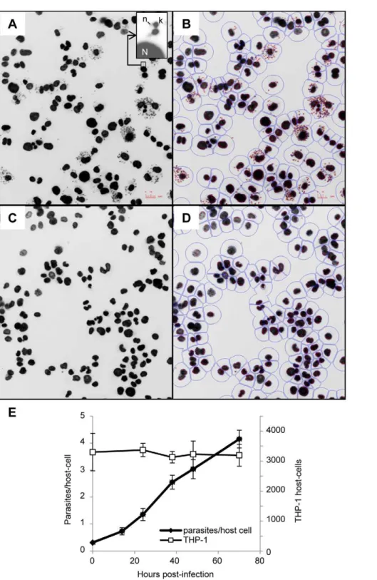
collected with an INCell Analyzer 1000 fluorescent microscope showed a significant size difference between host cell nuclei and parasite kinetoplasts. This feature was exploited for image segmentation and determination of the number of host cells and parasites (Figure 1A–D). The ratio between total number of parasites and total number of host cells was calculated for each well. In addition, counts of host cell nuclei were used as a quantitative measure of cell toxicity induced by the compounds. Incubation ofL. donovaniwith THP-1 for 4 hours at a ratio of 15 parasites per host cell led to an average infection of 4.1+/20.32 parasites per host cell after 72 h incubation, with an average of 30 +/29 percent of the cells infected and no change in the number of host cells (Figure 1E). Growth of parasite and host cells was not affected by 1% DMSO (Figure 2A–B). Amphotericin B, the first line drug used against leishmaniasis, was used as a positive control. At 2mM amphotericin B did not affect THP-1 host macrophages (Figure 2A) but significantly inhibited growth of intracellular L. donovani(Figure 2B) with an estimated GI50of 0.08mM (Figure 2C).
This is comparable to GI50values from previous reports [11,20].
Screening of a bioactive small molecule library againstL. donovaniintracellular amastigotes and free living
promastigotes
The intracellular amastigote imaging assay described above was used to screen a library of 909 bioactive small molecules (Iconix library). In the primary screen, compounds were assayed in duplicates at 10mM. The average Z’ value calculated per plate based on the positive and negative controls was 0.63, indicating a satisfactory robustness of the assay. Sixty percent parasite growth inhibition in at least one of the replicates was the cut-off arbitrarily determined for hit selection. This low threshold was purposely selected to evaluate sensitivity of the assay and guarantee identification of all active compounds. In addition, compounds toxic to the host cell, determined as inducing more than 20% reduction in THP-1 numbers, were excluded. A total of 27 compounds met these criteria and were selected for further analysis. This list of active chemicals included previously identified anti-leishmanials such as amphotericin B, pentamidine isothionate and tamoxifen citrate, thus validating the ability of the screen to identify molecules active againstLeishmania.
The Iconix library was screened in parallel againstL. donovani
free-living promastigotes. Promastigote viability was determined after 72 hours incubation with the compounds, using an ATP-bioluminescence assay previously described for high-throughput screening against Trypanosoma brucei [26]. This assay measures luminescence produced by luciferase in presence of cellular ATP; the intensity of light is proportional to the amount of ATP released and correlates with the number of viable parasites (data not shown) [27]. Amphotericin B at 2mM was used as a positive control and 1% DMSO as a negative control. In the primary screen, compounds were assayed at 10mM. The assay was robust with an average Z’ value of 0.72. Consistent with the image-based assay targeting intracellularL. donovani, 60% parasite growth inhibition was the cut-off used for active compound selection. Fifty-nine compounds were selected as hits for further validation.
The comparison of the results obtained for these screens showed that out of the 27 hits identified in the amastigote screen, 26 were also present in the promastigote screen. Only one compound, naloxonazine, showed complete specificity for the intracellular amastigote stage. Out of the 59 compounds identified in the promastigote screen, 19 were considered toxic to the THP-1 macrophage (Figure 3 and Table 1).
GI50values for these 60 hits (59 identified in the promastigote
established for both stages of the parasite. 15 compounds (25% of the hits) were equipotent against both stages of the parasite. 14 compounds (23%) were more potent against the intracellular
amastigotes while 13 compounds (22%) were more active against the promastigotes. The remaining compounds were toxic to the host cell (Table 1).
Figure 1. Infection of THP-1 withL. donovani: detection, segmentation and growth of host cell and parasite. A–D. Detection and segmentation of THP-1 host cell andL. donovaniintracellular amastigotes. Images obtained with the INCell Analyzer 1000 (20X) of THP-1 cells infected withL. donovaniand treated with 1% DMSO (A, B) or 2mM amphotericin B (C, D). Insert shows the relative fluorescence of DAPI-stained parasite kinetoplast (k) and nucleic DNA (n) and host cell nucleus (N). Segmentation of host cell nuclei and parasite kinetoplast using INCell developer toolbox software (B, D). Red outline: parasite kinetoplast, blue outlines: host cell nucleus and border representing the boundary of the host cell. E. Evolution of the number of parasites and THP-1 host cells in a 72 h time course. THP-1 andL. donovaniwere counted at several time points after infection using
the INCell 1000. White squares: average number of host nuclei per well (n = 8); black circles: average number of parasites counted per well divided by the total number of host nuclei per well (n = 8).
As axenic amastigotes have been considered to mimic the intracellular stage of the parasite, we analyzed their sensitivity to the 60 hits described above. This study indicated that compound activity against axenic amastigotes mostly correlated with promastigotes. The specific activity of naloxonazine against intracellular amastigotes was confirmed as this compound showed no activity against promastigotes or axenic amastigotes (Table 1).
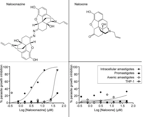
Differential activity of naloxone and naloxonazine, two m-opioid receptor antagonists, againstL. donovani
The Iconix collection contained two opioid receptor antago-nists, naloxone and naloxonazine. The first was not selected as a hit in any of the screens described above while the latter showed specific activity against the intracellular amastigote stage. To confirm these primary observations, the activity of both com-pounds was tested against promastigotes, intracellular and axenic amastigotes. Naloxonazine exhibited specific activity against intracellular amastigotes (GI50intracellular amastigote: 3.45mM;
GI50THP-1: 33.8mM; GI50promastigote:.50mM; GI50axenic
amastigote: .50mM), while naloxone was inactive against all parasite forms and not toxic to the host macrophage (Figure 4). At a curative concentration, the selectivity window of naloxonazine was reduced (GI90intracellular amastigote: 12.5mM; GI90THP-1:
50mM), limiting the possibility of using naloxonazine for treatment.
Discussion
Current chemotherapy for Leishmaniasis has several draw-backs, including cost, toxicities, route of administration, and the emergence of drug resistance. The pipeline for anti-leishmanial drugs therefore needs to be filled with new compounds. As the discovery of new and original leads suitable for optimization and drug development is dependent on the ability to screen many compounds, assays should be rapid, inexpensive and reproducible [14]. In addition, for pathogens displaying several life stages like
Leishmania, there is a need to determine the best parasite stage to target. In the case ofLeishmaniathere are three major options: first, Figure 2. Treatment of infected THP-1 with DMSO and amphotericin B.A. Number of infected THP-1 counted per well treated or not with 1% DMSO or 2mM amphotericin B. B. Number of parasites counted per well divided by the number of host nuclei per field. C. Dose response curve for amphotericin B plotting the percentage of parasite growth inhibition. Values are mean from at least 3 independent experiments.
doi:10.1371/journal.pntd.0001253.g002
Table 1.Host cell toxicity and efficacy of compounds againstL. donovaniintracellular amastigotes, promastigotes and axenic amastigotes.
Compounds
GI50THP-1
(mM)
GI50Intracellular
amastigote (mM)
GI50Promastigote
(mM)
GI50Axenic
amastigote (mM) Selectivitya Specificityb
Naloxonazine 33.8 3.49 .50 .50 9.68 14.32
Metergoline 37.14 0.78 15.08 .50 47.22 19.17
R(-)-Apomorphine .16.5 2.7 38.15 5.20 6.1 14.12
Clotrimazole .16.5 1.08 12.92 4.56 15.32 11.92
Cetylpyridinium chloride 9.16 0.41 3.30 0.92 22.34 8.05
Aminacrine 12.97 1.9 8.96 .16.5 6.82 4.71
4,49-Diethylaminoethoxyhexestrol 6.11 1.4 5 10.93 4.36 3.57
Hexachlorophene 16.5 4.54 15.88 14.76 3.62 3.49
Loperamide Hydrochloride .16.5 5.4 17.3 .50 3.02 3.15
Tamsulosin 27.97 6.3 20 .50 4.43 3.17
Salinomycin 16.09 0.92 2.71 0.06 17.30 2.92
Cycloheximide 21 1.15 2.95 0.39 18.13 2.55
Prazocin Hydrochloride 23.32 6.73 16.74 .50 3.46 2.48
Carvedilol 37.5 6.16 12.41 .16.5 6.07 2.01
Brilliant green 1.64 6 0.10 0.03 15.88 0.02
Antimycin A .50 2.15 0.1 0.01 500 0.05
BAY 11–7085 .50 .16,5 1.54 0.42 32.46 0.06
Haloprogin 20 6 1.05 0.56 19.04 0.17
Pentamidine Isethionate .50 9.55 1.69 1.64 29.49 0.17
Zinc Dibutyldithiocarbamate .50 .50 8.48 .50 5.89 0.17
Amlodipine .16.5 9.78 3.11 27 5.34 0.31
Nisoldipine .16,5 32.17 9.85 13.86 1.67 0.30
Parthenolide .16.5 .50 15.54 4.35 1.06 0.31
Pyrithione zinc 1.043 ND 0.07 0.01 14.9 ND
Thimerosal 12.81 ND 0.49 1.50 26.18 ND
Gramicidin 8.38 ND 0.68 5.76 12.19 ND
Digitonin 13.46 ND 4.376 13.81 3.075 ND
Emetine 1.28 0.082 0.03 1.62 15.58 0.42
Amphotericin B .50 1.12 1.61 5.42 44.52 1.43
Chlorhexidine .16,5 1.79 2.81 4.78 9.27 1.56
Oxiconazole .16,5 6.6 2.65 0.53 2.51 0.40
Bazedoxifene .16,5 4.8 6.52 .16.5 3.45 1.35
chlorquinaldol 39.3 5.5 6.3 1.72 7.14 1.14
Doxazocin .16,5 5 8.45 .16.5 3.32 1.69
Aclacynomycine a1 29.1 5.81 9.15 .16.5 5 1.57
Mebeverine .50 8.5 6.91 2.36 5.88 0.81
Miconazole .16,5 15.44 7.48 4.73 1.07 0.48
Terfenadine 39.03 14.4 11.92 0.61 2.71 0.82
Tamoxifen Citrate .16.5 20.79 10.22 .50 1.61 0.49
Auranofin 20.89 21.76 11.11 .50 1.88 0.51
Benzetonium chloride 40.18 10 11.34 10.7 3.54 1.13
Ciclopirox 42 22 15.25 .50 1.90 0.69
Sporidesmin A 0.02 ND 1.18 1.66 0.02 ND
targeting the extracellular living promastigote stage, second the axenic amastigote and third the intracellular amastigote stage. The first and second options meet the reproducibility, rapidity and low cost requirements for high-throughput screens, due to the ease in manipulating promastigotes or axenic amastigotesin vitro. This has been demonstrated by Siqueira-Netoet al. and Sharlowet al. who described screening 26,500 and 200,000 compounds against the
Leishmania promastigote stage, respectively [11,12]. These assays monitored parasite viability by measuring products from meta-bolically active cells (The Alamar Blue assay involves reduction of resazurin into fluorescent resorufin by live cells, while CellTiter-Glo luciferase catalyzes the production of luminescence in the
presence of cellular ATP). The axenic amastigotes offer the ability to screen easily the relevant-like stage of the parasite and allow testing the potency of compounds under low pH conditions. However, the major disadvantage of these two approaches is the absence of the host cell in the assays: the natural niche of the parasite is not taken into account and aspects of parasite biology such as host-parasite interactions or accessibility of the target are ignored.
The intracellular amastigote stage has been logically designated as the more relevant target for primary screening against
Leishmania, but previous methods were labor intensive and would not support automation [19,28]. Methods traditionally used to
Figure 4. Structure and activity of naloxonazine and naloxone.Lower panels: Dose response curve for naloxonazine (left) and naloxone (right) against intracellular amastigotes (black diamonds), promastigotes (black squares), axenic amastigotes (white diamonds) and THP-1 (white triangles) plotting the percentage of parasite growth inhibition.
doi:10.1371/journal.pntd.0001253.g004 Compounds
GI50THP-1
(mM)
GI50Intracellular
amastigote (mM)
GI50Promastigote
(mM)
GI50Axenic
amastigote (mM) Selectivitya Specificityb
3,39,49,5-tetrachlorosalicylanilide 0.32 ND 1.45 1.38 0.22 ND
Thiram 1.58 10.4 4.62 5.42 0.34 0.44
Idarubicin 1.89 ND 4.65 1.78 0.40 ND
Cerivastatin 7.15 .50 10.8 0.02 0.66 0.22
a
Selectivity is the ratio between parasite GI50and THP-1 GI50.
b
Specificity is the ratio between promastigote GI50and intracellular amastigote GI50. Specificity value.2 was the cut-off chosen to define a compound as more active
against the intracellular amastigote stage; while a specificity value,0.4 indicated a compound more active against promastigotes; compounds with specificity values between 0.4 and 2 were considered active against both stages.
doi:10.1371/journal.pntd.0001253.t001
detect and estimate the number of intracellularLeishmaniainclude Giemsa staining or the use of reporter gene-expressing parasites (green fluorescent protein, luciferase or beta-galactosidase) [29,30]. Giemsa staining is cumbersome as it needs manual counting. Reporter gene-expressing parasites are a powerful alternative, but stable recombinant parasite populations are required and they do not allow concomitant analysis of the host cell.
Here we describe a high content assay that allows the simultaneous visualization of both host cell nuclei and parasite kinetoplasts by using the DNA marker DAPI. The significant difference in the size of these two organelles facilitates discrimi-nation between host cells and parasites, and thus accurate counting of both entities. Reduction in the number of kinetoplasts gives a measure of inhibition of parasite growth, while a reduction in the number of host cell nuclei is indicative of compound cytotoxicity. Thus, this image-based assay allows the identification of leishmaniocidal as well as leishmaniostatic compounds. All steps of this assay are amenable to automation and could be reduced to 384-well format resulting in a robust high-throughput screening methodology.
To evaluate what differences might be obtained from screening against the extracellularversusthe intracellular parasite stages, we screened the same set of 909 compounds against bothL. donovani
promastigotes and intracellular amastigotes. We observed that the majority of the hits identified with the intracellular amastigote screen, defined as inducing 60% parasite growth inhibition at 10mM, were also found in the promastigote screen. One compound showed specific inhibition of intracellular amastigote and was completely inactive against promastigotes. Fifty-six percent of the hits from the promastigote screen were not found in the intracellular amastigote screen. These results indicated that a promastigote screen failed to identify all active compounds and led to 56% of compounds being likely false positives. Thus, while the promastigote stage appears suitable for high-throughput screening, a fraction of the hits would be missed; furthermore, a high rate of false positives is characteristic of primary screens against promastigotes, underlying the importance of evaluating compound activity against intracellular amastigotes at least in a secondary screen. This is in accordance with the findings of Siqueira-Netoet al.who found that only 4% of their hits identified in a promastigote primary screen were active in an intracellular context [11]. Advantages of the intracellular amastigote assay include cell-health information, very low cost of consumables and a reduced necessity for secondary assays. The importance of the host cell in the assay was also demonstrated by the dose response study against axenic amastigotes (i.e.amastigote-like stage obtained from differentiation of promastigotes in vitroin the absence of a host cell); although this parasite form should mimic the intracellular stage, the activity of compounds against axenic amastigotes mostly correlated with promastigotes rather than intracellular amastigotes.
A similar assay was also successfully developed for screening drugs against the intracellular stage of the related parasite
Trypanosoma cruzi [31]. Screening the Iconix library against intracellularT. cruziidentified 56 hits, among which 8 were also hits in the Leishmania screen presented here. Six of these were found to be more active against the intracellular amastigote stage ofL. donovanicompared to promastigotes, indicating inter-species activity of compounds only for the intracellular stages of these two different parasites.
Fifty percent of the compounds that were preferentially active against intracellular amastigotes are known to bind mammalian/ eukaryotic G protein coupled receptors (opioid receptors, serotonin or dopamine receptors and adrenergic receptors).
Heterotrimeric G proteins are absent in trypanosomatids [32] and we could not find convincing homologs of opioid receptors in the Leishmania genome. Compounds described as ligands of G protein coupled receptors may have different targets among parasitic proteins, leading to mechanisms of inhibition indepen-dent of the host cell. This is the case of a serotonin receptor agonist that interferes with P. falciparum growth by blocking a surface membrane channel [33], or a k-opioid agonist active against T. brucei whose target remains to be identified as no homolog of k-opioid receptor is found in the T. brucei genome [34]. However, the fact that the ligands of G protein coupled receptors identified in this study, showed a preferential activity towards the intracellular amastigote stage, also highlights the potential value of these host cell signaling pathways as targets. Previous reports demonstrated the involvement of such receptors in inhibition of infection by several intracellular pathogens includingLeishmania[35-37]. Targeting host factors essential for parasite development is an emerging drug discovery paradigm. It is assumed to be less likely to induce drug resistant pathogens and offers the possibility to repurpose drugs by exploiting compounds currently used for diseases unrelated to microbial infection [38,39].
Interestingly, one compound out of 909 was active against the intracellular amastigote stage but was completely inactive against promastigotes. This compound, naloxonazine, was also inactive against axenic amastigotes, indicating that its activity is dependent on a macrophage function. Naloxonazine is described as an irreversiblem1-opioid receptor antagonist [40]. There is evidence for the presence of opioid receptors on cells of the immune system [41,42] and it is known that opioids are involved in modulation of host resistance to infectious diseases [43,44]. The immune response of mice infected withL. donovani has been shown to be influenced by the opioid receptor agonist morphine, but the receptors involved and the mechanism leading to this immuno-modulation remain unknown [45]. Loperamide, a m-opioid receptor agonist, was also identified in this study as inhibiting parasite growth, and this compound was also more potent against the intracellular stage of the parasite. Anotherm-opioid receptor antagonist, naloxone, also present in the Iconix library, did not show any activity againstL. donovani. The differential selectivity of naloxone and naloxonazine for opioid receptor binding sites might explain their differential activity against intracellular L. donovani
[46]. Naloxone is monomer-like while naloxonazine appears as an inverted dimer (Figure 4). The presence of the macrophage appears to be essential for the activity of naloxonazine againstL. donovani, but the underlying cellular and molecular mechanisms remain to be elucidated. Although naloxonazine itself would not meet the requirement for a therapeutic drug due to the reduced selectivity window at a curative concentration, it would be interesting to analyze other compounds that target the same host cell pathway.
In summary, we report an automated screen against intracel-lular amastigotes ofL. donovani. It has the advantage of screening against the relevant stage of the parasite, taking into consideration crucial aspects of its biology, and giving the opportunity to identify host factors critical for the establishment of infection. This is essential for the identification of new, original and diverse lead compounds for anti-leishmanial therapy.
Acknowledgments
Author Contributions
Conceived and designed the experiments: GDM JCE JHM. Performed the experiments: GDM KKHA SC. Analyzed the data: GDM KKHA SC JCE
JHM. Contributed reagents/materials/analysis tools: MRA. Wrote the paper: GDM JCE MRA JHM.
References
1. Reithinger R, Dujardin JC, Louzir H, Pirmez C, Alexander B, et al. (2007) Cutaneous leishmaniasis. Lancet Infect Dis 7: 581–596.
2. Chappuis F, Sundar S, Hailu A, Ghalib H, Rijal S, et al. (2007) Visceral leishmaniasis: what are the needs for diagnosis, treatment and control? Nat Rev Microbiol 5: 873–882.
3. Handman E, Bullen DV (2002) Interaction of Leishmania with the host macrophage. Trends Parasitol 18: 332–334.
4. Croft SL, Seifert K, Yardley V (2006) Current scenario of drug development for leishmaniasis. Indian J Med Res 123: 399–410.
5. Kedzierski L, Sakthianandeswaren A, Curtis JM, Andrews PC, Junk PC, et al. (2009) Leishmaniasis: current treatment and prospects for new drugs and vaccines. Curr Med Chem 16: 599–614.
6. Singh S, Sivakumar R (2004) Challenges and new discoveries in the treatment of leishmaniasis. J Infect Chemother 10: 307–315.
7. Davis AJ, Kedzierski L (2005) Recent advances in antileishmanial drug development. Curr Opin Investig Drugs 6: 163–169.
8. Perez-Victoria FJ, Sanchez-Canete MP, Seifert K, Croft SL, Sundar S, et al. (2006) Mechanisms of experimental resistance of Leishmania to miltefosine: Implications for clinical use. Drug Resist Updat 9: 26–39.
9. Renslo AR, McKerrow JH (2006) Drug discovery and development for neglected parasitic diseases. Nat Chem Biol 2: 701–710.
10. Nwaka S, Hudson A (2006) Innovative lead discovery strategies for tropical diseases. Nat Rev Drug Discov 5: 941–955.
11. Siqueira-Neto JL, Song OR, Oh H, Sohn JH, Yang G, et al. (2010) Antileishmanial high-throughput drug screening reveals drug candidates with new scaffolds. PLoS Negl Trop Dis 4: e675.
12. Sharlow ER, Close D, Shun T, Leimgruber S, Reed R, et al. (2009) Identification of potent chemotypes targeting Leishmania majorusing a high-throughput, low-stringency, computationally enhanced, small molecule screen. PLoS Negl Trop Dis 3: e540.
13. St George S, Bishop JV, Titus RG, Selitrennikoff CP (2006) Novel compounds active againstLeishmania major. Antimicrob Agents Chemother 50: 474–479. 14. Fumarola L, Spinelli R, Brandonisio O (2004)In vitroassays for evaluation of
drug activity againstLeishmaniaspp. Res Microbiol 155: 224–230.
15. Doyle PS, Engel JC, Pimenta PF, da Silva PP, Dwyer DM (1991)Leishmania donovani: long-term culture of axenic amastigotes at 37 degrees C. Exp Parasitol 73: 326–334.
16. Bates PA, Robertson CD, Tetley L, Coombs GH (1992) Axenic cultivation and characterization ofLeishmania mexicanaamastigote-like forms. Parasitology 105 (Pt 2): 193–202.
17. Callahan HL, Portal AC, Devereaux R, Grogl M (1997) An axenic amastigote system for drug screening. Antimicrob Agents Chemother 41: 818–822. 18. Rochette A, Raymond F, Corbeil J, Ouellette M, Papadopoulou B (2009)
Whole-genome comparative RNA expression profiling of axenic and intracel-lular amastigote forms ofLeishmania infantum. Mol Biochem Parasitol 165: 32–47. 19. Buckner FS, Wilson AJ (2005) Colorimetric assay for screening compounds againstLeishmaniaamastigotes grown in macrophages. Am J Trop Med Hyg 72: 600–605.
20. Vermeersch M, da Luz RI, Tote K, Timmermans JP, Cos P, et al. (2009)In vitro
susceptibilities of Leishmania donovani promastigote and amastigote stages to antileishmanial reference drugs: practical relevance of stage-specific differences. Antimicrob Agents Chemother 53: 3855–3859.
21. Engel JC, Parodi AJ (1985)Trypanosoma cruzicells undergo an alteration in protein N-glycosylation upon differentiation. J Biol Chem 260: 10105–10110. 22. Goyard S, Segawa H, Gordon J, Showalter M, Duncan R, et al. (2003) Anin vitro
system for developmental and genetic studies ofLeishmania donovani phosphogly-cans. Mol Biochem Parasitol 130: 31–42.
23. Zhang JH, Chung TD, Oldenburg KR (1999) A Simple Statistical Parameter for Use in Evaluation and Validation of High Throughput Screening Assays. J Biomol Screen 4: 67–73.
24. Gebre-Hiwot A, Tadesse G, Croft SL, Frommel D (1992) Anin vitromodel for screening antileishmanial drugs: the human leukaemia monocyte cell line, THP-1. Acta Trop 51: 237–245.
25. Ogunkolade BW, Colomb-Valet I, Monjour L, Rhodes-Feuillette A, Abita JP, et al. (1990) Interactions between the human monocytic leukaemia THP-1 cell line and Old and New World species ofLeishmania. Acta Trop 47: 171–176. 26. Mackey ZB, Baca AM, Mallari JP, Apsel B, Shelat A, et al. (2006) Discovery of
trypanocidal compounds by whole cell HTS ofTrypanosoma brucei. Chem Biol Drug Des 67: 355–363.
27. Kangas L, Gronroos M, Nieminen AL (1984) Bioluminescence of cellular ATP: a new method for evaluating cytotoxic agentsin vitro. Med Biol 62: 338–343. 28. Sereno D, Cordeiro da Silva A, Mathieu-Daude F, Ouaissi A (2007) Advances
and perspectives inLeishmaniacell based drug-screening procedures. Parasitol Int 56: 3–7.
29. Neal RA, Croft SL (1984) Anin-vitrosystem for determining the activity of compounds against the intracellular amastigote form of Leishmania donovani. J Antimicrob Chemother 14: 463–475.
30. Dube A, Gupta R, Singh N (2009) Reporter genes facilitating discovery of drugs targeting protozoan parasites. Trends Parasitol 25: 432–439.
31. Engel JC, Ang KK, Chen S, Arkin MR, McKerrow JH, et al. (2010) Image-based high-throughput drug screening targeting the intracellular stage of
Trypanosoma cruzi, the agent of Chagas’ disease. Antimicrob Agents Chemother 54: 3326–3334.
32. El-Sayed NM, Myler PJ, Bartholomeu DC, Nilsson D, Aggarwal G, et al. (2005) The genome sequence ofTrypanosoma cruzi, etiologic agent of Chagas disease. Science 309: 409–415.
33. Locher CP, Ruben PC, Gut J, Rosenthal PJ (2003) 5HT1A serotonin receptor agonists inhibit Plasmodium falciparum by blocking a membrane channel. Antimicrob Agents Chemother 47: 3806–3809.
34. Jones DC, Hallyburton I, Stojanovski L, Read KD, Frearson JA, et al. (2010) Identification of a kappa-opioid agonist as a potent and selective lead for drug development against human African trypanosomiasis. Biochem Pharmacol 80: 1478–1486.
35. Singal P, Kinhikar AG, Singh S, Singh PP (2002) Neuroimmunomodulatory effects of morphine inLeishmania donovani-infected hamsters. Neuroimmunomo-dulation 10: 261–269.
36. Rahman MA, Azuma Y, Fukunaga H, Murakami T, Sugi K, et al. (2005) Serotonin and melatonin, neurohormones for homeostasis, as novel inhibitors of infections by the intracellular parasitechlamydia. J Antimicrob Chemother 56: 861–868.
37. Gets J, Monroy FP (2005) Effects of alpha- and beta-adrenergic agonists on
Toxoplasma gondiiinfection in murine macrophages. J Parasitol 91: 193–195. 38. Schwegmann A, Brombacher F (2008) Host-directed drug targeting of factors
hijacked by pathogens. Sci Signal 1: re8.
39. Jayaswal S, Kamal MA, Dua R, Gupta S, Majumdar T, et al. (2010) Identification of host-dependent survival factors for intracellularMycobacterium tuberculosisthrough an siRNA screen. PLoS Pathog 6: e1000839.
40. Hahn EF, Pasternak GW (1982) Naloxonazine, a potent, long-lasting inhibitor of opiate binding sites. Life Sci 31: 1385–1388.
41. Bidlack JM, Khimich M, Parkhill AL, Sumagin S, Sun B, et al. (2006) Opioid receptors and signaling on cells from the immune system. J Neuroimmune Pharmacol 1: 260–269.
42. Sharp BM, Roy S, Bidlack JM (1998) Evidence for opioid receptors on cells involved in host defense and the immune system. J Neuroimmunol 83: 45–56. 43. Roy S, Wang J, Kelschenbach J, Koodie L, Martin J (2006) Modulation of
immune function by morphine: implications for susceptibility to infection. J Neuroimmune Pharmacol 1: 77–89.
44. Pacifici R, Minetti M, Zuccaro P, Pietraforte D (1995) Morphine affects cytostatic activity of macrophages by the modulation of nitric oxide release. Int J Immunopharmacol 17: 771–777.
45. Singh PP, Singal P (2007) Morphine-induced neuroimmunomodulation in murine visceral leishmaniasis: the role(s) of cytokines and nitric oxide. J Neuroimmune Pharmacol 2: 338–351.