Variants in a Novel Epidermal Collagen
Gene
(COL29A1)
Are Associated with
Atopic Dermatitis
Cilla So¨derha¨ll1,2[, Ingo Marenholz1,2[, Tamara Kerscher1,2, Franz Ru¨schendorf2, Jorge Esparza-Gordillo1,2,
Margitta Worm3, Christoph Gruber1, Gabriele Mayr4, Mario Albrecht4, Klaus Rohde2, Herbert Schulz2, Ulrich Wahn1, Norbert Hubner2, Young-Ae Lee1,2*
1Department of Pediatric Pneumology and Immunology, Charite´ Universita¨tsmedizin Berlin, Germany,2Max-Delbru¨ck-Centrum for Molecular Medicine, Berlin, Germany,
3Department of Dermatology and Allergology, Charite´ Universita¨tsmedizin Berlin, Germany,4Max Planck Institute for Informatics, Saarbru¨cken, Germany
Atopic dermatitis (AD) is a common chronic inflammatory skin disorder and a major manifestation of allergic disease. AD typically presents in early childhood often preceding the onset of an allergic airway disease, such as asthma or hay fever. We previously mapped a susceptibility locus for AD on Chromosome 3q21. To identify the underlying disease gene, we used a dense map of microsatellite markers and single nucleotide polymorphisms, and we detected association with AD. In concordance with the linkage results, we found a maternal transmission pattern. Furthermore, we demonstrated that the same families contribute to linkage and association. We replicated the association and the maternal effect in a large independent family cohort. A common haplotype showed strong association with AD (p¼ 0.000059). The associated region contained a single gene, COL29A1, which encodes a novel epidermal collagen.
COL29A1 shows a specific gene expression pattern with the highest transcript levels in skin, lung, and the
gastrointestinal tract, which are the major sites of allergic disease manifestation. Lack ofCOL29A1expression in the outer epidermis of AD patients points to a role of collagen XXIX in epidermal integrity and function, the breakdown of which is a clinical hallmark of AD.
Citation: So¨derha¨ll C, Marenholz I, Kerscher T, Ru¨schendorf F, Esparza-Gordillo J, et al. (2007) Variants in a novel epidermal collagen gene(COL29A1)are associated with atopic dermatitis. PLoS Biol 5(9): e242. doi:10.1371/journal.pbio.0050242
Introduction
Atopic dermatitis (AD) is a chronic inflammatory skin disease that is characterized by intensely itchy skin lesions. AD is one of the most common chronic diseases in childhood affecting 10%–20% of children in industrialized societies [1], with a steady increase over the past decades [2,3]. Along with asthma and hay fever, AD is commonly associated with an abnormal immune response and the formation of allergy antibodies (IgE) against innocuous environmental allergens.
AD is often the first clinical manifestation of allergic disease. The onset of disease is typically observed during the first two years of life [4]. For the majority of affected children, AD heralds a lifetime of allergic disease. A susceptible child commonly passes a characteristic sequence of transient or persistent disease stages that is known as the‘‘atopic march’’ which begins with AD and food allergy in the young infant and continues with the development of allergic airways disease later in childhood and adulthood [5]. The close familial and intra-individual association of the allergic disorder strongly suggests shared genetic etiology.
A strong genetic component in allergic disorders has been recognized almost a century ago. Cooke and van der Veer first reported that the relatives of patients are at significantly increased risk of developing allergic disease [6]. Even today, a positive family history for allergic disorders is the single strongest predictor for the development of AD [7]. Additional evidence for the importance of genetic factors in atopic disease comes from twin studies. The concordance rate for AD among monozygotic twins of about 80% far exceeds the
concordance rate of 20% observed among dizygotic twins [8,9]. These data clearly indicate that the genetic contribution to the expression of AD is substantial. Furthermore, studies on the vertical transmission of AD and atopic disease show that children are more likely to inherit these disorders if the mother is affected (parent-of-origin effect) [10]. The pre-dominance of maternal inheritance may be due to environ-mental factors such as uterine milieu or breast feeding, but they may also arise due to genetic mechanisms such as parent-specific gene expression (genomic imprinting) [11].
AD and atopic disorders are regarded as multifactorial conditions, the onset and severity of which are influenced by both genetic and environmental factors. The data are consistent with an immune etiology shared by all allergic diseases and a congenital target organ defect, the penetrance of which is modified by multiple environmental factors
Academic Editor: Gonc¸alo Abecasis, University of Michigan, United States of America
ReceivedFebruary 9, 2007;AcceptedJuly 9, 2007;PublishedSeptember 11, 2007
Copyright:Ó2007 So¨derha¨ll et al. This is an open-access article distributed under the terms of the Creative Commons Attribution License, which permits unrestricted use, distribution, and reproduction in any medium, provided the original author and source are credited.
Abbreviations:AD, atopic dermatitis; cM, centiMorgan;COL29A1, collagen XXIX gene; ECM, extracellular matrix; LD, linkage disequilibrium; RT-PCR, reverse-transcriptase PCR; SNP, single nucleotide polymorphism; vWA, von Willebrand factor–type A domain
* To whom correspondence should be addressed. E-mail: [email protected]
during early childhood. The identification of genes under-lying AD and allergic disorders has the capacity to define primary physiologic mechanisms, thereby clarifying disease pathogenesis, identifying pathways and targets for therapeu-tic intervention.
Although several genome-wide linkage screens for AD have been conducted [12–14], there was no substantial overlap between the identified regions of highest linkage, and the underlying genes remained elusive [15]. We have previously mapped a major susceptibility locus for AD on Chromosome 3q21 [16]. Here we report the identification and character-ization of a novel epidermal collagen gene as the underlying disease gene.
Results
To narrow the candidate region spanning 12.75 centiMor-gans (cM) (13.5 Mb) on Chromosome 3q21 (Figure 1A), 96 additional microsatellite markers at an average distance of 140 kb were genotyped in 199 affected sibling families with AD from the original linkage scan. Linkage analysis yielded a 1-lod support interval of 5.4 Mb between the markers M3CS075 and M3CS233 (Figure 1B). Subsequently, we con-ducted an association scan of the 5.4-Mb region using 212 single nucleotide polymorphisms (SNPs) at an average distance of 25.5 kb (Table S1). The SNPs were selected primarily to cover known and predicted genes. Because we had observed a strong maternal effect in the linkage study [16], we chose a family-based association analysis that allowed us to search for risk alleles preferentially transmitted from the mother [17]. Two adjacent SNPs, rs5852593 (p¼0.0079) and rs1497309 (p¼0.016), located 36 kb apart, were associated with AD (Table S1 and Figure 1B). To define the critical region, we typed 16 additional SNPs. Eight of these markers showed association with AD and a maternal transmission pattern to affected children, which was consistent with our previous linkage results (Table 1). The strongest association with a single marker was observed for rs4688761 (pall¼0.0016,
pmaternal¼0.0006). We selected eight markers spanning 96 kb
that were associated with AD and carried nonredundant information based on the linkage disequilibrium (LD) in the region, and we performed haplotype analysis which confirmed the results (transmissions (T)all:non-transmissions (NT)all ¼
222:168,pall¼0.0076, Tmat:NTmat¼105:68,pmaternal¼0.0070). In addition, we assessed the significance of the difference in maternal-versus-paternal haplotype over transmissions em-pirically using the permutation procedure for parent-of-origin transmission disequilibrium test (TDT) implemented in PLINK (P_POOmaternal emp¼0.014) [18].
Next, we investigated whether the observed association accounted for the linkage in the region. We used the marker that had shown the strongest association, rs4688761, to identify 73 of the 199 families from the original linkage scan in whom the disease-associated allele had been transmitted to affected offspring. Nonparametric linkage analysis in the 73 associated families yielded significant evidence for linkage (Zall¼4.18 versus 4.31 in the complete cohort), demonstrating that the majority of the linkage signal was attributable to these families. The significance of this finding was assessed by performing nonparametric linkage analysis in 10,000 random selections of 73 families. An empirical significance level was calculated as the proportion of replicates for which the maximumZallscore was equal or greater than that obtained in the actual analysis. The probability of obtaining aZallscore of4.18 by chance in a random selection of 73 families was estimated by 10,000 simulations to be 0.027.
To confirm the association result, we used a large independent replication set consisting of 292 complete nuclear families including 481 children with AD. We genotyped the selected eight SNPs covering an interval of 96 kb that were associated with AD in the discovery dataset. We confirmed the association with AD across all markers with the strongest association with AD observed for marker A36637742 (p¼0.00038), which also showed a significant overtransmis-sion of the maternal allele (p ¼ 0.0013) (Table 2). The association remained significant after correction for multiple testing. For each marker, it was the more common allele that was overtransmitted. Haplotypes were constructed over the region, which confirmed the association with AD and showed that this phenotype is associated with the overtransmission of the most common haplotype (haplotype frequency 65%, Table 3), and of the maternal allele (pmaternal¼0.000025 for AD, P_POOmaternal emp¼0.033). Next, we compared the AD status among the parents: in the discovery cohort, significantly more mothers (n¼63) than fathers (n¼19) suffered from AD (odds ratio [OR] 4.39, 95% confidence interval [CI] 2.51 – 7.68,
p¼5.463108). Similarly, in the replication cohort, mothers (n¼83) were significantly more frequently affected with AD than fathers (n¼55) (OR 1.71, 95% CI 1.16 – 2.52,p¼8.33 103). To assess whether the parent-of-origin effect that was observed originated from the discrepancy in AD prevalence among the parents, we compared haplotype transmissions from affected and unaffected mothers. In the discovery cohort, the excess in maternal transmissions was not predominantly attributable to affected mothers (T:UT ¼
24:21,p¼0.14) compared to unaffected ones (T:UT¼81:47,
p¼0.0059). Similarly, affected mothers (T:UT¼25:17,p¼0.14) of the replication cohort did not contribute a larger trans-mission excess than the unaffected ones (T:UT¼70:29, p¼
0.00013). We conclude that the observed maternal
over-Author Summary
transmission pattern in both cohorts was not due to the higher AD prevalence among mothers.
A database search within the associated 96-kb interval revealed a single predicted gene,FLJ35880, extending 11.6 kb into the associated region. No other expressed sequence tag was detected. To identify any additional transcripts, we used putative exons predicted with The National Center for Biotechnology Information (NCBI) Modelmaker within and bordering the critical region to perform rapid amplification of cDNA ends (RACE) from human skin mRNA. We thus identified a single transcript of 9226 bp that consisted of 42 exons (Figure 1D) and included all eight exons ofFLJ35880. The corresponding gene spanned 139 kb of genomic sequence and completely encompassed the associated region. Pairwise LD measures (D9) of the genotyped markers indicated that the gene was contained within one 170-kb region of increased LD (Figure 2), whereas the neighboring genes,LOC440978andLOC131873, were located in separate blocks. The LD structure was consistent with the data for the European population in the HapMap database (Figure S1) [19]. The size of the transcript was confirmed by Northern blot hybridization with anFLJ35880-specific probe detecting
a single transcript of 9.6 kb in human skin mRNA (unpublished data), which was in good agreement with the RACE experiments.
The open reading frame yielded a protein of 2614 amino acids with an estimated molecular weight of 289.9 kDa. The predicted protein contained a collagenous domain in the central part and was therefore classified as a new member of the collagen superfamily, collagen XXIX. A BLAST search revealed the human collagen VI alpha 3 chain as its closest neighbor (32% identity). Homology with collagen VI alpha 3 was further strengthened by a similar domain architecture consisting of six N-terminal and three C-terminal von Willebrand factor–type A domains (vWAs) flanking a short collagen triple helix (Figure 1E), and an 18–amino acid secretion signal [20]. Sequence analysis of all 42 exons in 46 unrelated children with AD and 2 unaffected individuals revealed 13 common and six rare sequence variations (frequency,2%) predicted to cause nonsynonymous amino acid substitutions (Table S2). Four variants were located within the triple helix repeat of a collagen domain, however none of them changed the first glycine residue in the repeating sequence patterns (Gly-X-Y), which is critical for
Figure 1.Positional Cloning Strategy for the AD Disease Gene on Chromosome 3q21
(A) The candidate region spanned 12.75 cM between markersD3S1303andD3S1292. The y-axis depicts theGENEHUNTER nonparametric Zall as
previously reported [16].
(B) Fine mapping with 96 microsatellite markers narrowed the interval to 5.4 Mb between markersM3CS075andM3CS233. An association scan using 212 SNPs of the region revealed association of AD with two adjacent SNPs, rs5852593 and rs1497309. Genotyping of 16 additional SNPs refined the associated region.
(C) Genomic positions of the 42 exons ofCOL29A1are shown. The gene entirely encompasses the associated region. (D) TheCOL29A1mRNA consists of 9226 bp. Translation start site and stop codon are indicated.
(E) The predicted open reading frame encodes a protein of 2614 amino acids including a secretion peptide (SP), six N-terminal and three C-terminal vWAs, flanking a short collagen triple helix.
triple helix formation [21]. In addition, we performed in silico comparisons of the six variants located within a vWA with the crystal structure of the human vWF A3 domain. One variant, V669G, affects an amino acid within a highly conserved stretch of eight amino acids. The mutation of an adjacent amino acid within this conserved region of a vWA in the homologous geneCOLVIA1 has been reported to cause the monogenic muscle disorder Bethlem myopathy [22]. This variant, however was rare (allele frequency 1,3%). One additional variant (E455K) is located in a region predicted to be important for integrin–collagen interaction, and another one (M56T) is located in a helix near a rather conserved region that may also affect protein–protein
interaction. All coding SNPs were genotyped in the discovery cohort. Four of them showed a positive association with AD that did, however, not account for the observed haplotype association (Table S2).
Gene expression analysis in human tissues revealed a tissue-specific expression pattern of COL29A1. The highest expression was observed in the skin, but also in the lung, small intestine, colon, and testis (Figure 3A). Overall, COL29A1
expression is moderately low compared to more abundant epidermal transcripts such as keratin 10 (unpublished data). To specify the expression sites in the layers of the skin and to assess the role of collagen XXIX in AD, we performed in situ hybridization using aCOL29A1-specific cRNA-probe on
Table 1.Results of the ASPEX sib_TDT for SNPs in the Region ofCOL29A1for AD in the Discovery Cohort
Locus Position on Chromosome 3
Location in
COL29A1
MAF Major/ Minor Allele
All Transmissions Paternal Maternal
T NT v2 p-Value T NT v2 p-Value T NT v2 p-Value
rs1349747 131,543,410 59 18.0 A/G 129 99 3.95 n.s. 48 43 0.27 n.s. 72 47 5.25 n.s. rs1869319 131,547,433 exon 1 9.4 C/T 80 63 2.02 n.s. 36 32 0.24 n.s. 80 63 2.38 n.s. rs13095825 131,549,247 intron 1 36.6 T/C 222 167 7.78 0.0072 84 56 5.6 0.024 99 72 4.26 0.047 rs1453243 131,575,875 intron 2 30.5 G/A 187 149 4.3 n.s. 68 49 3.09 n.s. 91 72 2.21 n.s. rs5852593 131,580,335 intron 3 24.3 del/T 157 111 7.9 0.0079 71 45 5.83 0.026 67 47 3.51 0.045 rs16845861 131,581,039 exon 4 24.4 G/A 166 123 6.4 0.018 68 49 3.09 n.s. 79 55 4.3 0.037 rs1453242 131,586,229 intron 4 24.3 C/T 171 123 7.84 0.0099 75 51 4.57 0.045 79 55 4.3 0.036 rs10212372 131,586,896 exon 5 24.4 A/G 165 123 6.12 0.021 68 50 2.75 n.s. 78 54 4.36 0.037 A36603217 131,590,753 intron 6 22.6 A/G 161 118 6.63 0.015 73 56 2.24 n.s. 72 46 5.73 0.02 rs10934938 131,596,325 intron 7 22.0 T/G 156 114 6.53 0.013 71 54 2.31 n.s. 72 47 5.25 0.019 rs4688761 131,607,679 exon 16 28.6 T/C 187 128 11.05 0.0016 76 56 3.03 n.s. 79 40 12.78 0.0006 A36624476 131,612,012 exon 20 13.5 G/C 109 83 3.52 n.s. 50 44 0.38 n.s. 50 30 5 0.028 rs1497309 131,616,310 intron 22 28.7 C/T 185 137 7.16 0.016 76 57 2.71 n.s. 76 47 6.84 0.013 rs7628563 131,617,253 intron 23 15.1 A/G 123 97 3.07 n.s. 53 38 2.47 n.s. 53 42 1.27 n.s. A36637742 131.625.270 intron 28 27.8 del/T 184 146 4.4 0.05 75 62 1.44 n.s. 75 53 4.43 0.040 rs1566136 131,634,657 intron 33 28.4 A/G 191 154 3.97 n.s. 85 64 2.96 n.s. 76 60 1.88 n.s. rs9883988 131,645,085 exon 36 6.3 G/A 56 35 4.85 0.049 31 18 3.45 n.s. 19 11 2.13 n.s. rs322113 131,670,507 exon 38 7.8 C/T 46 52 0.37 n.s. 27 25 0.08 n.s. 17 25 1.52 n.s. rs322115 131,672,716 intron 39 18.2 A/T 116 99 1.34 n.s. 48 45 0.1 n.s. 52 38 2.18 n.s. rs819091 131,678,260 intron 40 23.7 G/A 153 118 4.52 n.s. 67 55 1.18 n.s. 62 39 5.24 0.025 rs7624712 131,686,432 39 11.6 A/G 75 74 0.01 n.s. 33 38 0.35 n.s. 37 31 0.53 n.s.
MAF, minor allele frequency. T, number of transmissions. NT, number of nontransmissions. n.s., not significant.
doi:10.1371/journal.pbio.0050242.t001
Table 2.Association Analysis of Single Markers for Atopic Dermatitis in the Replication Dataset
Locus Major/ Minor
Allele
All Transmissions Paternal Maternal
T NT v2 p-Valuea T NT v2 p-Value T NT v2 p-Value
rs13095825 T/C 218 174 4.94 0.028 81 66 1.53 0.18 95 66 5.22 0.022
rs16845861 G/A 177 119 11.36 0.00077* 68 46 4.25 0.038 82 46 10.12 0.0014* rs10212372 A/G 174 117 11.16 0.00093* 68 46 4.25 0.035 81 46 9.65 0.0017*
A36603217 A/G 180 118 12.9 0.00040* 81 52 6.32 0.013 77 44 9 0.0023*
rs10934938 T/G 182 122 11.84 0.00063* 83 54 6.14 0.019 76 45 7.94 0.005*
rs4688761 T/C 208 145 11.24 0.00082* 94 67 4.53 0.03 88 52 9.26 0.0031*
A36637742 del/T 204 139 12.32 0.00038* 84 57 5.17 0.028 89 51 10.31 0.0013*
rs9883988 G/A 75 43 8.68 0.0019* 37 23 3.27 0.055 37 19 5.79 0.0091
a
p-values of the ASPEX sib TDT for atopic dermatitis for all, paternal, and maternal transmissions for the major allele. None of the paternal transmissions were significant after simulation.* p-values significant after correction for multiple testing.
skin biopsies of five patients with AD and five healthy patients (controls). In normal skin,COL29A1was exclusively expressed in the epidermis with the strongest staining in the suprabasal viable layers. In contrast, the skin of patients with AD revealed a striking absence ofCOL29A1mRNA staining in the
most differentiated upper spinous and granular layers (Figure 3B and Figure S2). No significant difference in the expression of COL29A1 was observed comparing patients with AD to controls (1.28-6 0.53-fold down-regulation in AD patients,
p ¼ 0.41) using quantitative Taqman reverse-transcriptase
Figure 2.Pairwise LD Values (D9) Between 28 SNPs Based on Genotypes of the Founders in the Discovery Cohort
Boxes contain the LD values (D9) between the respective markers indicated on top. Higher LD values correspond to a darker shade of red. Positions on Chromosome 3 are given in Mb; 131.547 denotes the start and 131.686 the end ofCOL29A1on the genomic sequence. Boxes on the horizontal bar represent the 42 exons ofCOL29A1.
doi:10.1371/journal.pbio.0050242.g002
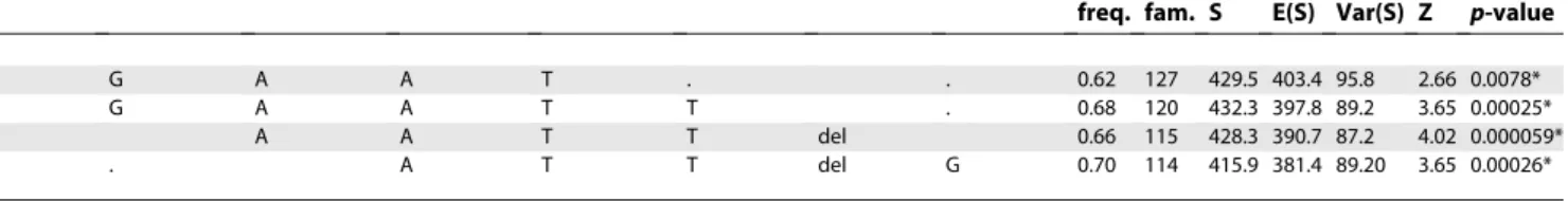
Table 3.Haplotype Analysis for AD in the Replication Dataset
rs13095825 rs16845861 rs10212372 A36603217 rs10934938 rs4688761 A3667742 rs9883988 Atopic Dermatitis
freq. fam. S E(S) Var(S) Z p-value
T G A A T . . 0.62 127 429.5 403.4 95.8 2.66 0.0078*
. G A A T T . 0.68 120 432.3 397.8 89.2 3.65 0.00025*
A A T T del 0.66 115 428.3 390.7 87.2 4.02 0.000059*
. . A T T del G 0.70 114 415.9 381.4 89.20 3.65 0.00026*
fbat statistic (Z) for 5-marker haplotype transmissions in the replication set adjusted for the correlation among siblings, reporting haplotype frequency (freq.) the number of informative families (fam.), the distribution (S), the expected distribution (E(S)), and the variance (Var(S)). *p-values significant after correction for multiple testing.
(RT)-PCR. These results indicate that while differences in mRNA quantity were not detected, AD patients show a distinct abnormal cellular distribution pattern ofCOL29A1
expression in the differentiated outer epidermis.
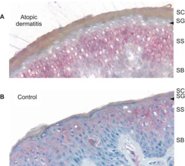
We generated a polyclonal antibody to visualize the collagen XXIX protein in the skin of five patients with AD and five normal controls, including four and three of each group, respectively, in whom in situ analysis was performed. Consistent with the in situ findings, we observed collagen XXIX staining in the differentiated suprabasal layers of the epidermis in normal human skin and a remarkable absence of staining in the most differentiated upper spinous and granular layers (Figure 4 and Figure S3).
Discussion
In a whole-genome linkage scan for AD, we previously identified a susceptibility locus on human Chromosome 3q21. The candidacy of this chromosomal region was further supported by Bradley et al., who mapped a locus for AD severity in close proximity (3q14) in a Swedish population [13]. By positional cloning, we have now identified the disease-causing gene, COL29A1, which encodes a novel epidermal collagen.
The disease gene was located in a two-staged investigation consisting of systematic linkage and association scanning of the region and subsequent confirmation of the association in a large, independent, replication dataset. In the first stage, we genotyped additional microsatellite markers in the candidate region, which narrowed the initial 12.75-cM linkage interval to 5.4 Mb. The association analysis with an average marker distance of 25.5 kb, finally, revealed an association that was confined to a haplotype block of 170 kb, which included a single gene, COL29A1. Pairwise LD measures indicated LD across the entire gene and defined two subblocks of increased LD. The strongest association was observed within the 96-kb subblock encoding the collagenous domain and the C terminus of collagen XXIX. A rapid decay of LD at the borders of the COL29A1 haplotype block and lack of association of the SNPs located within the neighboring genes clearly limited the association to COL29A1. In addition, we demonstrated in the discovery dataset that the families that contributed to the association of SNP rs4688761 with AD also accounted for most of the linkage signal. This finding corroborated that variants in COL29A1 explained the
Figure 3.Gene Expression Analysis ofCOL29A1
(A) RT-PCR analysis of collagen XXIX in human tissues. (B) In situ gene expression analysis ofCOL29A1in AD skin. In situ hybridization results of aCOL29A1-specific antisense probe on cryostat sections (5lm thick) of an AD skin biopsy (left) and a normal human control (right) are shown.
COL29A1mRNA detected by the digoxigenin-labelled probe is stained in blue (BCIP/NBT staining). The arrows point the different gene expression in the upper spinous and granular layer of the epidermis of an AD patient and a normal human control. Stratum basale (SB), stratum spinosum (SS), stratum granulosum (SG), and stratum corneum (SC) are indicated.
doi:10.1371/journal.pbio.0050242.g003
Figure 4.Immunohistochemical Analysis of Collagen XXIX Expression in AD Skin
Cryostat sections (5lm thick) of an AD skin biopsy (A) and a normal human control (B) are shown. In normal, human skin, collagen XXIX is expressed in the epidermis. In patients with atopic dermatitis, a striking lack of collagen XXIX staining was observed in the viable outermost spinous and granular layers of the epidermis (arrow). Stratum basale (SB), stratum spinosum (SS), stratum granulosum (SG), and stratum corneum (SC) are indicated. Collagen XXIX was stained with fuchsin (red). Sections were counterstained with hematoxylin (blue).
previously reported linkage of AD to 3q21 [16]. Finally, we confirmed association in a large independent family cohort, making COL29A1 the first AD susceptibility gene, to our knowledge, that is identified by positional cloning.
Consistent with the linkage analysis, we found a maternal transmission pattern to affected offspring in both family cohorts. Although the sexes were equally represented among the affected children of both cohorts, we observed a marked maternal preponderance in AD status among the parents in both cohorts. This finding clearly supports the notion of a maternal effect. It may, however, also raise the question whether the maternal overtransmission pattern observed for theCOL29A1haplotype was due to the different prevalence of AD in mothers and fathers. This is unlikely to be the case, because the analytical tools used in this study, nonparametric linkage and TDT, do not take into account the parental phenotype. Furthermore, we showed that the observed maternal effect was not predominantly attributable to trans-missions from affected rather than unaffected mothers. Although parent-of-origin effects have not previously been reported for genes from Chromosome 3q21, they have been observed at other loci influencing allergic disease [23]. Tissue-specific imprinting of genes encoding extracellular matrix (ECM) proteins has been reported in the mouse [24], and their disruption has been shown to impair skin structure and function [25]. Interestingly,COL29A1is expressed in human placenta, an organ of embryonic origin. Apart from classic genomic imprinting mechanisms, maternal effects may be due to an interaction of the child’s genotype with the maternal environment during prenatal life.
Sequencing of the exons and promoter region revealed 19 nonsynonymous coding SNPs, six of which were located within a vWA. In silico analysis of these variants revealed only one rare mutation altering a highly conserved amino acid, but all of them may affect protein–protein interaction. None of the nonsynonymous coding SNPs explained the observed association on its own. It has been demonstrated in other complex diseases that multiple independent variants may occur in a single disease gene [26]. It is therefore possible that several variants or combinations thereof which are associated with the most common haplotype ofCOL29A1are involved in the disease pathogenesis. The functional influence of the associated variants remains to be determined.
Involvement ofCOL29A1in AD is further supported by its tissue- and cell-specific expression pattern. LikeCOL29A1, a growing number of collagens recently identified show a restricted expression pattern. These are not mainly found in fibrous connective tissue, but rather in the ECM of more specialized tissue structures pointing to a distinctive function of these proteins [27]. Highest COL29A1 expression was observed in the skin, but also in other epithelial tissues such as the lung, small intestine, and colon, which are the main manifestation sites of allergic disorders, including asthma and food allergies. This gene expression pattern might indicate a role of collagen XXIX in a wider spectrum of allergic diseases and suggests a molecular link between AD, respiratory airways disease, and food allergies, which are epidemiologi-cally closely associated [28,29].
In human skin, collagen XXIX was detected throughout the viable layers of the epidermis with an increase toward the differentiated cells of the granular layer. Comparative expression analysis ofCOL29A1by in situ hybridization and
immunohistochemistry in skin biopsies of patients with AD and healthy controls revealed a distinct lack of COL29A1
mRNA and protein in the outer viable layers of the epidermis. The process of epidermal stratification is tightly regulated by an increasing gradient of extracellular Ca2þ
concentration and a specific special and temporal expression pattern of transcriptional regulators [30]. Our findings indicate that the specific cellular milieu acquired during terminal epidermal differentiation affect the regulation or degradation of
COL29A1 mRNA in the outer epidermis. However, our
findings do not allow us to distinguish between these two processes.
Lack of collagen XXIX in the outer epidermis of AD patients indicates that a defective ECM may give rise to the disease, proposing a new pathomechanism for AD. Collagens are the most abundant ECM proteins in vertebrates and play a crucial role in maintaining tissue integrity. Their impor-tance for tissue function has been highlighted by the wide spectrum of human diseases caused by mutations in collagen genes [31]. Although a large number of collagens in the connective tissue–rich dermis have been characterized, little is known about collagens in the ECM of the epidermis [32,33]. Collagen XXIX belongs to the vWA containing collagens. They form filaments with globular domains containing the vWA motifs, which are involved in protein–ligand interac-tions for the organization of tissue architecture and cell adhesion [34]. It is therefore conceivable that collagen XXIX plays an important role in keratinocyte cohesion. Lack of collagen XXIX may facilitate antigen penetration through the skin, which may explain the association found between
COL29Avariants and allergic sensitization, a common feature in AD patients [35]. Recent findings indicate that structural and functional integrity of the epidermis is a key factor in the development of AD [36] and in the disease progression to allergic airways disease [37].
Furthermore, ECM collagens influence the migration of epidermal antigen-presenting Langerhans cells and T cells [38,39] and may thus play an important role in the initiation and maintenance of cutaneous immune responses. In addition, ECM collagens participate in immune regulation by binding to inhibitory immune receptors [40], rendering collagen XXIX an interesting novel susceptibility gene for AD. Impairment of the immune defense function of the skin is a clinical hallmark of AD. Patients with AD show a diminished resistance against microbes resulting in the colonization of nonlesional skin withStaphylococcus aureusin nearly 90% of patients and an increased susceptibility to bacterial and viral skin infections [41]. Our findings led to the identification of collagen XXIX as a novel component of the epidermal ECM and propose a new disease mechanism in the etiology of atopic dermatitis and allergies.
Materials and Methods
Human subjects. The diagnosis of AD was made according to
participating centers approved the study protocol and informed consent was obtained from all probands or their legal guardians.
One discovery study sample and one replication sample were investigated. All families were of European origin. The discovery data set consisted of 199 complete affected sibling families composed of 427 children with AD that were studied in the original genome scan. [16] The replication set consisted of 292 families including 481 children with AD with an age of onset2 y and moderate to severe disease expression. Among AD patients in the discovery and the replication cohorts, the proportion of boys was 52% and 50.9%, and the proportion of children with allergic sensitization was 74% and 72.1%, respectively.
Punch biopsies of human skin were obtained from six patients with AD and 7 healthy donors aged 24–55 y with written informed consent. Specimens were prepared for in situ hybridization and immunohistochemistry as described below.
Microsatellite genotyping. In the first stage, fine mapping with
microsatellite markers was performed in the discovery dataset. 96 short tandem repeat markers were selected within the interval betweenD3S1303 (126.07 cM) and D3S1292 (138.82 cM) from the Genethon (http://www.genethon.fr/) and Marshfield (http://research. marshfieldclinic.org/genetics/) databases, or were identified by screen-ing human genome sequence data for short tandem repeats. Fluorescence-based semi-automated genotyping was performed as previously described [16]. Primer sequences, amplification condi-tions, and allele size are available on request.
Association scan with SNPs.We performed an association scan of
the 5.4-Mb region using 212 SNPs at an average distance of 25.5 kb. The SNPs were selected from the NCBI database (http://www.ncbi. nlm.nih.gov/projects/SNP/) to cover known and predicted genes. To determine the allele frequency of the polymorphisms, we amplified 600 to 800 bp surrounding each SNP by PCR for resequencing in 31 unrelated Caucasian individuals. Markers with a minor allele frequency (MAF) of.5% were selected for genotyping. To identify functional variants in collagen XXIX, we sequenced all 42 exons including the exon–intron boundaries and 5.1 kb of the promoter region in 46 unrelated patients with AD and two controls. Sequencing was performed on an ABI3730 DNA sequencer (Applied Biosystems; http://www.appliedbiosystems.com) using standard procedures.
We carried out SNP genotyping using TaqMan allelic discrim-ination, with probes and primers designed and synthesized by the supplier (Applied Biosystems), or by pyrosequencing using PSQ HS 96A (Pyrosequencing AB; http://www.biotagebio.com/). The averages genotyping success rate was 97.9%. All primer and probe sequences are available on request.
Statistical analysis.We performed linkage analysis of the
micro-satellite data using Genehunter V. 2.1 [42]. Each SNP was checked for compliance with Hardy-Weinberg equilibrium (HWE) in the parent population using av2test, and those markers that were not in HWE were excluded from the analysis. We calculated pairwise LD between each marker pair using the D9statistic.
In view of the strong imprinting effect at our locus, we conducted family-based association tests, because this strategy allows us to determine the parental origin of an associated allele. In the affected sibling families, we used the sib_TDT of the ASPEX software that performs a permutation procedure to calculate empiricalp-values that are independent of linkage within families [17]. Furthermore, to assess the significance of the maternal effect, we calculated empirical
p-values for the difference in maternal versus paternal haplotype transmissions using the parent-of-origin TDT implemented in PLINK [18].
The sib_TDT was also used in the analysis of eight markers in the replication dataset. The significance level of the replication results was assessed empirically. Using all pedigrees and all genetic markers used in the actual analysis, we generated 10,000 replicates using Allegro V1.2c [43] and conducted an association analysis as in the original dataset. An empirical significance level was calculated as the proportion of replicates for which the maximumv2score was greater than that obtained in the real dataset. All p-values are two-sided, significance was defined as statistical evidence expected to occur 0.05 times at random in the analysis. For multipoint analysis, we used the FBAT tools package [44] to generate haplotypes and performed family-based association tests for five marker haplotypes using the empirical variance option to adjust for correlation among sibling genotypes. To evaluate parent-of-origin effects in the multipoint analysis, we estimated haplotypes using MERLIN, recoded the haplotypes as alleles, and performed the sib_TDT using ASPEX.
In silico protein analysis.Protein sequences were retrieved from
the UniProt (http://www.uniprot.org) and Ensembl (http://www. ensembl.org) databases. The domain architecture of the collagen
XXIX protein was retrieved from the NCBI conserved domain search website (http://www.ncbi.nlm.nih.gov:80/Structure/cdd/wrpsb.cgi). The following domains were found in collagen XXIX and analyzed further: cd01472 (vWA_collagen), cd01450 (vWFA_subfamily_ECM), cd01470 (vWA_complement_factors), cd01465 (vWA_subgroup), and Pfam01391 (collagen triple helix repeat). To predict the 3D structure of the vWF protein domains in collagen XXIX, we explored the structure prediction results returned by the web servers GenTHREADER (http://bioinf.cs.ucl.ac.uk/psipred/) and FFAS03 (http://ffas.ljcrf.edu). Based on their very similar predictions, the human vWF A3 domain was chosen as the structural template to analyze the position of COL29A1 SNPs in 3D and to predict their potential effect on protein function.
Exon identification and characterization of the collagen XXIX
gene.A 2447-bp sequence from a human testis cDNA library which
covered eight exons was the starting point for the characterization of
COL29A1. Using rapid amplification of cDNA ends together with the
Model Maker of NCBI, we identified a total of 42 exons and determined the transcription start site as well as the 39end of the
COL29A1 transcript in cDNA from human skin. The complete
sequence of the transcript was confirmed by cloning and sequencing of the full-length cDNA. To explore the potential gene function of
COL29A1, the protein sequence was predicted (http://us.expasy.org/
tools/dna.html), a domain search was performed (http://www.sanger. ac.uk/Software/Pfam/search.shtml) [45], and the presence and location of signalling peptides was analyzed (http://www.cbs.dtu.dk/services/ SignalP/) [46].
Gene expression analysis.We examined tissue-specific expression
of COL29A1 using oligo(dT)-primed cDNA of 17 different human
tissues. cDNA samples of 16 tissues were from the human MTC Panels I and II, which are standardized for the expression of GAPDH (BD Biosciences; http://www.bdbiosciences.com). In addition, human skin poly(A)þRNA (BD Biosciences) was transcribed into cDNA using the Transcriptor First Strand cDNA Synthesis Kit (Roche Diagnostics; http://www.roche.com). PCR was performed using COL29A1-specific primers 59-GTTCTAACCAGAATGTATAATCATC (forward) and 59-TAATTCCCAAGAACATCTCTGGT (reverse), yielding a product of 694 bp, and the GAPDH control primers supplied with the MTC panels.
For in situ hybridization, we generated a plasmid by cloning a
COL29A1-specific PCR product amplified from human skin cDNA
[59-ACCTTAGGAGACAGGGTTGCT (forward); 59 -AGTTC-CAATCTGGCTTGTGG (reverse)] into the pCRII vector (Invitrogen; http://www.invitrogen.com). We synthesized antisense and sense digoxigenin-labeled riboprobes using the Dig RNA Labeling Kit (Roche Diagnostics). Punch biopsies of human skin were obtained from five AD patients and five healthy donors, immediately fixed in 4% paraformaldehyde for 4 h, cryoprotected in 30% sucrose overnight, and embedded in Tissue-Tek (Sakura; http://www.sakura.com) for cryosectioning. 10lm cryosections were mounted on slides and dried for 15 min at 508C. Sections were postfixed in 4% paraformaldehyde for 7 min at 48C and acetylated with 0.25% acetic acid for 10 min. Sections were prehybridized for 3 h and hybridized overnight at 508C with digoxigenin-labeled riboprobes. After hybridization, sections were washed twice with 23SSC at 538C and once with 0.13SSC at 58
8C. For detection of the hybridized probe, slides were incubated with BCIP/NBT staining solution (Roche Diagnostics) for 4 d according to the manufacturer’s recommendations.
To quantify gene expression in skin specimens, total RNA was isolated from 160lm cryosections of skin biopsies using the RNeasy Mini Kit (Qiagen; http://www.qiagen.com). RNA was reverse tran-scribed into cDNA with random hexamer primers using the Tran-scriptor First Strand cDNA Synthesis Kit (Roche Diagnostics). Taqman real-time PCR was performed with iTaq SYBR Green (BioRad; http:// www.biorad.com) and gene-specific PCR products were detected on the ABI PRISM 7900 sequence detection system (Applied Biosystems). All measurements were performed in duplicate.COL29A1expression was normalized for 18S rRNA expression. Differences in gene expression were calculated using theDDct method and were expressed as fold change. Gene-specific primers were as follows; COL29A1 -forward, 59-CCACCCTCTGGATCATCACT, COL29A1-reverse, 59-GTTTTCTGTGCCACCGTTCT, KRT10-forward, 59 -CTGAAACC-GAGTGCCAGAAT, KRT10-reverse, 59-GCCTCCGGAACTTCCCTCT, 18S rRNA-forward, 59-GGATGCGTGCATTTATCAGA, 18S rRNA-reverse, 59-GATCAGCCCGAGGTTATCTA. The sizes of the PCR products were confirmed by gel electrophoresis and the specificity of the reaction was confirmed by melting curve analysis of the PCR products. For statistical analysis, the unpairedt-test was used.
Antibodies and immunohistochemistry. A polyclonal antibody
rabbits with a collagen XXIX specific peptide (SLGSTRKDDMEELAC, residues 2115–2128) (Eurogentec; http://www.eurogentec.com). The specificity of the antibodies purified by affinity chromatography was tested by comparing their reactivity against recombinant proteins by Western blotting and by blocking experiments.
For immunohistochemistry, freshly isolated skin specimens from five AD patients and five healthy individuals were embedded in Tissue-Tek. Cryosections of 5lm thickness were prepared and fixed with acetone. Sections were incubated with anti-collagen XXIX antibodies followed by dextran-coupled anti-rabbit antibody, an alkaline phosphatase labelled amplification polymer (DAKO EnVi-sion System; http://www.dako.com) and detection with fuchsin (DAKO). Nuclei were counterstained with Mayer’s hematoxylin solution (Sigma-Aldrich; http://www.sigmaaldrich.com). Omission of primary antibody and preincubation with equimolar amount of peptide used for generation of the antibody in rabbits were used as negative controls for parallel sections. The results were consistent among the AD patients on one hand, and among the control biopsies on the other.
Supporting Information
Figure S1. Hapmap LD Data for theCOL29A1Region on Human
Chromosome 3
LD plot for the CEPH population (Utah residents with ancestry from northern and western Europe) is shown [19]. The intensity of the color of the red boxes represents the pairwise LD values (r2) between two markers. Positions of the markers genotyped by the HapMap Project are indicated by vertical dashes on the horizontal bar. The scale indicates the physical position on Chromosome 3 in kb. Gene loci were taken from the human genome NCBI build 36.2 and correspond to the largest mRNA identified (FLJ35880 and LOC131873) or were predicted (LOC646300).
Found at doi:10.1371/journal.pbio.0050242.sg001 (1.3 MB TIF).
Figure S2.Gene Expression Analysis
Gene expression analysis ofCOL29A1in the epidermis of AD patients (A) and of non-affected individuals (B). Cryosections (5lm thick) of skin biopsies of five AD patients (AD1 to AD5) and of five normal human control individuals (NC1 to NC5) were hybridized with a digoxigenin-labeled antisense probe specific forCOL29A1mRNA (on the left) and with a digoxigenin-labeled sense probe as a negative control (on the right). Staining of the detected transcripts was performed with the BCIP/NBT staining solution (blue). No staining was observed in the upper spinous and granular layer (indicated by arrows) of AD patients. The mRNA expression pattern was in good agreement with the results of the immunohistochemical staining of the protein. Sections were photographed at 2003magnification (AD5, 1003).
Found at doi:10.1371/journal.pbio.0050242.sg002 (15 MB TIF).
Figure S3.Immunohistochemical Analysis
Immunohistochemical analysis of collagen XXIX expression in the epidermis of AD patients (A) and of non-affected individuals (B). Cryosections (5lm thick) of skin biopsies of five AD patients (AD1– AD4, AD6) and of five normal human control individuals (NC1–NC3, NC6, NC7) were incubated with a rabbit polyclonal antibody against ColXXIX epitope and stained with fuchsin (red). Counterstaining was performed with hematoxylin (blue). Negative controls (on the right)
were lacking the primary antibody. In normal, human skin, collagen XXIX is expressed in the epidermis with a slight increase towards the more differentiated layers. AD patients lack the protein in the upper spinous and granular layer as indicated by arrows. Sections were photographed at 2003magnification (AD5, 1003).
Found at doi:10.1371/journal.pbio.0050242.sg003 (18 MB TIF).
Table S1.SNPs Used in the Association Scan in the 5.4-Mb Region
Between MarkersM3CS075andM3CS233
The average typing rate was97.9%
Found at doi:10.1371/journal.pbio.0050242.st001 (564 KB DOC).
Table S2.Nonsynonymous Coding SNPs inCOL29A1
vWA, von Willebrand factor domain; Col, collagen domain Found at doi:10.1371/journal.pbio.0050242.st002 (113 KB DOC).
Table S3.Results of the ASPEX sib_TDT for SNPs in the Region of
COL29A1for Allergic Sensitization in the Discovery Cohort
Found at doi:10.1371/journal.pbio.0050242.st003 (112 KB DOC).
Table S4. Association Analysis of Single Markers for Allergic
Sensitization in the Replication Dataset
Found at doi:10.1371/journal.pbio.0050242.st004 (58 KB DOC).
Table S5. Haplotype Analysis for Allergic Sensitization in the
Replication Dataset
Found at doi:10.1371/journal.pbio.0050242.st005 (49 KB DOC).
Accession Numbers
The GenBank (http://www.ncbi.nlm.nih.gov/Genbank/index.html) ac-cession numbers for genes and proteins discussed in this paper are: AK093199(FLJ35880),NM 153264(FLJ35880), and NP 476507 (human collagen VI alpha 3 chain),
The Protein Databank (http://www.pdb.com) accession number for the human vWF A3 domain is 1atz.
Acknowledgments
We thank the patients with atopic dermatitis and their families for their participation; Monika Schwarz, Jenny Pech, Christina Flachme-ier, and Sabine Enigk for technical assistance; and Dr. Sylke Schneider and Dr. Claudia Rasche for performing the skin biopsies.
Author contributions.YAL designed the experiments and directed
the study. CS, IM, TK, JEG, and NH performed the experiments. FR, KR, and HS performed the statistical analyses. GM and MA performed the in silico protein analysis. CG, UW, MW, and YAL were responsible for family recruitment, clinical phenotyping, and taking of the human skin biopsies. CS, IM, NH, and YAL wrote the paper.
Funding. This work was supported by grants from the German
Ministry for Education and Research through the Clinical Research Group for Allergy and the German National Genome Research Network‘‘Environmental Diseases’’and the‘‘DNA Platform’’to YAL and NH. Jorge Esparza-Gordillo is supported by an EMBO long-term fellowship (LTF 522-2006).
Competing interests.The authors have declared that no competing
interests exist.
References
1. The International Study of Asthma and Allergies in Childhood (ISAAC) Steering Committee (1998) Worldwide variation in prevalence of symptoms of asthma, allergic rhinoconjunctivitis, and atopic eczema: ISAAC. Lancet 351: 1225–1232.
2. Kay J, Gawkrodger DJ, Mortimer MJ, Jaron AG (1994) The prevalence of childhood atopic eczema in a general population. J Am Acad Dermatol 30: 35–39.
3. Taylor B, Wadsworth J, Wadsworth M, Peckham C (1984) Changes in the reported prevalence of childhood eczema since the 1939–45 war. Lancet 2: 1255–1257.
4. Rajka G (1960) Prurigo Besnier (atopic dermatitis) with special reference to the role of allergic factors. Acta Derm Venereol (Stockh) 40: 285–306. 5. Wahn U (2000) What drives the allergic march? Allergy 55: 591–599. 6. Cooke RA, Vander Veer A (1916) Human sensitization. J Immunol 1: 201–305. 7. Zutavern A, Hirsch T, Leupold W, Weiland S, Keil U, et al. (2005) Atopic dermatitis, extrinsic atopic dermatitis and the hygiene hypothesis: results from a cross-sectional study. Clin Exp Allergy 35: 1301–1308.
8. Larsen FS, Holm NV, Henningsen K (1986) Atopic dermatitis. A genetic-epidemiologic study in a population-based twin sample. J Am Acad Dermatol 15: 487–494.
9. Schultz Larsen F (1993) Atopic dermatitis: A genetic-epidemiologic study in a population-based twin sample. J Am Acad Dermatol 28: 719–723. 10. Chavanas S, Bodemer C, Rochat A, Hamel-Teillac D, Ali M, et al. (2000)
Mutations in SPINK5, encoding a serine protease inhibitor, cause Nether-ton syndrome. Nat Genet 25: 141–142.
11. Wilkins JF, Haig D (2003) What good is genomic imprinting: The function of parent-specific gene expression. Nat Rev Genet 4: 359–368.
12. Cookson WO, Ubhi B, Lawrence R, Abecasis GR, Walley AJ, et al. (2001) Genetic linkage of childhood atopic dermatitis to psoriasis susceptibility loci. Nat Genet 27: 372–373.
13. Bradley M, Soderhall C, Luthman H, Wahlgren CF, Kockum I, et al. (2002) Susceptibility loci for atopic dermatitis on chromosomes 3, 13, 15, 17 and 18 in a Swedish population. Hum Mol Genet 11: 1539–1548.
Atopic dermatitis—a total genome-scan for susceptibility genes. Acta Derm Venereol 84: 346–352.
15. Morar N, Willis-Owen SA, Moffatt MF, Cookson WO (2006) The genetics of atopic dermatitis. J Allergy Clin Immunol 118: 24–34.
16. Lee YA, Wahn U, Kehrt R, Tarani L, Businco L, et al. (2000) A major susceptibility locus for atopic dermatitis maps to chromosome 3q21. Nat Genet 26: 470–473.
17. Hinds DA, Risch N (1996) The ASPEX package: Affected sib-pair exclusion mapping. Available: http://aspex.sourceforge.net/. Accessed 31 July 2007. 18. Purcell S, Neale B, Todd-Brown K, Thomas L, Ferreira MAR, et al. (2007)
PLINK: A tool set for whole genome association and population-based linkage analyses. Am J Hum Genet. E-pub ahead of print.
19. International HapMap Consortium (2005) A haplotype map of the human genome. Nature 437: 1299–1320.
20. Bendtsen JD, Nielsen H, von Heijne G, Brunak S (2004) Improved prediction of signal peptides: SignalP 3.0. J Mol Biol 340: 783–795. 21. Persikov AV, Ramshaw JA, Brodsky B (2005) Prediction of collagen stability
from amino acid sequence. J Biol Chem 280: 19343–19349.
22. Pan TC, Zhang RZ, Pericak-Vance MA, Tandan R, Fries T, et al. (1998) Missense mutation in a von Willebrand factor type A domain of the alpha 3(VI) collagen gene (COL6A3) in a family with Bethlem myopathy. Hum Mol Genet 7: 807–812.
23. Cox HE, Moffatt MF, Faux JA, Walley AJ, Coleman R, et al. (1998) Association of atopic dermatitis to the beta subunit of the high affinity immunoglobulin E receptor. Br J Dermatol 138: 182–187.
24. Mizuno Y, Sotomaru Y, Katsuzawa Y, Kono T, Meguro M, et al. (2002) Asb4, Ata3, and Dcn are novel imprinted genes identified by high-throughput screening using RIKEN cDNA microarray. Biochem Biophys Res Commun 290: 1499–1505.
25. Danielson KG, Baribault H, Holmes DF, Graham H, Kadler KE, et al. (1997) Targeted disruption of decorin leads to abnormal collagen fibril morphology and skin fragility. J Cell Biol 136: 729–743.
26. Hugot JP, Chamaillard M, Zouali H, Lesage S, Cezard JP, et al. (2001) Association of NOD2 leucine-rich repeat variants with susceptibility to Crohn’s disease. Nature 411: 599–603.
27. Cookson W (2004) The immunogenetics of asthma and eczema: A new focus on the epithelium. Nat Rev Immunol 4: 978–988.
28. Spergel JM, Paller AS (2003) Atopic dermatitis and the atopic march. J Allergy Clin Immunol 112: S118–S127.
29. Sicherer SH, Sampson HA (1999) Food hypersensitivity and atopic dermatitis: Pathophysiology, epidemiology, diagnosis, and management. J Allergy Clin Immunol 104: S114–S122.
30. Koster MI, Roop DR (2007) Mechanisms regulating epithelial stratification. Annu Rev Cell Dev Biol
31. Myllyharju J, Kivirikko KI (2004) Collagens, modifying enzymes and their mutations in humans, flies and worms. Trends Genet 20: 33–43. 32. Edfors-Lubs ML (1971) Allergy in 7000 twin pairs. Acta Allergol 26: 249–
285.
33. Koch M, Veit G, Stricker S, Bhatt P, Kutsch S, et al. (2006) Expression of type XXIII collagen mRNA and protein. J Biol Chem 281: 21546–21557. 34. Whittaker CA, Hynes RO (2002) Distribution and evolution of von
Willebrand/integrin A domains: Widely dispersed domains with roles in cell adhesion and elsewhere. Mol Biol Cell 13: 3369–3387.
35. Leung DY, Boguniewicz M, Howell MD, Nomura I, Hamid QA (2004) New insights into atopic dermatitis. J Clin Invest 113: 651–657.
36. Palmer CN, Irvine AD, Terron-Kwiatkowski A, Zhao Y, Liao H, et al. (2006) Common loss-of-function variants of the epidermal barrier protein filaggrin are a major predisposing factor for atopic dermatitis. Nat Genet 38: 441–446.
37. Marenholz I, Nickel R, Ruschendorf F, Schulz F, Esparza-Gordillo J, et al. (2006) Filaggrin loss-of-function mutations predispose to phenotypes involved in the atopic march. J Allergy Clin Immunol 118: 866–871. 38. Gunzer M, Friedl P, Niggemann B, Brocker EB, Kampgen E, et al. (2000)
Migration of dendritic cells within 3-D collagen lattices is dependent on tissue origin, state of maturation, and matrix structure and is maintained by proinflammatory cytokines. J Leukoc Biol 67: 622–629.
39. Dustin ML, de Fougerolles AR (2001) Reprogramming T cells: The role of extracellular matrix in coordination of T cell activation and migration. Curr Opin Immunol 13: 286–290.
40. Lebbink RJ, de Ruiter T, Adelmeijer J, Brenkman AB, van Helvoort JM, et al. (2006) Collagens are functional, high affinity ligands for the inhibitory immune receptor LAIR-1. J Exp Med 203: 1419–1425.
41. Mempel M, Lina G, Hojka M, Schnopp C, Seidl HP, et al. (2003) High prevalence of superantigens associated with the egc locus inStaphylococcus aureusisolates from patients with atopic eczema. Eur J Clin Microbiol Infect Dis 22: 306–309.
42. Kruglyak L, Daly MJ, Reeve-Daly MP, Lander ES (1996) Parametric and nonparametric linkage analysis: A unified multipoint approach. Am J Hum Genet 58: 1347–1363.
43. Gudbjartsson DF, Jonasson K, Frigge ML, Kong A (2000) Allegro, a new computer program for multipoint linkage analysis. Nature Genet 25: 12–13. 44. Horvath S, Xu X, Lake SL, Silverman EK, Weiss ST, et al. (2004) Family-based tests for associating haplotypes with general phenotype data: application to asthma genetics. Genet Epidemiol 26: 61–69.
45. Bateman A, Coin L, Durbin R, Finn RD, Hollich V, et al. (2004) The Pfam protein families database. Nucleic Acids Res 32: D138–D141.