Orthodontics as risk factor for temporomandibular
disorders: a systematic review
Eduardo Machado*, Patricia Machado**, Paulo Afonso Cunali***, Renésio Armindo Grehs****
Introduction: The interrelationship between orthodontics and temporomandibular disorders (TMD) has attracted an increasing interest in dentistry in the last years, becoming subject of discussion and controversy. In a recent past, occlusion was considered the main etiological factor of TMD and orthodontic treatment a primary therapeutical measure for a physiological reestablishment of the stomatognathic system. Thus, the role of orthodontics in the preven-tion, development and treatment of TMD started to be investigated. With the accomplish-ment of scientific studies with more rigorous and precise methodology, the relationship be-tween orthodontic treatment and TMD could be evaluated and questioned in a context based on scientific evidences. Objective: This study, through a systematic literature review had the purpose of analyzing the interrelationship between orthodontics and TMD, verifying if the orthodontic treatment is a contributing factor for TMD development. Methods: Survey in re-search bases MEDLINE, Cochrane, EMBASE, Pubmed, Lilacs and BBO, between the years of 1966 and 2009, with focus in randomized clinical trials, longitudinal prospective nonrandom-ized studies, systematic reviews and meta-analysis. Results: After application of the inclusion criteria 18 articles were used, 12 of which were longitudinal prospective non-randomized studies, four systematic reviews, one randomized clinical trial and one meta-analysis, which evaluated the relationship between orthodontic treatment and TMD. Conclusions: Accord-ing to the literature, the data concludes that orthodontic treatment cannot be considered a contributing factor for the development of temporomandibular disorders.
Abstract
Keywords: Temporomandibular joint dysfunction syndrome. Temporomandibular joint disorders. Craniomandibular disorders. Temporomandibular joint. Orthodontics. Dental occlusion.
* Specialist in Temporomandibular Disorders (TMD) and Orofacial Pain, Federal University of Paraná (UFPR). Dental Degree, Federal Univer-sity of Santa Maria (UFSM).
** Specialist in Prosthetic Dentistry, Pontiical Catholic University of Rio Grande do Sul (PUCRS). Dental Degree, UFSM.
*** Ph.D. in Sciences ,Federal University of São Paulo (UNIFESP). Professor of Graduate and Post-graduate Course in Dentistry, Federal Uni -versity of Paraná (UFPR). Coordinator of the Specialization Course in TMD and Orofacial Pain, UFPR.
IntROduCtIOn
In recent years a considerable increase in the prevalence of signs and symptoms of temporomandibular disorders (TMD) has been observed.44 Several theories have been
proposed to determine the etiology of TMD, but a single and specific factor was not de-tected.44,47 The etiology of TMD has a
multi-factorial nature and is associated with muscle hyperactivity, trauma, emotional stress, mal-occlusion and other predisposing, precipitat-ing or perpetuatprecipitat-ing factors of this condition.47
Due to the etiological complexity and variety of signs and symptoms that may, generally, also represent other conditions, recognition and differentiation of temporomandibular disorders can present itself in a very unclear way to the professional.5
Epidemiological studies show that the signs and symptoms of TMD are commonly found in children and adults,9,32 and may reach up
to 31% of the population42 and it affects more
than 10 million people in the USA.41 Usually
the signs and symptoms are milder in child-hood and increases in adolescence both in prevalence and severity.49
Some studies have attempted to evaluate the possible effect of occlusal factors on the development of TMD. The results of these studies indicate that occlusal factors have small etiological importance in relation to pain and to the functional alterations of the stomatognathic system, but the role of occlu-sion in the etiology of TMD is still a subject of discussion.17
Thus, the role of orthodontics in the de-velopment, prevention and treatment of TMD remains controversial. This study aimed to analyze by a systematic literature review the inter-relationship between orthodontic treat-ment and TMD and specifically verify if orth-odontic treatment is a contributing factor to the development of TMD.
MAtERIAL And MEtHOdS
A computerized search in MEDLINE, Co-chrane, EMBASE, PubMed, Lilacs and BBO was performed for the period from 1966 through January 2009. The research descriptors used were “orthodontics”, “orthodontic treatment”, “temporomandibular disorder,” “temporoman-dibular joint”, “cranioman“temporoman-dibular disorder”, “TMD”, “TMJ”, “malocclusion” and “dental oc-clusion”, which were crossed in search engines. The initial list of articles was submitted to re-view by two rere-viewers, who applied inclusion criteria to determine the final sample of arti-cles, which were assessed by their title and ab-stract. If there was any disagreement between the results of the reviewers, a third reviewer would be consulted after reading the full ver-sion of the article.
Inclusion criteria for article selection were: » Studies that evaluated orthodontics in re-lation to its role in the development of TMD and in which orthodontic treatment is already finished in the samples.
» Randomized clinical trials (RCTs), longi-tudinal prospective non-randomized studies, systematic reviews and meta-analysis. Clinical trials should present control group.
» Clinical trials in which clinical examina-tion in patients were performed and at least one clinical evaluation was done after the end of orthodontic treatment. Studies based only on nuclear magnetic resonance imaging (MRI), computed tomography (CT), electromyog-raphy, cephalometry and conventional radio-graphs were excluded.
» Studies written in English, Spanish and Portuguese and published between 1966 and January 2009.
4 1 1
12
RESuLtS
After applying the inclusion criteria 18 ar-ticles were selected: 12 longitudinal prospective non-randomized studies, 4 systematic reviews, 1 randomized clinical trial and 1 meta-analysis, as shown in Figure 1.
The final sample of selected articles was di-vided into two groups: 1) clinical trials, in which clinical evaluations were performed and 2) sys-tematic reviews and meta-analysis, as presented in Tables 1, 2 and 3.
FIGURE 1 - Design of included studies.
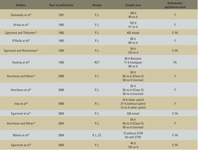
TABLE 1 - Design of clinical trials.
P: prospective; L: longitudinal; RCT: randomized clinical trial; CC: case-control; tt: treatment; F: fixed appliances; FA: functional appliances; NS: not specified.
Authors Year of publication Design Sample size Orthodontic
appliances used
Sadowsky et al52 1991 P, L 160 tt
90 no tt F
Hirata et al24 1992 P, L 102 tt
41 no tt F
Egermark and Thilander13 1992 P, L 402 mixed F, FA
O’Reilly et al46 1993 P, L 60 tt
60 no tt F
Egermark and Ronnerman12 1995 P, L 50 tt
135 no tt F, FA
Keeling et al26 1995 RCT
60 tt Bionator 71 tt headgear
60 no tt
FA
Henrikson and Nilner21 2000 P, L
65 tt 58 no tt (Class II) 60 no tt (normal)
F
Henrikson et al22 2000 P, L
65 tt 58 no tt (Class II) 60 no tt (normal)
F
Imai et al25 2000 P, L
18 tt (after splint) 27 tt (without splint) 13 no tt (after splint)
F
Egermark et al11 2003 P, L 320 mixed F, FA
Henrikson and Nilner23 2003 P, L
65 tt 58 no tt (Class II) 60 no tt (normal)
F
Mohlin et al40 2004 P, L, CC 72 without DTM
62 with DTM F, FA
Egermark et al10 2005 P, L 40 tt
135 no tt F, FA
Prospective longitudinal non-randomized studies Design of included studies
Systematic reviews
Randomized clinical studies
TABLE 2 - Results of clinical trials.
TABLE 3 - Systematic reviews and meta-analysis.
Tt: Treatment; MMO: Maximum mouth opening; MM: Mandibular movement; NE: not evaluated.
SR: systematic review; MA: meta-analysis; F: fixed appliances; FA: functional appliances; OC: orthognathic surgery.
Authors Time of assessment Diagnostic criteria
for TMD
Relationship between extractions and TMD
Relationship between Orthodontics and TMD
Sadowsky et al52 After tt TMJ sounds no no
Hirata et al24 1.2 years during tt Questionnaire, MMO, TMJ
sounds, deviations NE no
Egermark and Thilander13 10 years Questionnaire,
Helkimo index NE Improvement
O’Reilly et al46 During, just after tt Lateral movement,
TMJ sounds, tenderness no no
Egermark and Ronnerman12 Before, during and after tt Questionnaire,
Helkimo index no Improvement
Keeling et al26 Follow-up of 2 years TMJ sound, TMJ pain,
muscle pain NE no
Henrikson and Nilner21 2 years after 1st evaluation Symptoms (headache,
TMJ sounds, pain) NE Improvement
Henrikson et al22 2 years after 1st evaluation Signs (MM, pain,
TMJ sounds) NE Improvement
Imai et al25 Initial, after splint, after tt,
1 year after tt
TMJ sounds, pain,
restriction NE no
Egermark et al11 20 years after 1st evaluation Questionnaire,
Helkimo index NE no
Henrikson and Nilner23 Beginning, after 1 and 2 years of tt
and 1 year after the end of tt Signs and symptoms no no
Mohlin et al40 Performed at 19 and 30 years old Questionnaire, clinical
assessment, psychological status no no
Egermark et al10 Before, during, after tt and 15-18
years after the end of tt
Questionnaire,
Helkimo index NE no
Authors Year of publication Design included studiesNumber of appliances usedOrthodontic orthodontics and TMDRelationship between
McNamara and Turp37 1997 SR 21 F, FA no
Kim et al27 2002 MA 31 F, FA no
Popowich et al50 2003 SR 5 Herbst appliance Insufficient
evidences
Mohlin et al39 2007 SR 30 F, FA no
Abrahamsson et al1 2007 SR 3 OC Insufficient
dISCuSSIOn
Considerations about the subject should al-ways be performed through a critical reading of the methodology used by different authors. The use of the basic research principles allows the researchers to try to control as best as possible the biases of the study generating higher levels of evidence. Thus, the sample size calculation becomes important, so that the sample becomes representative and the results can be extrapo-lated to the studied population. Moreover, the calibration intra and inter-examiners should be performed to assure the reliability of diagnostic criteria, as well as adoption of randomization and blinding criteria. Likewise, careful match-ing for age and sex between the test and control groups should also be observed.53
Within this context of an evidence-based dentistry, it appears that the most common types of studies published in Brazilian journals correspond to studies of low potential for di-rect clinical applications: in vitro studies (25%), narrative reviews (24%) and case reports (20%). The low number of studies with greater strength of evidence shows the necessity to expand the knowledge of evidence-based methods among Brazilian researchers.45
The supposed relationship between ortho-dontics and temporomandibular disorders has attracted the interest of the orthodontic class in last years. Despite significant advances in di-agnostic capability due to advanced techniques such as nuclear magnetic resonance imaging, 3D computed tomography, volumetric Cone-Beam tomography and application of more sophisti-cated clinical procedures, this possible relation-ship remains unclear. A reflection of this con-troversy is the way that orthodontic treatment is considered in several publications. If, for some authors, orthodontic correction may be the cure for TMJ dysfunction, for others it may predis-pose patients to pain and dysfunction of the sto-matognathic system.5
For the establishment of a risk factor, it must fill out several methodological criteria to qual-ify as a true risk factor. Thus, the factor should be identified with the outcome in longitudinal studies, must be present before the establish-ment of the disease and show a biological plau-sibility with the disease. Moreover, the factor remains associated after being controlled for other risk factors, a dose-response relationship must exist, that is, the higher the risk factor, the higher the outcome and this factor must be identified in different populations.2
Cross-sectional or retrospective studies allow the study of associations that identify risk indi-cators and generate hypotheses. Subsequently, these hypotheses need to be tested in longitudi-nal studies to identify true risk factors, because only longitudinal studies can be used as gen-erators of cause and effect evidence due to its temporal component.54 Therefore, the clinical
trials included in this systematic review show longitudinal design, whereas it should be in this point of view that the interrelationship between orthodontics and TMD must be considered.
There is a difference in the quality of the designs of clinical studies before and during the 80s, and the most recent ones.35 Studies of
cross sectional and observational nature, meth-odological errors—such as lack of information about randomization, blinding, sample size calculation, calibration and control of factors— and inadequate quality of study designs com-promised the power for generating scientific evidence. Furthermore, the heterogeneity of results in published studies makes an adequate meta-analysis more difficult to be obtained. Added to this, there is a lack of a standardized classification system for TMD diagnosis. Thus, you can always find a scientific article to prove a point of view.27
the diagnostic criteria adopted by the authors. Due to the lack of a universal classification sys-tem and validated for TMD, in this syssys-tematic review various diagnostic methods used by the authors of the included studies can be found: Helkimo index,18,19 craniomandibular index,15,16
as well as adaptations of these or other ques-tionnaires. This fact complicates the comparison and analysis of results obtained in the studies evaluated in this systematic review.
In order to standardize the diagnostic cri-teria and facilitate future clinical trials, the Research Diagnostic Criteria for Temporoman-dibular Disorders (RDC/TMD) was formulat-ed, which examined jointly the physical and psychosocial aspects of TMD, in the axis I and II, respectively.8 This diagnostic method has
been translated, culturally adapted and vali-dated in Brazil.31,48 Thus, future clinical
stud-ies may use a standardized and universal index, which will facilitate comparison of study re-sults. It is important to be noted that none of the studies evaluated in this systematic review used the RDC/TMD as a diagnostic criteria.
Studies also analyzed the relationship be-tween TMJ sounds and its morphology. Sounds can be associated with various pathologies and the presence of clicks and crackles does not necessarily indicate a TMJ with abnormal mor-phology.14 It becomes important to
acknowl-edge situations of disk displacement with and without reduction, as well as the presence of crackles as indicative of osteoarthritis. It is im-portant to emphasize that the absence of TMJ sounds, not necessarily characterized a situa-tion of normality.14,52
There are many factors that may cause or ag-gravate TMD. A review of the literature did not find a positive association between orthodontic treatment in children and adolescents and future risk of TMD development. In addition, orthodon-tic mechanotherapy performs gradual changes in a system that has a great capacity of adaptation.51
Similar results were obtained in other studies, and worsening of signs and symptoms of TMD pre-treatment were not observed.20
In a critical review of the literature, a low as-sociation between occlusal factors that charac-terize TMD was noted. Moreover, orthodontic treatment performed during adolescence usual-ly does not increase or decrease the probability of developing TMD in the future. Some occlu-sal factors, such as skeletal anterior open bite, overjet greater than 6-7 mm, retruded cuspal position/intercuspal position slides greater than 4 mm, unilateral posterior crossbite and absence of five or more posterior teeth may be associ-ated with specific diagnosis of TMD.36
In another critical review, it was found that the signs and symptoms of TMD can occur in healthy individuals, increasing with age, particu-larly during adolescence, until menopause, and that the TMD that begin during orthodontic treatment may not be related to the treatment. Moreover, there is no risk for TMD associated with any type of orthodontic mechanics and there is no evidence that a stable occlusion, as ideal objective of orthodontic treatment, pre-vents signs and symptoms of TMD. Still, the extraction of teeth as part of orthodontic treat-ment plan does not increase the risk for devel-opment of TMD.35
Current studies, within a context of an evidence-based Dentistry, such as randomized clinical trials, longitudinal prospective non-randomized studies, systematic reviews and meta-analysis, through the use of more rigor-ous methodological criteria and adequate de-signs, evaluated more precisely the interaction between orthodontic treatment and temporo-mandibular disorders.
or absence of extractions during orthodon-tic treatment did not increase the prevalence or worsened signs and symptoms related to TMD.11,23 Randomized clinical trials26 and
lon-gitudinal prospective non-randomized stud-ies,10,11,21,23,25 as well as meta-analysis27 and
sys-tematic review,39 besides presenting more
rig-orous methodologies, generate a greater power of scientific evidence. Moreover, the correct occlusal relationship between the teeth did not cause a change in the physiological position of the condyles and articular discs in TMJ when examined MRIs and CT.3,28,29
Reviewing the literature in search of ran-domized clinical trials—studies that generate a high level of scientific evidence—about the in-terrelation of orthodontic treatment and TMD, there is only one study in the evaluated period in this systematic review.26 This fact occurs due
to difficulties in the accomplishment of ran-domized clinical trials evaluating orthodontic treatment and TMD, due to ethical and practic-es reasons.27 Difficulties are also present when
assessing other forms of irreversible therapies such as TMD treatment protocols. An example of this situation is the occlusal adjustment, from 1966 to 2002, there are only 6 RCTs evaluating the occlusal adjustment as treatment and pre-vention option for TMD in a systematic review published in Cochrane Library.30
Regarding to the role of orthognathic surgery and orthodontic treatment with the Herbst ap-pliance in relation to TMD, the literature analy-sis shows that there is a necessity for a higher number of longitudinal studies, controlled and randomized, to obtain more precise conclusions about the role of these treatment modalities in relation to TMD. Systematic reviews that at-tempted to assess both therapeutics and their relationship with signs and symptoms of TMD were inconclusive, due to small number of sig-nificant scientific evidences.1,50 In relation to the
role of therapy with Bionator26 and headgear,26
it appears that they have no association with the development of TMD. It is important to be not-ed that the use of chincup4,6,7 and facial mask43
shows weak or nonexistent associations in rela-tion to TMD, but studies with this conclusions were not included due to the methodological criteria of this systematic review.
Before the beginning of orthodontic treat-ment, in asymptomatic patients, a full history and physical examination on signs and symp-toms of TMD should be performed by the or-thodontist.34 Studies evaluating the attitude of
orthodontists to TMD show that this interrela-tionship is viewed differently as the possibility of orthodontic treatment increase the probabil-ity of developing of TMD.33,34
Assessing the attitudes and beliefs of ortho-dontists regarding TMD, in a cross-sectional study, the authors obtained results as the ma-jority of respondents did not feel secure about the diagnosis, therapeutic decision and assess-ment of treatassess-ment outcomes of TMD. The vast majority of respondents reported believing that orthodontic treatment does lead to a higher in-cidence of TMD and orofacial pain, but believe that it can be a form of prevention and treat-ment of these disorders. It is important to noted that most participants reported knowledge at a basic level or no knowledge about TMD and orofacial pain during their postgraduate course in orthodontics.38
On the other hand, the results of a research examining the attitudes of Chinese orthodon-tists, regarding orthodontic treatment and TMD, using a questionnaire, showed that most ortho-dontists think that an inadequate orthodontic treatment could increase the development of TMD and an adequate orthodontic treatment could prevent it.33
irreversibly, such as orthodontic treatment and occlusal adjustment, should be indicated in a conscious and precise way. Furthermore, this decision should be based on significant scien-tific evidences.
COnCLuSIOnS
» Many of the available studies in literature have limitations in their designs and methodol-ogies, as well as heterogeneity of results, which reduces the power of evidence generated. Cur-rent studies, with rigorous methodological crite-ria and adequate designs, present more precise evidences of the interrelationship of the orth-odontic treatment and TMD.
» The systematic literature review shows that there is no increased in prevalence of TMD due to traditional orthodontic treatment, either with protocols for extractions or not, with significant
scientific evidences, such as longitudinal con-trolled randomized and non-randomized trials, systematic reviews and meta-analysis, conclud-ing for a tendency of no association. However, it is necessary to perform further randomized clinical trials, with standardized diagnostic cri-teria for TMD for the determination of more accurate causal associations.
1. Abrahamsson C, Ekberg E, Henrikson T, Bondemark L. Alterations of temporomandibular disorders before and after orthognathic surgery: a systematic review. Angle Orthod. 2007 Jul;77(4):729-34.
2. Beck JD. Risk revisited. Community Dent Oral Epidemiol. 1998 Aug;26(4):220-5.
3. Carlton KL, Nanda RS. Prospective study of posttreatment changes in the temporomandibular joint. Am J Orthod Dentofacial Orthop. 2002 Nov;122(5):486-90.
4. Deguchi T, Uematsu S, Kawahara Y, Mimura H. Clinical evaluation of temporomandibular joint disorders (TMD) in patients treated with chin cup. Angle Orthod. 1998 Feb;68(1):91-4.
5. Delboni MEG, Abrão J. Estudo dos sinais de DTM em pacientes ortodônticos assintomáticos. Rev Dental Press Ortod Ortop Facial. 2005 jul-ago;10(4):88-96.
6. Dibbets JM, Van Der Weele LT. Extraction, orthodontic treatment, and craniomandibular dysfunction. Am J Orthod Dentofacial Orthop. 1991 Mar;99(3):210-9.
7. Dibbets JM, Van Der Weele LT. Long-term effects of orthodontic treatment, including extraction, on signs and symptoms attributed to CMD. Eur J Orthod. 1992 Feb;14(1):16-20.
8. Dworkin SF, Leresche L. Research diagnostic criteria for temporomandibular disorders: review, criteria, examinations and speciications, critique. J Craniomandib Disord. 1992 Fall;6(4):301-55.
9. Egermark-Eriksson I. Mandibular dysfunction in children and individuals with dual bite. Swed Dent J Suppl. 1982(10):1-45. 10. Egermark I, Carlsson GE, Magnusson T. A prospective
long-term study of signs and symptoms of temporomandibular disorders in patients who received orthodontic treatment in childhood. Angle Orthod. 2005 Jul;75(4):645-50.
11. Egermark I, Magnusson T, Carlsson GE. A 20-year follow-up of signs and symptoms of temporomandibular disorders and malocclusions in subjects with and without orthodontic treatment in childhood. Angle Orthod. 2003 Apr;
73(2):109-15.
12. Egermark I, Ronnerman A. Temporomandibular disorders in the active phase of orthodontic treatment. J Oral Rehabil. 1995 Aug; 22(8):613-8.
13. Egermark I, Thilander B. Craniomandibular disorders with special reference to orthodontic treatment: an evaluation from childhood to adulthood. Am J Orthod Dentofacial Orthop. 1992 Jan;101(1):28-34.
14. Eriksson L, Westesson PL, Rohlin M. Temporomandibular joint sounds in patients with disc displacement. Int J Oral Surg. 1985 Oct;14(5):428-36.
15. Fricton JR, Schiffman EL. The reliability of a craniomandibular index. J Dent Res. 1986 Nov;65(11):1359-64.
16. Fricton JR, Shiffman EL. The craniomandibular index: Validity. J Prosthet Dent. 1987 Aug;58(2):222-8.
17. Greene CS. The etiology of temporomandibular disorders: implications for treatment. J Orofac Pain. 2001 Spring;15(2):93-105.
18. Helkimo M. Studies on function and dysfunction of the masticatory system. II. Index for anamnestic and clinical dysfunction and occlusal state. Sven Tandlak Tidskr. 1974 Mar;67(2):101-21.
19. Helkimo M. Studies on function and dysfunction of the masticatory system. III. Analyses of anamnestic and clinical recordings of dysfunction with the aid of indices. Sven Tandlak Tidskr. 1974 May;67(3):165-81.
20. Henrikson T. Temporomandibular disorders and mandibular function in relation to Class II malocclusion and orthodontic treatment. A controlled, prospective and longitudinal study. Swed Dent J. Suppl. 1999;134:44.
21. Henrikson T, Nilner M. Temporomandibular disorders and need of stomatognathic treatment in orthodontically treated and untreated girls. Eur J Orthod. 2000 Jun;22(3):283-92. REfEREnCES
22. Henrikson T, Nilner M, Kurol, J. Signs of temporomandibular disorders in girls receiving orthodontic treatment. A prospective and longitudinal comparison with untreated Class II malocclusions and normal occlusion subjects. Eur J Orthod. 2000 Jun;22(3):271-81.
23. Henrikson T, Nilner M. Temporomandibular disorders, occlusion and orthodontic treatment. J Orthod. 2003 Jun;30(2):129-37.
24. Hirata HR, Heft MW, Hernandez B, King GT. Longitudinal study of signs of temporomandibular disorders (TMD) in orthodontically treated and non-treated groups. Am J Orthod Dentofacial Orthop. 1992 Jan;101(1):35-40.
25. Imai T, Okamoto T, Kaneko T, Umeda K, Yamamoto T, Nakamura S. Long-term follow-up of clinical symptoms in TMD patients who underwent occlusal reconstruction by orthodontic treatment. Eur J Orthod. 2000 Feb;22(1):61-7. 26. Keeling SD, Garvan CW, King GJ, Wheeler TT, McGorray S.
Temporomandibular disorders after early Class II treatment with bionators and headgears: results from a randomized controlled trial. Semin Orthod. 1995 Sep;1(3):149-64. 27. Kim MR, Graber TM, Viana MA. Orthodontics and
temporomandibular disorder: a meta-analysis. Am J Orthod Dentofacial Orthop. 2002 May;121(5):438-46.
28. Kinzinger G, Roth A, Gulden N, Bucker A, Diedrich P. Effects of orthodontic treatment with fixed functional orthopaedic appliances on the condyle-fossa relationship in the temporomandibular joint: a magnetic resonance imaging study (Part I). Dentomaxillofac Radiol. 2006 Sep;35(5):339-46.
29. Kinzinger G, Roth A, Gulden N, Bucker A, Diedrich P. Effects of orthodontic treatment with fixed functional orthopaedic appliances on the disc-condyle relationship in the temporomandibular joint: a magnetic resonance imaging study (Part II). Dentomaxillofac Radiol. 2006 Sep;35(5):347-56.
30. Koh H, Robinson PG. Occlusal adjustment for treating and preventing temporomandibular joint disorders (Cochrane Review). In: The Cochrane Library. Oxford: Update Software; 2008.
31. Kosminsky M, Lucena LBS, Siqueira JTT, Pereira Jr FJ, Góes PSA. Adaptação cultural do questionário “Research Diagnostic Criteria for Temporomandibular Disorders: Axis II” para o Português. J Bras Clín Odontol Integr. 2004 jan-fev; 8(43):51-61.
32. Luther F. Orthodontics and the temporomandibular joint: where are we now? Part 1. Angle Orthod. 1998 Aug;68(4):295-304.
33. Mao Y, Duan XH. Attitude of chinese orthodontists towards the between orthodontic treatment and temporomandibular disorders. Int Dent J. 2001 Aug;51(4):277-81.
34. Martins DR, Janson GRP, Touno JLA. Avaliação das disfunções temporomandibulares no exame ortodôntico inicial. Rev Dental Press Ortod Ortop Facial. 2000 jan-fev;5(1):12-6. 35. McNamara JA. Orthodontic treatment and
temporomandibular disorders. Oral Surg Oral Med Oral Pathol Oral Radiol Endod. 1997 Jan;83(1):107-17. 36. McNamara JA Jr, Seligman DA, Okeson JP. Occlusion,
orthodontic treatment, and temporomandibular disorders: a review. J Orofac Pain. 1995 Winter;9(1):73-90.
37. McNamara JA Jr, Türp JC. Orthodontic treatment and temporomandibular disorders: is there a relationship? Part 1: clinical studies. J Orofac Orthop. 1997;58(2):74-89. 38. Moana Filho EJ. Levantamento das atitudes e crenças dos
ortodontistas com relação à disfunção temporomandibular e dor orofacial. Rev Dental Press Ortod Ortop Facial. 2005 jul-ago;10(4):60-75.
Contact address
Eduardo Machado
Rua Francisco Trevisan, no. 20, Bairro Nossa Sra. de Lourdes CEP: 97.050-230 - Santa Maria / RS
E-mail: machado.rs@bol.com.br
Submitted: February 2009 Revised and accepted: August 2009 40. Mohlin BO, Derweduwen K, Pilley R, Kingdon A, Shaw WC,
Kenealy P. Malocclusion and temporomandibular disorder: a comparison of adolescents with moderate to severe dysfunction with those without signs and symptoms of temporomandibular disorder and their further development to 30 years of age. Angle Orthod. 2004 Jun;74(3):319-27. 41. National Institute of Health. Management of
temporomandibular disorders. NIH Technology Assessment Conference. Bethesda: NIH; 1996.
42. Nekora-Azak A, Evlioglu G, Ordulu M, Issever H. Prevalence of symptoms associated with temporomandibular disorders in a Turkish population. J Oral Rehabil. 2006 Feb;33(2):81-4. 43. Ngan PW, Yiu C, Hagg U, Wei SH, Bowley J. Masticatory
muscle pain before, during, and after treatment with orthopedic protraction headgear: a pilot study. Angle Orthod. 1997;67(6):433-7.
44. Okeson JP. Orofacial pain. Guidelines for assessment. Diagnosis and management. Chicago: Quintessence;1996. p. 113-84.
45. Oliveira GJ, Oliveira ES, Leles CR. Tipos de delineamento de pesquisa de estudos publicados em periódicos odontológicos brasileiros. Rev Odonto Ciênc. 2007 jan-mar;22(55):42-7. 46. O’Reilly MT, Rinchuse DJ, Close J. Class II elastics and
extractions and temporomandibular disorders: a longitudinal prospective study. Am J Orthod Dentofacial Orthop. 1993 May;103(5):459-63.
47. Parker MW. A dynamic model of etiology in
temporomandibular disorders. J Am Dent Assoc. 1990 Mar;120(3):283-90.
48. Pereira Jr FJ, Favilla EE, Dworkin S, Huggins K. Critérios de diagnóstico para pesquisa das disfunções temporomandibulares. Tradução oicial para a língua portuguesa. J Bras Clín Odontol Integr. 2004 out-dez; 8(47):384-95.
49. Pilley JR, Mohlin B, Shaw WC, Kingdon A. A survey of craniomandibular disorders in 500 19-year-olds. Eur J Orthod. 1997 Feb;19(1):57-70.
50. Popowich K, Nebbe B, Major PW. Effect of Herbst treatment on temporomandibular joint morphology: a systematic literature review. Am J Orthod Dentofacial Orthop. 2003 Apr;123(4):388-94.
51. Sadowsky C. The risk of orthodontic treatment for producing temporomandibular mandibular disorders: a literature overview. Am J Orthod Dentofacial Orthop. 1992 Jan;101(1):79-83.
52. Sadowsky C, Theisen TA, Sakols EI. Orthodontic treatment and temporomandibular joint sounds - a longitudinal study. Am J Orthod Dentofacial Orthop. 1991 May;99(5):441-7. 53. Susin C, Rosing CK. Praticando odontologia baseada em
evidências. 1ª ed. Canoas: Ulbra; 1999.
54. Susin C, Rosing CK. A importância do treinamento,