Dental Press J Orthod 14 2010 Nov-Dec;15(6):14-7
Moving teeth faster, better and painless.
Is it possible?
The history has shown attempts to correct crowded or protruding teeth since 3000 year ago. Egyptian mummies have been found with crude metal bands wrapped around individual teeth, and primitive and surprisingly well-de-signed orthodontic appliances have also been
found with Greek and Etruscan artifacts.1
From Pierre Fauchard, passing through Ben Kingsley, Calvin Case, and finally to Edward H. Angle, we have seen technology evolved. The modern era of orthodontics has initiated its history around 1900 and has gone from metal bands adjusted around the teeth to bonded braces on the buccal and the lingual sides, as well as clear aligners, mini-implants/ mini-plates, self-ligating brackets, digital mod-els, lasers and so on. Thus, the continuing quest for improvements on materials and techniques leads us to the desire to treat patients faster, better, and totally painless.
Today, many people receive orthodontic treatment which brings about better occlu-sion, improved oral function and harmonized facial appearance. However, two perplexing challenges have not been solved in clinical or-thodontics, i.e. long treatment time (on aver-age 2-3 years) and iatrogenic root resorption. Figuring out these challenges will dramatically improve the quality of orthodontic care.
By nature, orthodontic tooth movement (OTM) is a process of mechanically-induced bone modeling wherein new bone formed on the ten-sion side and resorbed on the compresten-sion side of the periodontal ligament (PDL). Historically, it has been found that when forces are applied, three distinct phases of tooth movement can be observed, namely the 1st strain phase in which the PDL is squeezed (less than 5 seconds), the 2nd lag phase in which tooth movement pauses due to hyalinization formed in the PDL (as long as 7-14 days), and the 3rd move phase in which the tooth moves readily with significant undermining re-sorption of the adjacent alveolar bone.2 Therefore, it is logical to assume that if the 2nd phase (hyalin-ization in the PDL) can be avoided or minimized, the tooth can move smoothly and faster.
From a clinical standpoint, force application owns features of magnitude, frequency and dura-tion. For years, studies on the magnitude and du-ration of forces have been emphasized, resulting in most of the solid scientific findings in today’s literature. In brief, if light forces are applied, it seems that the second phase is not present and the tooth moves much more atraumatically (no hyalinization) through the alveolar bone, which is obviously ideal. The problem with heavy force application is that although the tooth moves ulti-mately through the alveolar bone, the tooth root WH A T’S NE W I N DE N T I S T R Y
Jose A. Bosio*, Dawei Liu**
* Assistant Professor – Postgraduate Clinic Director – Department of Developmental Sciences/ Orthodontics - Marquette University School of Dentistry, Milwaukee, WI.
** Assistant Professor – Undergraduate Program Director and Research Director – Department of Developmental Sciences/ Orthodontics - Marquette Uni-versity School of Dentistry, Milwaukee, WI.
Bosio JA, Liu D
Dental Press J Orthod 15 2010 Nov-Dec;15(6):14-7
surface will be resorbed due to the long duration of contacting the wall of the alveolar socket.3 Clin-ically, lighter forces are considered to be proper, however the hyalinization still cannot absolutely be prevented per se due to the irregular surfaces of the root and the wall of alveolar socket.4
With regard to the frequency of force applica-tion which has rarely been studied, all the cur-rently available orthodontic appliances can only apply static forces. Therefore, it can be hypoth-esized that if a light alternating force is applied on teeth, the tooth movement will be faster and root resorption risks reduced due to the possible absence of hyalinization delay.
But, how can we achieve a light alternating (pulsating, cyclical) orthodontic force? One of the possible means is to impose mechanical vibration to the conventionally applied static orthodontic force. Are there any scientific evidences supporting our hypothesis? Yes. In recent years, whole body weight-bearing bones have been shown to be sensitive to low-level mechanical vibrations.5,6 With less than 50µm of displacement and as little as 5 minutes per day, the mechanical vibration signals can promote bone formation, enhance bone morphology, in-crease bone strength, and attenuate the negative ef-fects associated with catabolic stimuli.6 In dentistry, Kusano et al7 found that both ultrasonic (1.6MHz) and vibratory (141Hz) toothbrush mechanisms increased the proliferation and collagen synthesis of gingival fibroblasts in dogs. More importantly, Nishimura et al8 reported that the resonance vibra-tion could increase tooth movement rate in rats.In clinical orthodontics, Marie found vibration to be possible to reduce pain in orthodontic patients, but without looking at the vibratory stimulation effect on OTM.9 These findings strongly encourage the researchers to investigate the possibility of using mechanical vibration to enhance orthodontic tooth movement and reduce root resorption.
As one of the pioneers focusing on this issue, Liu has reported that when mechanical vibration (4Hz, 20µm displacement, 5 min/day) is applied
to help orthodontically move teeth for 4 weeks in mice, compared with the non-vibrated tooth movement group, the tooth movement rate under vibration is increased by about 50%.10 However, cautions should be taken when extrapolating the experimental findings and conclusions from ani-mals to human being.
With the advancement of research, a new orthodontic company “OrthoAccel” founded in 2007 brought his brand generation of dental vi-brator named “AcceleDent” (Fig 1B) into the mar-ket in 2009. To explore the clinical effects of this device, Kau et al11 conducted a clinical trial in which 14 orthodontic patients were recruited and instructed to use the device for 20 minutes daily for a period of 6 consecutive months. As a result, it was found that the total rate of movement for the mandibular crowding was 2.1 mm per month and for the maxillary arch was 3.0 mm per month, which apparently is faster than the traditional finding as of about 1.0 mm per month.12 The patient compliance was 67% with good patient perception. It was thus concluded that the Ac-celeDent device is a useful adjunct to orthodontic treatment. If used appropriately, it can acceler-ate routine orthodontic tooth movement.11 Cur-rently, the “AcceleDent” device is marketed in the European Union and Australia, while the opening to the US market will not take place until the out-come of an ongoing clinical trial being conducted at the University of Texas Health Science Center San Antonio gets approved by the US Food and Drug Administration (FDA).
A B
What´s new in dentistry
Dental Press J Orthod 16 2010 Nov-Dec;15(6):14-7
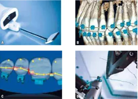
Another “new” orthodontic system has also been present in the literature since 2002. It is called SureSmile®. In this system, the or-thodontist needs to scan the teeth and asso-ciated structures 3-dimensionally and send the records over to the company through the internet, with the doctor’s prescriptions and preferences for brackets, for treatment plan-ning and fabrication of the appliance. The or-thodontist only has to follow the track set by the company to finish the case and possibly to retain as well.13
By looking back in our profession, we re-alize that traditionally, the orthodontists have relied heavily on a standard prescription de-signed into the bracket for the first half of the treatment cycle. In the second half, the doc-tor focuses on correcting errors resulting from improper diagnosis, limitations of the standard bracket prescription and placement. This stage of the treatment is considered a highly reactive phase. The frequency of patient visits increases substantially, and the demands on doctor time increase.14 SureSmile is designed to facilitate a proactive care delivery model. It enables the orthodontist to provide personalized and tar-geted therapeutics using robotically fabricated prescription archwires. The robot is driven by input from the doctor. In simple words, impres-sions are not taken anymore because teeth are
scanned with special intraoral scanner and a digital model is produced, the doctor then sees a malpositioned tooth, changes the position in the computer, the information is sent auto-matically to the company which activates the robot to produce a pre-adjusted wire. This, in turn, will be sent back to the participant ortho-dontist to be delivered to the patient mouth.
Dr. Saschdeva states that “the treatment-planning software has many functional ponents: 3D visualization, measurement, com-munication, decision making with simulation, bracket placement, setup and archwire design, quality and outcome assessment, and SureS-mile patient management. Each of these utili-ties used either singularly or in combination enables the doctor to make better informed decisions and design the targeted prescription archwire”.14 According to his statements, it will take a motivated and experienced orthodontist a minimum of 2 years and the completion of at least 100 patients to develop competency in treating with SureSmile. However, we be-lieve that the orthodontic community would be interested to see unbiased strong level of evidence studies showing that teeth can be moved faster, better, and more efficiently with SureSmile technology.
A
C D
B
Bosio JA, Liu D
Dental Press J Orthod 17 2010 Nov-Dec;15(6):14-7
25 minutes to take a full mouth impression, 2) clinical chair time is reduced but computer organizing time is greater, 3) initial cost with
FIGURE 2 - A) Intraoral Scanner; B) 3-D individualized model; C) Robotic wire bending; D) Individual-ized tooth wire bending.
the equipment set up is still very high. A chal-lenging technology will show to our orthodon-tic community its efficacy in the near future.
1. Wahl N. Orthodontics in 3 millennia. Chapter 2: entering the modern era. Am J Orthod Dentofacial Orthop. 2005 Apr;127(4):510-5.
2. Reitain K. Some factors determining the evaluation of forces in orthodontics. Am J Orthod. 1957;43:32-45.
3. Profit W. Contemporary Orthodontics. 4th ed. St. Louis: Mosby Year Book; 2007. cap. 9, p. 331-40.
4. Cattaneo PM, Dalstra M, Melsen B. Moment-to-force ratio, center of rotation, and force level: a inite element study predicting their interdependency for simulated orthodontic loading regimens. Am J Orthod Dentofacial Orthop. 2008 May;133(5):681-9.
5. Rubin C, Turner AS, Bain S, Mallinckrodt C, McLeod K. Anabolism. Low mechanical signals strengthen long bones. Nature. 2001 Aug 9;412(6847):603-4.
6. Xie L, Rubin C, Judex S. Enhancement of the adolescent murine musculoskeletal system using low-level mechanical vibrations. J Appl Physiol. 2008 Apr;104(4):1056-62. 7. Kusano H, Tomofuji T, Azuma T, Sakamoto T, Yamamoto T,
Watanabe T. Proliferative response of gingival cells to ultrasonic and/or vibration toothbrushes. Am J Dent. 2006 Feb;19(1):7-10. 8. Nishimura M, Chiba M, Ohashi T, Sato M, Shimizu Y, Igarashi K, et
al. Periodontal tissue activation by vibration: intermittent stimulation by resonance vibration accelerates experimental tooth movement in rats. Am J Orthod Dentofacial Orthop. 2008 Apr;133(4):572-83. 9. Marie SS, Powers M, Sheridan JJ. Vibratory stimulation as a
method of reducing pain after orthodontic appliance adjustment. J Clin Orthod. 2003 Apr;37(4):205-8.
REFERENCES
10. Liu D. Acceleration of orthodontic tooth movement by mechanical vibration. Access: 2009 Jan 12. Available from: http://iadr.confex. com/iadr/2010dc/webprogram/Paper129765.html.
11. Kau CH, Jennifer TN, Jeryl D. The clinical evaluation of a novel cyclical-force generating device in orthodontics. Orthodontic Practice US. 2010;1(1):43-4.
12. Mandall N, Lowe C, Worthington H, Sandler J, Derwent S, Abdi-Oskouei M, et al. Which orthodontic archwire sequence? A randomized clinical trial. Eur J Orthod. 2006 Dec;28(6):561-6. 13. Mah J, Sachdeva R. Computer assisted orthodontic treatment:
The SureSmile process. Am J Orthod Dentofacial Orthop. 2001 Jul;120(1):85-7.
14. Scholz RP, Sachdeva RCL. Interview with an innovator: SureSmile Chief Clinical Oficer Rohit C. L. Sachdeva. Am J Orthod Dentofacial Orthop. 2010 Aug;138(2):231-8.
Contact address