Artigo Original
Sandro Júnior Henrique Lima1 Leandro de Araújo Pernambuco2 Aline de Lima Lins3 Lucas Carvalho Aragão Albuquerque1 Hilton Justino da Silva4
Descritores
Rinite Respiração Bucal Sistema Estomatognático Fala Mandíbula Keywords
Rhinitis Mouth Breathing Stomatognathic System Speech Jaw
Correspondence address: Sandro Júnior Henrique Lima Rua Professor Artur de Sá, s/n. Cidade Universitária,Recife (PE), Brasil, CEP: 50740-520.
E-mail: [email protected] Received: 08/18/2014
Accepted: 03/01/2015
Study carried out at Hospital das Clínicas da Universidade Federal de Pernambuco – UFPE – Recife (PE), Brasil. (1) Graduate Program in Neuropsychiatry and Behavioral Sciences, Universidade Federal de Pernambuco – UFPE – Recife (PE), Brasil.
(2) Graduate Program in Collective Health, Universidade Federal do Rio Grande do Norte; Department of Speech Language Pathology and Audiology, Universidade Federal do Rio Grande do Norte – UFRN – Recife (PE), Brasil.
(3) Speech Language Pathology and Audiology course, Universidade Federal de Pernambuco – UFPE – Recife (PE), Brasil.
(4) Graduate Program in Nutrition, Universidade Federal de Pernambuco; Department of Speech Language Pathology and Audiology, Universidade Federal de Pernambuco – UFPE – Recife (PE), Brasil.
in children with allergic rhinitis
Movimentos mandibulares na fala
em crianças com rinite alérgica
ABSTRACT
Introduction: Allergic rhinitis can cause changes in stomatognathic functions, which may alter the mandibular dynamics. Electrognathography is used in the recording of jaw movements, making it valid for analysis of movements in speech. Purpose: To characterize the amplitude and velocity of jaw movements during speech in children with and without allergic rhinitis. Methods: The sample consisted of 32 children aged 7–12 years, treated at a university hospital, divided into two groups: one with rhinitis and the other without rhinitis. To capture the jaw movements during speech, we used an electrognathography with the aid of a list of phonetically balanced igures. For the analysis of data, we used, in addition to descriptive statistics, nonparametric tests, Spearman correlation coeficient and the Mann-Whitney test, with a signiicant value of p=0.05. Results: No signiicant difference was observed in jaw movements between groups, with values of p equals to 0.175, 0.650, and 0.462 for amplitude and jaw opening and closing velocity, respectively. However, a strong correlation was observed between the variables velocity and amplitude of mouth opening, being slightly higher in the group of children with allergic rhinitis. Conclusion: The amplitude and velocity of jaw movements are found to be similar in children with and without allergic rhinitis, and a correlation exits between these variables. In addition, they were more heterogeneous in the group without allergic rhinitis.
RESUMO
INTRODUCTION
Allergic rhinitis is dei ned as an inl ammatory response of the nasal mucosa to exposure to allergen factors mediated by immunoglobulin E, which may result in chronic or recurrent symptoms(1). The consequences of this clinical entity are changes on quality of life and reduction in academic and professional performance, causing economic repercussions(2).
It is noted in scientii c literature that patients with allergic rhinitis have modii cations in the functions of chewing, swal-lowing, and breathing(3) — the latter being mainly related to nasal obstruction, which is the predominant symptom in this pathology and directly relates to changes in the stomatognathic system(4-6).
Some authors say that with the appearance of nasal obstruc-tion and possibly opening of the mouth to breathe, there are structural alterations that cause imbalance in orofacial func-tions(7). These changes appear by a neuromuscular response that induces the adjustment of various structures of the stomato-gnathic system, generating physiologically inappropriate mus-cular activity. Therefore, skeletal, dental, and muscle changes are caused, while hypotrophy, hypotonia, and hypofunction of the mandibular elevator muscles may occur(8,9).
It is also noted that the production of speech depends on the mobility of the phonoarticulatory system, such as tongue, lips, and cheeks, as well as the positioning of teeth, tongue, and jaws(10). Problems in this function are associated with the occurrence of alterations in mobility, tone, and posture of the phonoarticulatory system and orofacial disorders(11). Therefore, jaw movements may be modii ed during speech production in the occurrence of nasal obstruction.
A method that records the mandibular dynamics is elec-trognathography, which works through a computerized system of three-dimensional analysis of jaw movements. In addition to simplifying the recording of jaw movements, electrogna-thography is a test that may help in diagnosis, monitoring, and development of stomatognathic alterations(12). In the evaluation of the jaw movements during speech through electrognathography, the so-called structured situations may be used, which allow the nonoccurrence of pauses and mini-mize alterations in velocity, better showing the performance of motor mechanisms of speech production(13). Examples of such situations are repetition of phonemes or even repeti-tion of words.
Thus, the objective of this study was to characterize the amplitude and velocity of jaw movements during the speech of children with and without allergic rhinitis.
METHODS
This study was observational, descriptive, and transver-sal. The nonprobabilistic sample comprised 32 children, 18 female and 14 male, aged 7–12 years, treated at the Clinics of Allergy, Immunology and Pediatrics at a university hospital, between April and June 2013. The selection of participants
was through a convenience criteria related to the demand for care in the clinics.
Participants were divided into groups consisting of 16 chil-dren with a medical diagnosis of allergic rhinitis (8 females) and a control group with the same number of volunteers, con-sisting of children without allergic rhinitis (10 females).
The children were selected through the consultation of med-ical records of the clinics mentioned, considering the criteria for inclusion and exclusion of this study.
Children aged 7–12 years and having been diagnosed with allergic rhinitis were included in the study group. The control group had children without rhinitis, aged 7–12 years.
Children presenting craniofacial malformations; using braces; lacking dental elements that could compromise speech; presenting neurological syndromes; and presenting any com-municative, neurological, cognitive, and sensorial dei cits were excluded from both the groups.
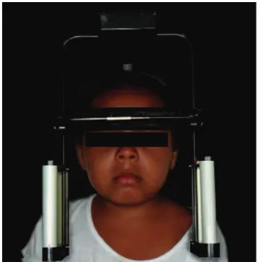
For registration of jaw movements, we used the elec-trognathography (Bio-Research® Associates and the BioPak Program™ software), with a notebook computer. In the exams, the participant was instructed to remain seated in a chair and then a magnetic sensor (magnet) was i xed in the oral mucosa, below the lower central incisors, with Corega® tape. Soon after, the electrognathography was attached to the participant’s head, and adjustments were made to position the equipment in rela-tion to the anatomical features of the child (Figure 1).
Then a printed picture of i gures of easy appointment(14,15) was presented, containing all phonemes of Portuguese. The par-ticipant was asked to appoint, in sequence, all i gures, to capture the jaw movements during speech (Figure 2). The route of the
jaw during the appointment of igures was recorded using the software for further data analysis (Figure 3).
For descriptive statistical analysis, we considered the mean, standard deviation, and the conidence interval. For comparison between case and control groups, the Mann-Whitney nonpara-metric test was adopted; to verify the correlation between the
amplitude of jaw opening and the velocity during opening and closing of the mouth, the Spearman correlation test was used (signiicance level was 95%).
Data collection was initiated after approval of the research eth-ics committee of the institution (CAAE: 10733513.4.0000.5208; opinion no 257 940), and parents signed an informed consent before the proceedings were initiated.
RESULTS
The sample comprised 56.25% of female children and 43.75% male, showing a homogeneous inclusion of partici-pants. In addition, the rhinitis group had half the participants belonging to each of the sexes, whereas the control group was composed of 62.5% female.
Table 1 shows a higher mean of amplitude of jaw opening in the control group, with a 2.23 mm difference compared to the case group, but with no statistical signiicance. In addition, in the data related to jaw opening velocity, in both groups, a dif-ference of 10.31 mm/s is observed between the means, and the speed of this movement is larger in the control group; however, no statistically signiicant difference was observed between these results. A difference of 12.31 mm/s was also observed between the means of case and control groups in the variable jaw closing velocity, and the control group had a faster clos-ing, although it was not a signiicant difference. From the con-idence interval calculation, a lower dispersion was evcon-idenced
Figure 2. Frame with phonetically balanced figures for Brazilian Portuguese
Figure 3. Record of the velocity (blue) and jaw movements during speech in the frontal (red) and sagittal (green) in a volunteer
Table 1. Comparison between the amplitude of jaw opening and jaw opening and closing velocity in millimeters during speech in volunteers of case and control groups
Variable Case (n=16) Control (n=16) p-value
Mean±SD CI Median Median Mean±SD CI Min.–Max. Median
Amplitude 13.18±4.09 11.00–15.37 16.5 16.5 15.75±6.32 12.38–19.12 5.60–26.10 18 0.175
Opening
velocity 78.00±28.18 62.98–93.02 97.5 97.5 88.31±52.20 60.49–116.13 10–200 111 0.650
Closing
velocity 89.94±40.76 68.21–111.66 104.5 104.5 102.25±52.71 74.16–130.34 26–227 132 0.462
Mann-Whitney nonparametric test – p<0.05
in the case group, which points to a more accurate estimate of the value of individuals with allergic rhinitis. These values indicate that there are no differences between the control group and the group with allergic rhinitis, considering the variables of the mandibular movements studied: jaw opening amplitude, jaw opening velocity, and jaw closing velocity during speech.
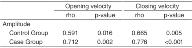
Table 2 shows a positive correlation between amplitude and jaw opening velocity, and this was slightly higher in the case group, whereas the correlation between amplitude and jaw clos-ing velocity was positive and strong in both groups. Considerclos-ing these indings, it is possible to note that the numerical values of the amplitude and jaw opening and jaw closing velocity are directly proportional. Such analyses of the Spearman corre-lation coeficient were made considering the distance of the actual values of the perfect coeficient of reference, represented by the number one.
this research, considering the ratings of standard deviation, both in control and in the case group, although the study focused on adults.
Another study of adults using a similar electromagnetic analysis showed an average opening of the mouth of 14.5 mm during speech, which suggests, in addition to discrete values for this variable, small differences between asymptomatic chil-dren and young adults(19). In this regard, the literature showed, in studies of measurement of mandibular movements through the caliper, that the maximum amplitude of mouth increases according to the age(20,21). However, we can infer by these ind-ings that possibly a little broad nature of these movements and the few changes seen in the comparison between age groups would be associated to the complex nature of motor control during speech. The amplitude of jaw movement in this func-tion does not have to be proporfunc-tional to the maximum open-ing of the mouth.
Furthermore, other studies showed lower values than the above, ranging from 2 to 8.6 mm(22-24). One possible expla-nation for the cause of this discrepancy would be associated with the language, which highlights the importance of studies that characterize the mandibular movements in Portuguese(25).
The resulting data show differences, but not statistically sig-niicant, in the values observed among the investigated groups; however, such changes may indicate that possible orofacial alterations in the stomatognathic system, present in the rhini-tis group, justify such indings.
The data show high values of velocity during the opening and closing of jaw in speech, both in the control and in the case group, with a higher speed during closing. Considering this, in one study(17) with asymptomatic adults, velocities of 88.65 and 89.90 mm/s were observed during the opening and clos-ing movements of the jaw, respectively, which shows a small increase in the mouth closing speed. It can be inferred from these results that this speed increase in mandibular closure is related to masticatory function, as it depends on an integrated complex of muscles that raise and lower the jaw, and the efi-ciency of this function is directly related to the contraction force of these muscles, especially the jaw elevators(26).
Also in relation to the velocity of the mandibular move-ment, it can be noted, from the means, that the values for both opening and closing of jaw were lower in the group with rhi-nitis. According to Murdoch(27), the jaw velocity depends, among other factors, on the action of the muscles and liga-ments of the stomatognathic system. Considering this, it is noted that the literature indicates a large prevalence of oral breathing in subjects with allergic rhinitis, and neuromuscu-lar changes in the stomatognathic system found in the mouth breathing that cause hypofunction and muscular hypotonia, especially in the jaw elevator muscle(28,29). Thus, it is possible that the decrease in jaw velocity in the group of children with allergic rhinitis is related to alterations in the respiratory mode caused by nasal obstruction.
Regarding the comparison between the amplitude and the jaw opening and closing velocities, a study(17) also mentioned
Table 2. Correlation between the amplitude of jaw movements and jaw opening and closing velocity during speech in volunteers of case and control groups
Opening velocity Closing velocity
rho p-value rho p-value
Amplitude
Control Group 0.591 0.016 0.665 0.005
Case Group 0.712 0.002 0.776 <0.001
p<0.05
Caption: rho = correlation coefficient of Spearman’s rank
DISCUSSION
Analysis of the mandibular dynamics is pointed in some studies as an important mean of evaluation of the functional state of the stomatognathic system, as well as an auxiliary tool for the correct diagnosis of possible alterations(16,17). It is observed in the literature that the research conducted to ver-ify the jaw movement presents a variety of clinical situations studied, which also shows the reliability of electrognathogra-phy in obtaining clinical data on the speed and scale of these movements(12).
Although no statistically signiicant differences were observed between the measurements of mandibular movement in children with and without rhinitis, some considerations are possible.
Initially considering the data related to the distribution of sexes in the groups studied, there was a homogeneous alloca-tion of participants. Therefore, in a study with the objective of evaluating the mandibular movements of children through the use of the caliper, no differences were found in the jaw open-ing amplitude in the comparison between male and female, supporting the indings of this study(18).
that there is a direct relationship between these variables. In this study, a statistically signiicant positive correlation was observed in both groups, which is slightly higher in chil-dren with rhinitis. Although this difference is small, we can infer that this result is related to the fact that the numerical values of the control group are more heterogeneous, possi-bly by the existence of physiological conditions of the sto-matognathic system not addressed in the exclusion criteria and that can modify muscle condition, such as facial type. One fact that can strengthen this conjecture are the values of the conidence interval of the index, which presented less dispersion in the group of children with rhinitis and higher in the control group in all analyses, showing greater range of values in the latter group, considering the minimum and maximum values in millimeters.
In addition, one might think, with these data, that possi-bly the correlation between the opening amplitude variables and the opening and closing jaw velocities increases in the occurrence of discrete values in these variables, which was observed in the group with allergic rhinitis. The indings may also be related to some limiting factors of the research, such as the usage of a nonprobabilistic sample and a small number of participants, in addition to no prior evaluation of stomato-gnathic changes of the patients, which would be relevant in future studies involving allergic rhinitis. We also suggest, in future studies, an investigation of the presence of signs and symptoms of temporomandibular dysfunction, considering evidence that show possible alterations in jaw movements in speech under that condition.
CONCLUSION
The results showed no significant statistical difference between the jaw movements in the presence and absence of allergic rhinitis, considering the variables studied, showing that the amplitude of jaw opening and the velocity of man-dibular movements during speech have similar values among children with and without allergic rhinitis. Nevertheless, sig-niicant correlation was found between these variables of the jaw movement, with a greater level of correlation in the group of children with allergic rhinitis.
It was also observed that children without allergic rhinitis had greater heterogeneity in the values of variables than the ones with allergic rhinitis.
These results may indicate the intimate relationship between the mandibular dynamics and the balance of the stomatognathic system, and provide additional information to clinical practice on orofacial functional changes secondary to allergic rhinitis.
*SJHL participated in data collection, analysis of collected data, statistical calculations, and writing of the article; LAP participated in the implementation of the statistical calculations and revision of the article; ALL participated in data collection and revision of the article; LCAA participated in the analysis of statistical calculations; HJS was responsible for scientiic guidance and participated in the revision and writing of the article.
REFERENCES
1. Ibiapina CC, Sarinho ESC, Camargos PAM, Andrade CR, Cruz Filho AAS. Rinite alérgica: aspectos epidemiológicos, diagnósticos e terapêuticos. J Bras Pneumol. 2008;34(4):230-40.
2. Weckx LL, Lopes CP, Naspitz N, Naspitz CK. Apostila do respirador bucal. São Paulo: Unifesp; 2001. p. 1-24.
3. Lemos CM, Wilbelmsen NSW, Mion OG, Mello-Júnior JF. Alterações funcionais do sistema estomatognático em pacientes com rinite alérgica: estudo caso-controle. Braz J Otorhinolaryngol. 2009;75(2):268-74. 4. Luzzi V, Lerardo G, Viscogliosi A, Fabbrizi M, Consoli G, Vozza I, et al.
Allergic rhinitis as a possible Rick factor for malocclusion: a case-control study in children. Int J Paediatr Dent. 2013;23(4):274-8.
5. Campanha SMA, Fontes MFJ, Camargos PAM, Freire LMS. The impact of speech therapy on asthma and allergic rhinitis control in mouth breathing children and adolescents. J Pediatr (Rio J). 2010;86(3):202-8. 6. Wandalsen GF, Mendes AI, Solé D. Correlação entre resistência nasal e diferentes parâmetros da rinometria acústica em crianças e adolescentes com e sem rinite alérgica. Braz J Otorhinolaryngol. 2012;78(6):81-6. 7. Cunha DA, Silva GAP, Silva HJ. Repercussões da respiração oral no
estado nutricional: por que acontece? Arquivos Int Otorrinolaringol. 2011;15(2):223-30.
8. Köhler GI, Köhler JFW. A importância do enfoque terapêutico multidisciplinar nas inadequações morfofuncionais da face. Ortodont Paranaen. 1992;13(1):17-39.
9. Marchesan IQ, Krakauer. A importância do trabalho respiratório na terapia miofuncional. In: Marchesan IQ, Bolafi C, Gomes ICD, Zorzi JL. Tópicos em Fonoaudiologia. São Paulo: Lovise; 1995. p. 15-60. 10. Camargo MEPS, Respiração: movimento de vida. Rev Temas Sobre
Desenvolv. 2004;13(77):27-36.
11. Nishimura CM, Gimenez SRML. Peril da fala do respirador oral. Rev CEFAC. 2010;12(3):505-8.
12. Pinheiro PF Jr, Cunha DA, Filho MG, Caldas AS, Melo TM, Silva HJ. The use of electrognathography in jaw movement research: a literature review. Cranio. 2012; 30(4): 293-303.
13. Wertzner HF, Silva LM. Velocidade de fala em crianças com e sem transtorno fonológico. Pró-fono. 2009;21:19-24.
14. Marchesan IQ. Avaliação e terapia dos problemas da respiração. In: Marchesan IQ. Fundamentos em Fonoaudiologia: aspectos clínicos da motricidade oral. Rio de Janeiro: Guanabara Koogan; 1998. p. 23-36.
15. Bianchini EMG, Andrade CF. A model of mandibular movements during speech: normative pilot study for the Brazilian Portuguese Language. Cranio. 2006;24(3):197-206.
16. Okeson JP. História e exame das desordens têmporomandibulares. In: Okeson JP. Tratamento das desordens temporomandibulares e oclusão. 6ª ed. São Paulo: Elsevier Editora Ltda.; 2008. p. 173-28.
17. Anelli W. Atuação fonoaudiológica na desordem temporomandibular. In: Lopes FOC. Tratado de fonoaudiologia. São Paulo: Editora Roca; 1997. p. 821-8.
18. Hamazaki CM, Kawaura R, Bianchini EMG, Assencio-Ferreira VJ. Veriicação da amplitude dos movimentos mandibulares em crianças. Rev CEFAC. 2002;4(1):35-40.
19. Nielsen IL, Marcel T, Chun D, Miller AJ. Patterns of mandibular movements in subjects with craniomandibular disorders. J Prosthet Dent.1990; 63(2): 202-17.
20. Machado BCZ, Medeiros APM, Felício CM. Limite de movimentos mandibulares em crianças. Pró-fono. 2009;21(3):189-94.
21. Leles CR, Neto JJSM, Giro EMA, Compagnoni MA. Valores normais da amplitude do movimento mandibular em crianças. Pós-Grad Rev Fac Odontol. 2000;3(2):121-6.
22. Peraine M, Salsench J, Torrent J, Noqueras J, Samso J. Study of mandibular movements during speech. Cranio. 1990;8(4):324-31. 23. Howell PG. Incisal relationships during speech. J Prosthet
24. B u r n e t t C A . R e p r o d u c i b i l i t y o f t h e s p e e c h e nve l o p e a n d interocclusal dimensions in dentate subjects. Int J Prosthodont. 1994;7(6):543-8.
25. Bianchini EMG, Paiva G, Andrade CRF. Mandibular patterns during speech in subjects with temporomandibular disorders and in asyntomatic individuals. Cranio. 2008;26(1):50-8.
26. Nascimento GKBO. Eletromiografia de superfície do músculo masseter durante a mastigação: Uma revisão sistemática. Rev CEFAC. 2012;14(4):725-31.
27. Murdoch BE. In: Murdoch BE. Desenvolvimento da fala e distúrbios da linguagem: uma abordagem neuroanatômica e neuroisiológica. Rio de Janeiro: Editora Revinter; 1997. p. 67-89.
28. Barros JRC, Becker HMG, Pinto JA. Evaluation of atopy among mouth breathing pediatric patients referred for treatment to a tertiary care center. J Pediatr (Rio J.). 2006;82(6): 458-64.