Potenciais evocados auditivos em indivíduos acima de 50 anos
de idade*****
Auditory evoked potentials in individuals over 50 years
*Fonoaudióloga. Doutora em Distúrbios da Comunicação Humana pela Universidade Federal de São Paulo - Escola Paulista de Medicina. Docente do Curso de Fonoaudiologia da Faculdade de Medicina da Universidade de São Paulo. Endereço para correspondência: Av. Divino Salvador, 107, Apto 32 São Paulo -SP - CEP 04078-010
(cgmatas@usp.br).
**Fonoaudióloga. Doutoranda pelo Programa de Pós-Graduação em Ciências da Reabilitação da Faculdade de Medicina da Universidade de São Paulo.
***Fonoaudióloga. Mestranda em Distúrbios da Comunicação Humana pela Universidade Federal de São Paulo - Escola Paulista de Medicina. ****Fonoaudióloga. Especialização em Audiologia Clínica pelo Curso de Fonoaudiologia do Departamento de Fisioterapia, Fonoaudiologia e Terapia Ocupacional da Faculdade de Medicina da Universidade de São Paulo. *****Trabalho Realizado no Departamento de Fisioterapia, Fonoaudiologia e Terapia Ocupacional da Faculdade de Medicina da Universidade de São Paulo.
Artigo de Pesquisa
Artigo Submetido a Avaliação por Pares Conflito de Interesse: não
Recebido em 09.08.2005.
Revisado em 13.09.2005; 29.05.2006; 06.09.2006.
Aceito para Publicação em 26.10.2006. Carla Gentile Matas*
Valdete Alves Valentins dos Santos Filha** Melissa Mitsue Cunha Pires Okada*** Juliana Reis Resque****
Abstract
Background: auditory evoked potentials. Aim: to describe the results of brainstem auditory evoked potentials (PEATE), middle latency auditory evoked potentials (PEAML) and cognitive potential (P300) in individuals over 50 years. Method: this study was developed at the Speech and Hearing Investigation Laboratory in Auditory Evoked Potentials of the Speech-Language and Hearing Course of the Department of Physiotherapy, Speech-language and Hearing Sciences and Occupational Therapy of FMUSP. Twenty four subjects (45 ears) were evaluated through PEATE and P300, and only 18 of these subjects (36 ears) were evaluated through PEAML. All subjects had ages between 51 and 74 years and were divided in three groups: GI (50-59 years), GII (60-69 years) and GIII (70-79 years). All subjects presented either normal hearing or neurossensorial hearing loss of a moderate-severe level in the PEATE, and of a moderate level in the PEAML and in the P300. The frequency range evaluated in the PEATE and in the PEAML varied from 3000 to 6000Hz, while in the P300 it varied from 1000 to 1500Hz. For the statistical analyses of the data, the Kruskal-Wallis test, the Mann-Whitney test and the two proportion equality test were used. Results: significant statistical differences were simultaneously observed between the groups for the interpeak I-V in the PEATE and for the Na wave latency in the PEAML - in the PEATE the difference was caused by GIII and in the PEAML it was caused by GI. A statistically significant difference between the groups was observed for the latency of the P300 component. Considerable alterations were also found regarding the quality of the responses of the auditory evoked potentials, indicating a strong correlation between the deterioration of the responses and the increase in age. Conclusion: the aging process of the auditory system progressively affects the auditory pathways throughout the brainstem and temporal lobe.
Key Words: Auditory Evoked Potentials; P300 Event-Related Potentials; Aging. Resumo
Tema: potenciais evocados auditivos. Objetivo: descrever os resultados dos potenciais evocados auditivos de tronco encefálico (PEATE), potenciais evocados auditivos de média latência (PEAML) e potencial cognitivo (P300) em indivíduos acima de 50 anos de idade. Método: este estudo foi desenvolvido no Laboratório de Investigação Fonoaudiológica em Potenciais Evocados Auditivos do Curso de Fonoaudiologia, do Departamento de Fisioterapia, Fonoaudiologia e Terapia Ocupacional da FMUSP. Foram avaliados 24 pacientes (45 orelhas) por meio do PEATE e do P300, sendo que apenas 18 destes pacientes (36 orelhas) foram avaliados por meio do PEAML. Todos os indivíduos encontravam-se na faixa etária de 51 a 74 anos de idade, divididos em três grupos:GI (50 - 59 anos), GII (60 - 69 anos) e GIII (70 a 79 anos) e apresentavam audição normal ou até perda auditiva neurossensorial de grau moderadamente severo no PEATE e de grau moderado no PEAML e no P300. A faixa de frequências avaliadas no PEATE e no PEAML abrangeu 3000 a 6000 Hz, enquanto que no P300 a faixa foi de 1000 a 1500Hz. Para a análise estatística dos dados foram utilizados os testes estatísticos de Kruskal-Wallis, Mann-Whitney e igualdade de duas proporções. Resultados: observaram-se diferenças estatisticamente significantes entre os grupos simultaneamente para o interpico I -V no PEATE e para a latência da onda Na no PEAML, sendo que no PEATE foi o GIII que provocou a diferença e no PEAML foi o GI. Evidenciou-se diferença estatisticamente significante entre os grupos simultaneamente para a latência do componente P300. Verificou-se, também, alterações consideráveis em relação à qualidade dos traçados dos potenciais evocados auditivos, indicando uma forte correlação entre piora na qualidade do traçado e aumento da idade. Conclusão: o processo de envelhecimento do sistema auditivo afeta progressivamente as vias auditivas ao longo do tronco encefálico e lobo temporal. Palavras-Chave:Potenciais Evocados Auditivos; Potencial Evocado P300; Envelhecimento.
Referenciar este material como:
Introduction
Technological, sanitary and control disease advances associated with better life quality allowed the increase of general population longevity, specially in individuals above the 5th life decade,
according to data of the World Health Organization. This way, it became essential to comply with the health necessities of elderly individuals (Bess et al., 2001). It is important to note that by 2025 Brazil will have the 6th largest elderly population of the
world.
The auditory loss due to degenerative changes related to the aging process of the human organism, called Presbyacusis, may be mentioned as one of the most common causes of sensorineural hearing loss in adults. This is probably one of the most frustrating sensorial losses to with the adult individual goes through in aging process. This loss or deficiency of one of the senses symbolizes, to the elderly, a series of worries about aging and the symptoms and limitations related to it (Tanaka et al., 2002; Soncini et al., 2003).
With organic aging several structural changes of the auditory nerve occur along the central pathway of the brainstem and the temporal lobe. The retrocochlear cells degeneration is associated with the loss of synchrony of the central auditory pathways (Bess et al., 2001). These changes may be evidenced by the auditory process disorder as well as by cognitive involvement leading to learning, attention, memory and cognition impairments (Calero e Navarro, 2004).
Specialized literature shows that, due to these impairments, deficits in central auditory tests and electrophysiological anomalies in Auditory Brainstem Response (ABR), Middle Latency Response (MLR) and Late Latency Response(LLR) may be found in individuals with presbyacusis (Bess et al., 2001).
Auditory Brainstem Responses (ABR) are registered as waves and observed in the first 10 milliseconds (ms) after the acoustic stimulus presentation, starting in the acoustic nerve and auditory pathways of the brainstem (Durrant e Ferraro, 2001).
Middle Latency Response (MLR) and Late Latency Response (LLR) consist of a series of positive and negative waves that follow the ABR, present respectively in 60 to 80 ms and 100 to 500 ms after acoustic stimulation (Musiek e Lee, 2001). The MLR starts in the primary areas of the auditory cortex and helps in the diagnosis of auditory process disorder. The P300, the late
latency potential most used in clinic, starts in the primary and secondary areas of the auditory cortex and is affected by sleep, sedation and attention to the acoustic stimulus, being therefore related to cognition and attention functions (Schochat, 2003). Assis et al. (2005) point out that the real influence of age in the results of Auditory Evoked Potential is still controversial.
According to these considerations the study of short, middle and late latency evoked potential is very important in the evaluation of individuals above 50 years of age, in order to detect possible central auditory disorders that may be present in this population, allowing the possibility of prevention and rehabilitation with these individuals.
This way, the aim of this study was to describe the results of Auditory Evoked Potentials (ABR, MLR, P300) in individuals above 50 years of age.
Method
This work was approved by the Ethical Committee of the Physiotherapy, Audiology, Speech and Language Pathology and Occupational Therapy Department of the School of Medicine, University of São Paulo with protocol number 039/ 05. All participants signed the consent form that described all the procedures, this way agreeing with the realization of the research and the publication of the results according to Resolution 196/96.
Subjects
This study was developed in the Audiology Research Laboratory in Auditory Evoked Potential of the Audiology, Speech and Language Pathology Graduation Course, of the Physiotherapy, Audiology, Speech and Language Pathology and Occupational Therapy Department of the School of Medicine of University of São Paulo.
compatible with the degree of the hearing loss and type A tympanometric curve. To avoid interferences of the hearing loss with the results obtained in the exams the ears with conductive, mixed or moderately severe (PEAML and P300), severe or profound (ABR, MLR and P300) hearing losses were excluded.
Subjects were divided in three groups: GI (50-59 years), GII (60-69 years), GIII (70-79 years) and data obtained with each group were compared between themselves. In ABR presence or absence of disorder was analyzed as well as the description of the type of disorders observed. In MLR the values of latencies of Na and Pa waves and the amplitude of Pa wave, considering the contralateral auditory pathway (Kimura, 1961). And in P300 the presence or absence of this potential in both ears (45 ears) as well as its latency.
Procedures
Initially subjects were interviewed to provide data about auditory complaints, presence of risk factors for hearing loss, complaints of otitis among other disturbances related to middle ear and external ear.
After that, the external acoustic meatus was inspected with the use of a Heine otoscope,
checking the conditions to proceed to conventional audiologic evaluation and electrophysiological exams.
Pure Tone Audiometry was realized in acoustic cabin and evaluated the frequencies 250 to 8000Hz by air-conduction and 500 to 4000Hz by bone-conduction (in frequencies with thresholds above 20dBNA by air way). The speech audiometry was also realized in acoustic cabin by Speech Reception Threshold (SRT) and Speech Discrimination. Both tests used a GSI 61 Grason-Stadler audiometer. This evaluation aimed to select participants and allow the interpretation of electrophysiological exams.
Acoustic Immitance Measurements included timpanometry with probe tone of 226Hz and evaluation of the acoustic reflex (ipsilateral and contralateral) in frequencies of 500, 1000, 2000 and 4000Hz. This last measure is not indicated to subjects with sound hypersensitivity specially when realized in levels above the subject’s discomfort threshold. So, it wasn’t realized with individuals with low sound tolerance. To these measures the GSI 33 Grason-Stadler middle ear analyzer was used.
To the evaluation of the evoked auditory
potentials the portable system Bio-Logic Traveler Expresswas used. Initially the skin was cleaned
with abrasive cream and the electrodes fixed on the individual’s skin with an electrolytic cream and adhesive tape (micropore) in pre-determined positions. The values of electrode impedance were determined and should be under 5 Kohms. The acoustic stimulus was presented by a pair of headphones TDH-39, eliciting the responses.
To the ABR the acoustic stimulus used was a rarefaction click presented monaurally at 80dBNA, with a presentation speed of 19.0 clicks per second, with 0.1 milliseconds of duration and a total of 2000 stimulus used. The electrodes were placed on the vertex (Cz) and on right and left ears (A2 and A1). The absolute latencies of waves I, III and V and interpeaks I-III, III-V and I-V were determined.
To the determination of MLR the electrodes were positioned on the right and left ears (A2 and A1), on right and left temporal-parietal joints (C4 and C3) and on the vertex (Cz) according to the IES 10-20 (International Electrode System). The
stimulus used was the click presented monaurally at 70 dBNA, with a presentation speed of 10 clicks per second, with a total of 1000 stimulus. MLR results were analyzed based on latency and amplitude of Pa wave in contralateral modalities (C3/A2 and C4/A1) because, according to the specialized literature the contralateral modality is the most indicated to analyze the studied variables (Kimura, 1961).
To obtain the P300 the electrodes were positioned on the right and left ears (A2 and A1), on the vertex (Cz) and on the front (Fz) according to the IES 10-20 (International Electrode System).
The acoustic stimulus used was the tone-burst at 75 dBNA in the frequencies of 1000 Hz (frequent stimulus) and 1500Hz (rare stimulus) randomically presented by the computer. The rare stimulus occurred in 15 to 20% of the total 300 stimulus. The presence or absence of this potential was registered as well as its latency when present.
Statistical analysis
GI GII GIII GI GII GIII GI GII GIII GI GII GIII GI GII GIII GI GII GIII
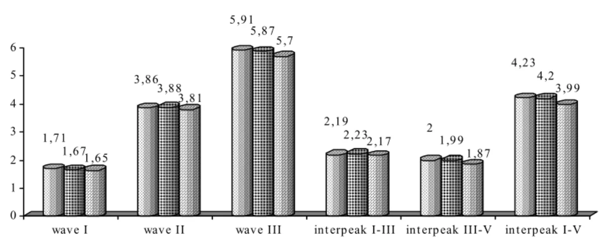
Mean 1.71 1.67 1.65 3.86 3.88 3.81 5.91 5.87 5.70 2.19 2.23 2.17 2.00 1.99 1.87 4.23 4.20 3.99
p.value 0.388 0.727 0.170 0.607 0.392 0.075*
Results
ABR
The results show a statistically significant mean difference (p-value=0.075) simultaneously between groups to the interpeak I-V (Kruskal-Wallis Test) (Table 1 and Figure 1). Besides, they proved that group GIII produced the difference to both the other groups (p-value 0,029 to group GI and 0.036 to GII when compared to GIII) – Mann Whitney Test – Comparison of two groups (Table 2).
In group GI 36% of the analyzed ears presented disorders on ABR where the most frequent was the absence of wave I and presence of waves III and V with latencies within the normal range (40%). In group GII 58% of the analyzed ears presented disorders on ABR where the most frequent was the presence of waves I and V with absolute latencies within normality and delayed absolute latency of wave III (42,85%). In group III 85% of the analyzed ears presented disorders on ABR, where the absence of waves I, III and V was the most frequent (33,33%). (Figure 2).
MLR
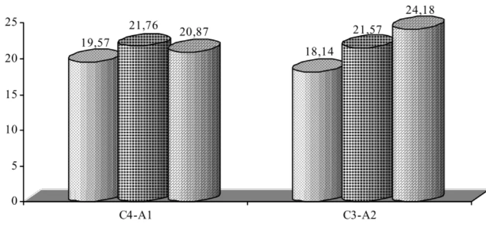
Results show statistically significant mean difference (p-value = 0.071) simultaneously between groups on wave Na latency in the contralateral modality C3-A2 (Kruskal-Wallis Test) (Table 3 and Figure 3). Besides it proved in the
same modality that group GI produced a difference for both the other groups (p-value 0.064 to group GII and 0.065 to group GIII when compared to group GI) – Mann-Whitney Test – comparison of two
groups at a time. (Table 4).
It wasn’t observed statistically significant mean differences simultaneously between groups to Pa wave latency in contralateral modality (C4-A1 and C3A2) in Table 5 and Figure 4 (Kruskal-Wallis Test).
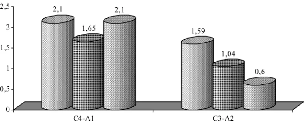
It wasn’t observed statistically significant mean differences simultaneously between groups to amplitude Na-Pa in contralateral modality (C4-A1 and C3A2) in Table 6 and Figure 5 (Kruskal-Wallis Test).
P300
Initially to verify presence or absence of this potential the groups were not divided according to age. Using the Equality of Two Proportions Test the results show statistically significant mean difference (0.001) between presence and absence of P300 (Table 7 and Figure 6).
Considering only the presence of P300 the results show statistically significant mean difference (0.002) simultaneously between groups referring to the absolute latency of this component (Kruskal-Wallis Test) (Table 8 and figure 7).
TABLE 1. Mean values of absolute latencies of waves I, III, V and interpeaks I-III, III-V and I-V to groups GI,GII and GIII.
FIGURE 1. Mean values of absolute latencies of waves I, III, V and interpeaks I-III, III-V and I-V to groups GI,GII and GIII.
TABLE 2. Comparison of GI, GII and GIII groups two-by-two by Mann-Whitney Test.
Interpeak I-V GI GII
GII 0.612
GIII 0.029* 0.036*
p-values
FIGURE 2. Distribution of normal and disordered results on ABR to groups G I, G II and G III.
1,71 1,671,65
3,86 3 ,88
3,81
5,91 5,87
5,7
2,19
2,232,17 2 1,9 9 1,87
4,23 4,2
3,9 9
0 1 2 3 4 5 6
wave I wave II wave III int erp eak I-III int erp eak III-V in t erpeak I-V
64%
36% 42%
58%
15% 85%
0% 20% 40% 60% 80% 100%
group I group II group III
TABLE 3. Mean values of wave Na latency in groups GI, GII e GIII, in contralateral modalities C4-A1 e C3-A2.
C4-A1 C3-A2 Na
GI GII GIII GI GII GIII
Mean 19.57 21.76 20.87 18.14 21.57 24.18
p-value 0.472 0.071*
TABLE 5. Mean values to Pa wave latency to groups GI, GII e GIII, in contralateral modalities C4-A1 e C3-A2.
C4-A1 C3-A2 Pa
GI GII GIII GI GII GIII
Mean 31.53 32.60 36.08 29.38 33.07 31.79
p-value 0.385 0.203 FIGURE 3. Mean values of wave Na latency in groups GI, GII e GIII, in contralateral modalities C4-A1 e C3-A2.
TABLE 4. Comparison of groups GI, GII and GIII,, two at a time, with the Mann-Whitney Test.
C3-A2 GI GII
GII 0.064*
GIII 0.065* 0.333
p-values
19,57
21,76 20,87
18,14 21,57
24,18
0 5 10 15 20 25
C4-A1 C3-A2
TABLE 6. Mean values of amplitude Na-Pa in groups GI, GII and GIII, in contralateral modalities C4-A1 e C3-A2.
C4-A1 C3-A2 Amplitude
Na-Pa
GI GII GIII GI GII GIII
Mean 2.10 1.65 2.10 1.59 1.04 0.60
p-value 0.585 0.206
TABLE 7. Presence and absence of P300 in studied individuals.
P300 N % p-value
Present 40 88.90%
Absent 5 11.10%
<0.001*
TABLE 8. Mean values of P300 latency to groups GI, GII and GIII.
P300 P300
GI GII GIII
Mean 331.71 370.67 407.50
p-value 0.002*
FIGURE 4. Mean values to Pa wave latency to groups GI, GII e GIII, in contralateral modalities C4-A1 e C3-A2.
31,53 32,6
36,08
29,38 33,07
31,79
0 5 10 15 20 25 30 35 40
C4-A1 C3-A2
FIGURE 5. Mean values of amplitude Na-Pa in groups GI, GII and GIII, in contralateral modalities C4-A1 e C3-A2.
FIGURE 6. Presence and absence of P300 in studied individuals.
FIGURE 7. Mean values of P300 latency to groups GI, GII and GIII.
2,1
1,65 2,1
1,59
1,04
0,6
0 0,5 1 1,5 2 2,5
C4-A1 C3-A2
group I group II group III
88,90% 11,10%
present absent
331,71 370,67
407,5
0 100 200 300 400 500
Discussion
Several works in the literature show that in elderly individuals a larger number of disorders on ABR may occur, this fact is not observed in the ABR results of young adults (Bess et al., 2001; Burkard e Sims, 2002; Boettcher, 2002). These findings were also observed in the present study where it was verified an increasing number of disorders on ABR associated with increasing age, with the individuals between 70 and 79 years of age presenting 85% of the analyzed years with disorders on the ABR (Figure 2).
Considering the types of disorders that can be found in ABR of elder individuals it can be observed a great diversity in literature data. It involves the increase of the absolute latency of wave V (around 0.2ms) and a decrease of the amplitude of wave V (Jerger and Hall, 1980) and also a delay of the absolute latency of all the components of ABR, as described by Munhoz et al. (2003). On the other hand Burkard e Sims (2002) and Boettcher (2002) evaluating individuals with presbyacusis observed increased absolute latencies on ABR waves with preservation of interpeaks latency.
In the study of Jerger and Hall (1980) the authors identified increase on the absolute latency of wave V and therefore on the interpeak I-V in elder individuals. In the present research which simultaneously analyzed three groups it was observed statistically significant difference to the I-V interpeak, determining that the group of higher age produced this difference (Table 2), although the mean values obtained in this interpeak measure were smaller in this group (Table 1 and Figure 1). However, the separate analysis of the types of disorders observed on the ABR in the three groups verified that the most frequent disorder was the absence of responses on the group of 70 to79 years of age (Figure 2).
Boettcher (2002) stated in his research that the absolute latencies of waves on ABR present a tendency to increase along with increasing age, but not all studies found strong evidence that aging process and absolute latencies on ABR may be related.
In the study conducted by Freitas and Oliveira (2001) it was found an increase of latencies of waves I and V with stable interpeak I-V on individuals with presbyacusis, suggesting a peripheral disorder. It is important to stress that this kind of disorder, as well as ABR waves delay with normal interpeaks, also suggesting peripheral
disorder was not observed in the present study. This fact may be explained by the inclusion criteria used to determine the sample, which considered only the ears with normal hearing or with sensorineural hearing loss up to moderate level in tonal audiometry, excluding ears with conductive or mixed losses.
This diversity of results observed on ABR in elder individuals could be explained by the fact that, as an effect of brainstem auditory pathway degeneration, it is possible to observe auditory nerve atrophy of the basal cochlea gyrus due to peripheral hearing loss that would lead to disorders of the first ABR waves, or synaptic transmission delay, neural loss and changes of the permeability of the neural membrane (Johannsen e Lehn, 1984) that could generate a delay of absolute latencies as well as a decrease of ABR wave amplitudes.
Although some studies verified differences on MLR with increasing age – longer Pa wave and larger amplitude, changes in latencies of MLR waves (Gerken et al., 2001) in the present research it wasn’t observed statistically significant differences on Pa wave latencies (Table 5 and Figure 4) and on the amplitude Na-Pa (Table 6 and Figure 5) between the studied groups. Only wave Na latency in the contralateral modality C3-A2 determined statistically significant difference between groups simultaneously; with group GI of 50 to 59 years producing the difference for both groups (Table 3 and Figure 3). These data agree with Paludetti et al.’s study (1991) that considered the large acceptability range of the MLRs and the variability between-subjects as factors that directly interfere in the quantitative analysis of MLR waves’ latencies and amplitude.
evoked potentials, specially the cognitive potential (P300).
It is important to mention that even with the progressive increase of absolute latency of P300wave with progressive age the mean latencies found are within the normal values determined by McPherson (1996) to each age range studied.
Even though the qualitative aspects of the waves was not the object of this study, in several cases it was possible to observe considerable distortions of the trace quality of the auditory
evoked potential waves, indicating a strong correlation with the study of Jerger and Lew (2004) which also identified deterioration of trace quality of the evoked auditory potentials with age increase.
Conclusion
Auditory system aging process progressively affects the auditory pathways throughout the brainstem and the temporal lobe.
References
ASSIS, C. L.; SOUZA, F. C. R.; BARAKY, L. R.; BERNARDI, A. P. A. Estudo da audiometria de tronco encefálico em indivíduos de 20 a 30 anos com audição normal. R. Cefac, São Paulo, v. 7, n. 1, p. 87-92, jan.-mar. 2005.
BESS, F. H.; HEDLEY-WILLIAMS, A.; LICHTENSTEIN, M. J. Avaliação audiológica dos idosos. In: MUSIEK, F. E.; RINTELMANN, W. F. Perspectivas atuais em avaliação auditiva. Barueri: Manole, 2001. cap. 12, p. 343-370. BOETTCHER, F. A. Presbiacusis and auditory brainstem response. J. Speech Lang. Hear. Res., Rockville, v. 45, n. 6, p. 1249-1261, dec. 2002.
BURKARD, R. F.; SIMS, D. The human auditory brainstem response to high click rates: aging effects. Am. J. Audiol.,
Rockville, v. 11, n. 1, p. 53-61, dec. 2001. Erratum in:
Am. J. Audiol., Rockville, v. 11, n. 1, p. 12, jun. 2002. CALERO, M. D.; NAVARRO, E. Relationship between plasticity, mild cognitive impairment and cognitive decline.Arch. Clin. Neuropsychol., United States, v. 19, n. 5, p. 653-660, aug. 2004.
DURRANT, J. D.; FERRARO, J. A. Potenciais auditivos evocados de curta latência: eletrococleografia e audiometria de tronco encefálico. In: MUSIEK, F. E.; RINTELMANN, W. F. Perspectivas atuais em avaliação auditiva. Barueri: Manole, 2001. cap. 7, p. 193-238.
ELWAN, O.; MADKOUR, O.; ELWAN, F.; MOSTAFA, M.; ABBAS HELMY, A.; NASEER, M.; ABDEL-SHAFY, S.; EL FAIUOMY, N. Brain aging in normal egyptians: cognition, education, personality, genetic and immunological study. J. Neurol. Sci., Netherlands, v. 211, n. 1 e n. 2, p. 15-22, jul. 2003.
FREITAS, M. R.; OLIVEIRA, J. A. A. Audiometria de respostas evocadas de tronco cerebral em indivíduos idosos com e sem presbiacusia. R. Bras. Otorrinolaringol., São Paulo, v. 67, n. 2, p. 171-178, 2001.
GERKEN, G. M.; HESSE, P. S.; WIORKOWSKI, J. J. Auditory evoked responses in control subjects and in patients with problem-tinnitus. Hear. Res., Netherlands, v. 157, n. 1 e n. 2, p. 52-64, jul. 2001.
JERGER, J.; HALL, J. Effect of age and sex on the auditory brainstem response. Arch. Otolaryngol., Chicago, v. 106,
n. 7, p. 387-391, maio 1980.
JERGER, L.; LEW, H. L. Principles and clinical applications of auditory evoked potentials in the geriatric
population. Phys. Med. Rehabil. Clin. N. Am., Philadelphia, v. 15, n. 1, p. 235-250, feb. 2004. JOHANNSEN, H. S.; LEHN, T. The dependence of early acoustically evoked potentials on age. Arch. Otorhinolaryngol., Germany, v. 240, n. 2, p. 153-158, 1984. KIMURA, D. Some effects of temporal lobe damage on auditory perception. Can. J. Exp. Psychol., Canada, v. 15, p. 157-165, sep. 1961.
KNOTT, V.; BRADFORD, L.; DULUDE, L.; MILLAR, A.; ALWAHHABI, F.; LAU, T.; SHEA, C.; WIENS, A. Effects of stimulus modality and response mode on the P300 event-related potential differentiation of young and elderly adults. Clin. Electroencephalogr., United States, v. 34, n. 4, p. 182-190, oct. 2003.
KNOTT, V.; MILLAR, A.; DULUDE, L.; BRADFORD, L.; ALWAHHABI, F.; LAU, T.; SHEA, C.; WIENS, A. Event-related potentials in young and elderly adults during a visual spatial working memory task. Clin. EEG. Neurosci., Wheaten, v. 35, n. 4, p. 185-192, oct. 2004.
MAURITS, N. M.; ELTING J. W.; JAGER D. K.; VAN DER HOEVEN, J. H.; BROUWER, W. H. P300 component identification in auditory oddball and novel paradigms using source analysis techniques: reduced latency variability in the elderly. J. Clin. Neurophysiol., Netherlands, v. 22, n. 3, p. 166-175, jun. 2005. McPHERSON, D. Late Potentials of the auditory system. San Diego: Singular Publishing Group. 1996. p. 75-100. MUNHOZ, A. S. L.; SILVA, M. L. G.; CAOVILLA, H. H.; FRAZZA, M. M.; GANANÇA, M. G.; CÂMERA, J. L. Z. Respostas auditivas de tronco encefálico. In: MUNHOZ, M. S. L.; CAOVILLA, H. H.; SILVA, M. L. G.; GANANÇA, M. M. Audiologia clínica. São Paulo: Atheneu, 2003. cap. 12, p. 191-220.
MUSIEK, F. E.; LEE, W. W. Potenciais auditivos de média e longa latência. In: MUSIEK, F. E.; RINTELMANN, W. F. Perspectivas atuais em avaliação auditiva. Barueri: Manole, 2001. cap. 8, p. 239-267.
PALUDETTI, G.; MAURIZI, M.; D'ALATRI, L.; GALLI, J. Relationships between middle latency auditory responses (PEAML) and speech discrimination tests in the elderly.
Acta Otolaryngol. Suppl., Stockholm, v. 476, p.
105-109, 1991.
CARVALLO, R. M. M. Fonoaudiologia informação para a formação: procedimentos em audiologia. Rio de Janeiro: Guanabara Koogan, 2003. p. 57-70.
SONCINI, F.; COSTA, M. J.; OLIVEIRA, T. M. T. Influência do processo de envelhecimento no reconhecimento da fala em indivíduos normo-ouvintes. Pró-Fono R. Atual. Cient., Barueri, v. 15, n. 3, p. 287-296, set.-dez. 2003.