Study conducted at the Post-graduate Program in Health Human Communication, Universidade Federal de Pernambuco – UFPE – Recife (PE), Brazil, with a scholarship conceived by the REUNI assistance to teaching UFPE.
(1) Post-graduate Program in Health Human Communication (Masters Degree), Universidade Federal de Pernambuco – UFPE – Recife (PE), Brazil. (2) Departament of Neurology, Universidade Federal de Pernambuco – UFPE – Recife (PE), Brazil.
(3) Post-graduate Program in Neuropsychiatry and Behavioral Science, Universidade Federal de Pernambuco – UFPE – Recife (PE), Brazil.
(4) Department of Speech-Language Pathology and Audiology, Universidade Estadual de Ciências da Saúde de Alagoas – UNCISAL – Maceió (AL), Brazil. Conflict of interests: No
Author´s contribution: ATLC main researcher, research elaboration, chronogram elaboration, casebook, collecting, analysis of data, writing of the essay, sub-mission and proceedings form the essay; OGL advisers, research elaboration, chronogram elaboration, analysis of data, editing the article, final version approval; IAS advisers, research elaboration, chronogram elaboration, analysis of data, editing the article, final version approval; KCLA assistant researchers, casebook, data collection and writing the article; PLM advisers, research elaboration, chronogram elaboration, analysis of data, editing the article, final version approval. Correspondence adress: Aline Tenório Lins Carnaúba. Programa de Pós-graduação em Saúde da Comunicação Humana. R. Arthur de Sá, s/n, Cidade Univer-sitária, Recife (PE), Brazil, CEP: 50670-420. E-mail: [email protected]
Received: 3/17/2013; Accepted: 9/4/2013
Vestibular evoked myogenic potential and its
implications in the frequency domain
Potencial miogênico evocado vestibular e suas implicações no
domínio das frequências
Aline Tenório Lins Carnaúba1, Otávio Gomes Lins2, Ilka do Amaral Soares3, Kelly Cristina Lira de Andrade1,
Pedro de Lemos Menezes4
ABSTRACT
Purpose: Determine whether there is an association between VEMP in the time domain and the frequency domain. Methods: The sample was composed of 18 individuals (36 ears), six men and 12 women. In the VEMP test in the time domain, 200 tone burst stimuli were promediated at a frequency of 500 Hz, with 5.1 stimulations/s at an intensity of 95 dBNAn. This was followed by capture in the frequency domain, where 200 stimuli consisting of 500 Hz (carrier frequency) pure tones were promediated and modulated at a frequency of 40 Hz. Odds ratio was calculated and the chi-squared test was applied to compare the responses of two domains. This is a cross-sectional contemporary cohort study. Results: VEMP in the time domain was recorded by unilateral stimula-tion and capture, with adequate morphology, in 88.88% of the ears. In the frequency domain test, a steady-state peak was found in 30 of the 36 tests (83.33%). The chi-squared test and odds ratio calculation showed a strong relationship between the two domains. Conclusion: There is a significant association between the VEMP tests in the time and frequency domains, a finding that suppports its use in clinical practice.
Keywords: Function Tests; Motor Evoked Potential; Accoustic Stimula-tion; Sacculum and Utriculum; Labyrinth Vestibulum
RESUMO
Objetivo: Verificar a existência de associação entre o VEMP no do-mínio do tempo e no dodo-mínio das frequências. Métodos: A amostra foi composta por 18 indivíduos (36 orelhas), sendo seis do gênero masculino e 12 do gênero feminino. No exame de VEMP no domínio do tempo, foram promediados 200 estímulos tone burst na frequência de 500 Hz, com taxa de estimulação de 5,1 estímulos/s na intensidade de 95 dBNAn. Seguiu-se com a captação no domínio das frequências, onde foram promediados 200 estímulos constituídos por tons puros de 500 Hz (frequência portadora), modulados na frequência de 40 Hz. Foi realizado o cálculo do Odds ratio e aplicado o teste Qui-quadrado para a comparação entre as respostas dos dois domínios. Estudo de coorte con-temporânea com corte transversal. Resultados: O VEMP no domínio do tempo foi registrado por meio da estimulação e captação unilateral, com morfologia adequada, em 88,88% das orelhas. Na realização do exame no domínio das frequências, foi registrada a presença de pico de estado estável em 30 (83,33%) dos 36 exames. Quando realizado o teste Qui--quadrado e o cálculo do Odds ratio, foi possível observar a existência de forte relação entre os dois domínios. Conclusão: Conclui-se que existe forte associação entre os exames de VEMP no domínio do tempo e no domínio das frequências, o que habilita sua utilização na prática clínica.
INTRODUCTION
Ever since its first description in 1992(1), the vestibular
evoked myogenic potential (VEMP) became a complementary test of vestibular function. The VEMP is a muscle reflex, evoked by stimulation of the cervical vestibular pathway (normally with loud sound)(2-4) and recorded by electrodes placed on the
sternocleidomastoid muscle(5).
This test assesses the integrity of the saccule and inferior vestibular nerve, information not obtained by traditional ves-tibular assessment, such as vector eletronystagmography(6).
VEMP waves are highly reproducible, irrespective of ster-nocleidomastoid stimulation being unilateral or bilateral, or whether capture takes place at different moments. Thus, the VEMP can be used clinically, with numerous applications in the diagnosis of vestibular disorders(7).
The biological signals of VEMP are traditionally analyzed in the time domain. Most of the time recording is made in the form of a wave, which represents the variation in electrical potential over time. However, signal errors and abnormalities in this domain hinder interpretation and the test depends ex-clusively on the knowledge and experience of the examiner. In order to obtain more relevant information and eliminate interferences, signal processing techniques, such as the Fourier Transform, can be used to observe the event in the frequency rather than the time domain(8).
The promising steady-state technique assesses auditory potentials in the frequency domain. Modulating tones can be applied to both ears simultaneously, and the response analyzed by the Fast Fourier Transform, associated to statistical techni-ques(9,10). This is a much faster and more accurate test, in terms
of compromised frequencies(11).
Studies conducted on the use of the steady-state technique to detect the VEMP found that this test, as well as auditory evoked potential, can also be performed in the frequency domain(7,12). In
addition to early detection of inferior vestibular nerve disorders, it favors a more accurate diagnosis in a shorter time frame and independent of the examiner’s interpretation(7).
In the time domain, the response is recorded and analyzed by selecting peaks and assessing amplitudes and latencies. In the frequency domain, analysis is based on the amplitude and phase of the response for each peak of the frequency spectrum(13,14).
Thus, the aim of this study was to determine whether there is an association between the vestibular evoked myogenic potential in the time and frequency domains.
METHODS
The study was approved by the Research Ethics Committee of the Universidade Estadual de Ciências da Saúde de Alagoas (UNCISAL), under protocol no. 990/09, and all participants gave their informed consent.
The professors, students and employees of public and pri-vate universities in the city of Maceio, Brazil were informed of the study. The subjects appeared spontaneously at the study site and received no financial contribution for taking part in the experiment. However, they did receive a copy of the auditory and vestibular assessment.
A total of 18 individuals (36 ears) participated in the study, 6 men and 12 women, selected according to the following inclusion criteria: aged between 18 and 35 years and auditory threshold less than or equal to 20 dBNA, with frequency differ-ences between the ears less than or equal to 10 dB. Exclusion criteria were exposure to occupational or leisure noise, middle or inner ear surgery, more than three middle ear infections, use of ototoxic medication, presence of systemic alterations, such as diabetes and hypertension, that could contribute to vestibular-cochlear pathologies, hormonal alterations, ringing in the ears, vertigo, dizziness, or other vestibular-cochlear disturbances.
First, a questionnaire regarding general health and auditory and vestibular function was applied to screen the participants (Appendix 1). The following procedures were then carried out: otoscopy, pure-tone threshold audiometry and VEMP in the time and frequency domains.
The VEMP tests were carried out with a specific device, denominated the analyzer of evoked potentials in the time and frequency domains (AEPTFD). Developed at two public universities the APEDTF is composed of biological ampli-fiers, filters, electrical protection system and a logic system to investigate the VEMP.
Surface electrodes, placed on previously cleaned skin, the positive electrode on the middle third of the sternocleidomas-toid muscle (SCM) ipsilateral to stimulation, were used to record potentials. The negative electrode was positioned at the level of the tendon, just above the clavicle, and the ground electrode on the frontal midline. During recording of the SCM muscle, the patient remained sitting, with maximum lateral rotation of the head toward the side contralateral to the stimulus.
In VEMP testing in the time domain, 200 tone burst stimuli were promediated at a frequency of 500 Hz, stimulation rate of 40.8 stimuli/s at an intensity of 95 dBNAn, presented through ER3A earphones equipped with disposable foam tips. A pass-band filter, configured between 5 and 1000 Hz, showing from 10 to 25 µV per division, was used. The recordings were made in 40 ms windows, sufficient time to include all the responses(15,16).
To interpret the findings, the responses were analyzed by two authors/evaluators, using morphology and demarcating p13 and n23 waves by the latencies of the first positive and negative peaks. Discrepancies exhibited by the authors/evaluators were resolved by a third author/evaluator.
For VEMP analysis in the frequency domain, the frequency potencies acquired by Fast Fourier Transform were promediated 200 times and subsequently analyzed using the magnitude-squared coherence (MSC) statistical method in order to confirm the responses.
The choice of modulator for the test in the frequency do-main was based on the responses obtained in the assessment of steady-state auditory evoked potentials. For this test, the modulation rate of 40 Hz results in responses with higher wave amplitude(17). Moreover, this modulation frequency is
equivalent to an observation of 25 ms in the time domain, appropriate for assessing VEMP, which has a late wave com-ponent (≈ 23 ms)(6).
During VEMP in the frequency domain, the equipment exhibited the following characteristics for processing this type of signal: conversion of digital and analog signals at a sampling rate of 44.1 kHz and resolution of 16 bits; digital-analog conversion, conducted at an acquisition frequency of 2.75 kHz, exactly 1/16 of the signal generation rate in order to ensure fast Fourier transform of up to 1.3 kHz, also with a resolution of 16 bits.
Statistical method
The data were tabulated and processed by Predictive Analytics Software(PASW® STATISTIC), version 17.0. A tabular and graphical presentation of the average of standard deviations and percentages was used.
Odds ratio and the chi-squared test were applied to con-firm that the percentages of responses between domains were statistically similar.
RESULTS
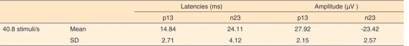
VEMP in the time domain was recorded using stimulation and unilateral capture, with adequate morphology, in 88.88% of the ears, and a stimulation rate of 40.8 stumuli/s, with tone burst stimulus at a frequency of 500 Hz.
In the mapping of the test, waves were marked, determining absolute latencies and amplitudes of p13 and n23 (Table 1).
During the test in the frequency domain, a steady-state peak was recorded in 30 (83.33%) of the 36 tests. One person (two tests) from this sample exhibited responses only in the time domain and two others (four tests) had no responses in any of the domains assessed.
The chi-squared test, conducted to compare the presence and absence of responses between the two domains, showed that the small numerical differences were significant; that is, when responses were present in the time domain, they were also present in the frequency domain (Tables 2 and 3).
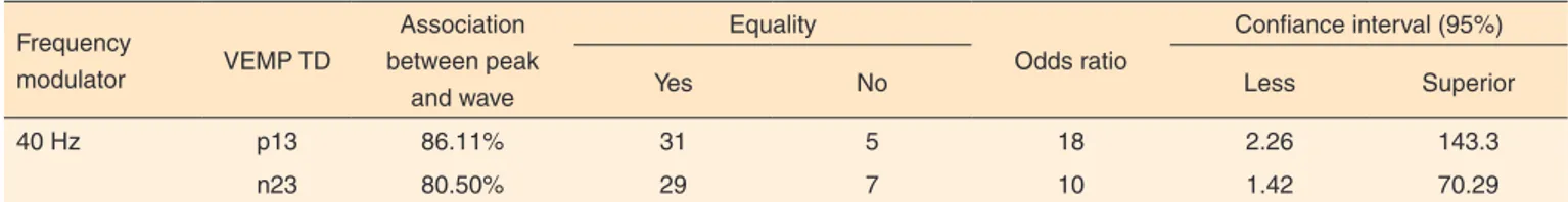
The odds ratio showed an association between the presence of p13 and n23 waves of the VEMP in the time domain and the presence of a steady-state peak of the VEMP in the frequency domain, with an odds ratio value of 18 and 10, respectively. In other words, with the presence of the wave in time (p13), a peak in the steady-state test is 18 times more likely, and in the presence of the n23 wave, there is ten times more probability of the steady-state peak (Table 4).
DISCUSSION
Analyses of the responses obtained in this sample demons-trated that it is possible to consistently record the p13 and n23 waves in the time domain, exhibiting similar results to other studies(15,18).
With respect to the absence of responses in the two domains,
Table 1. Latencies and amplitudes of VEMP in time domain
Latencies (ms) Amplitude (µV )
p13 n23 p13 n23
40.8 stimuli/s Mean 14.84 24.11 27.92 -23.42
SD 2.71 4.12 2.15 2.57
Note: VEMP = vestibular evoked myogenic potential, SD = standard deviation
Table 2. Relationship between the presence of p13 peak of VEMP in the time domain and the presence of the VEMP peak in the frequency domain
Presence of peak of VEMP in the FD
Presence Absent Total
Presence of peak 13
Presence 27 2 29
Absent 3 4 7
Total 30 6 36
p-value 0.008*
*Significant values (p≤0.05) – Chi-square test
Note: VEMP = vestibular evoked myogenic potential, DF = frequency domain
Table 3. Relationship between the presence of the n23 peak of VEMP in the time domain and the presence of the VEMP peak in the frequency domain
Presence of peak of VEMP in the FD
Presence Absent Total
Presence of peak n23
Presence 25 2 27
Absent 5 4 9
Total 30 6 36
p-value 0.024*
*Significant values (p≤0.05) – Chi-square test
observed in four tests, we can establish two hypotheses: the first is that the two subjects in question showed vestibular alterations during some part of the cervical-vestibular reflex trajectory(16,19);
the second considers the external factors involved in assess-ing the potential, such as the presence of errors that may have caused the absence of responses, or alterations in the wake state of the patient(9).
In relation to the association between the two domains, it can be inferred, based on the findings of the present study and those contained in the literature, that there is a strong relation-ship between the two domains, confirming that VEMP can be conducted in the frequency domain without losing the informa-tion required for vestibular assessment (10,11).
Establishing an analogy with the steady-state auditory evoked potential (SSAEP), the greatest advantage of the VEMP in the frequency domain is the objective way of assessing responses, which can be done automatically with software. This decreases the likelihood of diagnostic errors, especially at slightly lower intensities, since the subjective interpretation by a professional visually evaluating the results is no longer necessary(8,9). Furthermore, the use of this technique does not
require patients to be sedated and offers the possibility of simul-taneously exploring several frequencies, without significantly increasing assessment time(9,13).
A number of authors report that the use of VEMP in the time domain in individuals with normal hearing has limited clinical applicability, due to significant sound exposure during the test(11,20). Another contraindication would be that
individu-als complaining of ringing in the ears would not be submitted to the test, since it could worsen symptoms(11). However, the
VEMP in the frequency domain, a faster test, minimizes these contraindications. Moreover, it can assess several frequencies simultaneously, demonstrating its usefulness in evaluating sac-cular function, enabling early diagnosis of Ménière’s disease(21).
Finally, in VEMP assessment, the p13 and n23 peaks do not represent the trajectory of the potential through vestibular pathway structures, as occurs in the Brainstem Auditory Evoked Potentials (BAEP) with waves I to V in the corresponding pathway(16). Thus, the VEMP in the frequency domain does
not lose temporal information, which helps identify the origin of the interruption in nerve transmission(21).
CONCLUSION
There was a strong association between VEMP tests in the
time and frequency domains, which enables its use in clinical practice.
REFERENCES
1. Colebatch JG, Halmagyi GM. Vestibular evoked potentials in human neck muscles before and after unilateral vestibular deafferentation. Neurology.1992;42(8):1635-36.
2. Rosengren SM, Kingma H. New perspectives on vestibular evoked myogenic Potentials. Curr Opin Neurol. 2013;26(1):74-80. 3. Rocha MF, Azevedo DF, Russomano T, Figueira MV, Helegda
S. Mobile remote monitoring of biological signals. In: IEEE 2006 International Conference of the Engineering in Medicine and Biology Society, 2006, Nova York. IEEE 2006 International Conference of the Engineering in Medicine and Biology Society. NY, EUA: IEEE-EMBS, 2006;1:2057-59.
4. Rauch SD. Vestibular evoked myogenic potentials. Otol Neurotol. 2006;14(5):299-304.
5. Sazgar AA, Akrami K, Akrami S, Yazdi ARK. Recording of vestibular evoked myogenic potentials. Acta Med Iran. 2006;44(1):13-6.
6. Hall JW. New Handbook for auditory evoked responses. Boston: Pearson Education. 2006.
7. Barreto ACO, Colafêmina JF, Menezes PL. Saccular sensitivity function measured by vestibular evoked myogenic potential. Acta Otolaryngol. 2011;131(6):618-23.
8. Akin FW, Murnane OD, Panus PC, Caruthers SK, Wilkinson AE, Proffitt TM. The influence of voluntary tonic EMG level on the vestibular-evoked myogenic potential. J Rehabil Res Dev. 2004;41(3B):473-80.
9. Ramos EG, Zaeyen EJB, Simpsoni DM, Infantosi AFC. Detecção da resposta auditiva no EEG de crianças utilizando técnicas no domínio da frequência. Rev Bras Eng Biomed. 2000;16(3):127-37.
10. Menezes PL. Desenvolvimento e avaliação de um dispositivo capaz de registrar e analisar potenciais evocados auditivos no domínio do tempo e no domínio das frequências por meio de uma placa de som para computador pessoal [tese]. Ribeirão Preto: Universidade de São Paulo, Departamento de Física e Matemática; 2008.
11. Lins OG. Audiometria fisiológica tonal utilizando respostas de estado estável auditivas do tronco cerebral [tese]. São Paulo: Universidade Federal de São Paulo – Escola Paulista de Medicina, Departamento de Medicina; 2002.
12. Bell SL, Fox L, Id Bihi R. Vestibular evoked myogenic responses to amplitude modulated sounds (L). J Acoust Soc Am. 2010;128(2):559-62.
Table 4. Investigation on the association between the presence of a wave in the time and frequency domains of the VEMP examination
Frequency
modulator VEMP TD
Association between peak
and wave
Equality
Odds ratio
Confiance interval (95%)
Yes No Less Superior
40 Hz p13 86.11% 31 5 18 2.26 143.3
n23 80.50% 29 7 10 1.42 70.29
13. Stapells DR, Herdman A, Small SA, Dimitrijevic A, Hatton J. Current status of the auditory steady-state responser for estimating an infant`s audiogram. In: Seewald RC, Banckford J. (Eds.) A Sound Foundation Through Early Amplification. Chicago: Phonak, 2005. p. 43-59.
14. Picton TW, John MS, Dimitrijevic A, Purcell D. Human auditory steady–state response. Int J Audiol. 2003;42(4):177-219.
15. Basta D, Todt I, Ernst A. Normative data for P1/N1 latencies of vestibular evoked myogenic potencials induced by air or bone conducted tone bursts. Clin Neurophysiol. 2005;116(9):2216-9. 16. Sheykholeslami K, Habiby Kermany M, Kaga K. Frequency
sensitivity range of the saccule to bone conducted stimuli measured by vestibular evoked myogenic potencials. Hear Res. 2001;160(1-2):58-62.
17. Dimitrijevic A, John MS, Picton TW. Auditory steady-state responses
and word recognition scores in normal hearing and hearing impaired adults. Ear Hear. 2004;25(1):68-84.
18. Wu CH, Murofushi T. The effect of click repetition rate on vestibular evoked myogenic potenctial. Acta Otolaryngol. 1999;119(1):29-32. 19. Sheykholeslami K, Kermany MH, Kaga K, Megerian CA, Arnold
J. Vestibular-evoked myogenic potencials in infancy and early childhood. Laryngoscope. 2005;115(8):1440-4.
20. Rauch SD, Silveira MB, Zhou G, Kujawa SG, Wall C 3rd, Guinan JJ,
et al. Vestibular evoked myogenic potentials versus vestibular test battery in battery in patients with Menière’s disease. Otol. Neurotol. 2004;25(6):981-6.
21. Rosengren SM, Welgampola MS, Colebatch JG. Vestibular evoked myogenic potentials: past, present and future. Clin Neurophysiol. 2010;121(5):636-51.
Appendix 1. Questionnaire on general health history and auditory and vestibular function
Identification
Name:___________________________________________ Protocol n°.:
Date: __________ Age: _____ Gender: __
Otologic history
Ear infections (more than three episodes/year): Y ( ) N ( )
Use of ototoxic medication: ...Y ( ) N ( )
Ear surgery: Y ( ) N ( ) Type: _____________________
Ringing in the ears, vertigo, dizziness or other vestibular-cochlear disorders: Y ( ) N ( )
Hereditary deafness: ...Y ( ) N ( )
Exposure to occupational or leisure noise:... Y ( ) N ( )
If yes, minimum weekly exposure time: ______________________
Minimum auditory rest of 14 hours:...S ( ) N ( )
General health status
Diseases: ________________________________________________________
Medications: _____________________________________________________
Hormonal alterations: Y ( ) N ( )
Inspection of external auditory canal...