J Bras Pneumol. 2007;33(1):101-104
101
CASE REPORT
Idiopathic tracheal stenosis. A report of four cases*
Carolina Rossi1, Fernanda Colombari1, Alda Losi Guembarowsky2,
Olavo Franco Ferreira Filho3, João Carlos Thomson4
Abstract
Idiopathic tracheal stenosis is uncommon. Herein, we report four cases, all presenting a similar clinical profile: diagnosed through bronchos-copy and having a history of being treated unsuccessfully for bronchospasm. Three of the patients were treated with dilatation and an oral corticosteroid. One of those three underwent tracheoplasty. In the remaining patient, the stenosis was more extensive (2 cm, with a 70% reduction in the size of the lumen), and dilatation was not an option. Therefore, that patient underwent laryngotracheal resection. In all four patients, the evolution was favorable. Idiopathic tracheal stenosis should be contemplated in cases of ‘bronchitis’ that are not resolved using conventional treatments. Bronchoscopy and dilatation have provided satisfactory results. Occasionally, laryngotracheal reconstruction is necessary.
Keywords: Trachea/surgery; Constriction; Pathologic; Case reports [publication type].
* Study carried out at the Universidade Estadual de Londrina (UEL, Londrina State University) – Londrina (PR) Brazil. 1. Medical student at the the Universidade Estadual de Londrina (UEL, Londrina State University) – Londrina (PR) Brazil.
2. Adjunct Professor of Pathological Anatomy at the Universidade Estadual de Londrina (UEL, Londrina State University) – Londrina (PR) Brazil.
3. Adjunct Professor of Pulmonology in the Clinical Medicine Department of the Universidade Estadual de Londrina (UEL, Londrina State University) – Londrina (PR) Brazil.
4. Coordinator of the Masters and PhD Programs in Medicine and Health Sciences at the the Universidade Estadual de Londrina (UEL, Londrina State University) – Londrina (PR) Brazil.
Correspondence to: João Carlos Thomson. Rua Júlio César Ribeiro, 204, CEP 86039-200, Londrina, PR, Brasil. E-mail: [email protected]
Submitted: 22/1/06. Accepted, after review: 7/4/06.
Introduction
Idiopathic tracheal stenosis (ITS) was first described in 1972 by Brandenburg, who monitored three cases of indefi-nite etiology over a ten-year period. Although similar cases have since been reported, there have been few studies, and the etiology of the disease remains unclear.(1) It is a
rare disease, characterized by the formation circumferen-tial, dense fibrous tissue, which is usually located in the subglottic region, although it can extend to the vocal folds or to the upper trachea.(2) The most important characteristic
is the fact that patients do not present predisposing factors for the onset of ITS. Patients with ITS present progres-sive dyspnea, respiratory sounds and dry cough, and are frequently treated for ‘bronchitis’. The duration of symptoms ranges from four months to fifteen years, and most patients report having had symptoms for one to three years.(2,3)
The diagnosis should be made after ruling out other causes of tracheal stenosis, such as prolonged orotracheal intubation, tracheostomy, chemical burns, or external trauma with laryngotracheal fracture. Rarer causes, such as Wegener’s granulomatosis, collagenosis, sarcoidosis, tuberculosis, and chronic atrophic polychondritis, should be considered.(3-5)
In addition to the above-mentioned clinical profile, conventional radiological techniques can reveal the location and the extent of the lesion. However, computed tomog-raphy and its evolved form, virtual bronchoscopy, as well as bronchoscopy itself (rigid or flexible), show the lesion, its site and extent, making it possible to plan the most appro-priate treatment.(6-8)
After diagnosis, the options for treatment range from simple dilatation of the lesion to the use of a carbon dioxide laser, treatment with corticosteroids (local or systemic), and even surgical resection.(5,6,9,10-15)
102 Rossi C, Colombari F, Guembarowsky AL, Ferreira Filho OF, Thomson JC
J Bras Pneumol. 2007;33(1):101-104
Only one of the patients underwent surgical resec-tion of the trachea. All four patients presented laryngotracheal stenosis.
Case report
Case 1
A 38-year-old, white female patient, who was a nonsmoker, reported that she had been experiencing dry cough and ‘wheezing’, as well as tiring easily upon exertion, for two years, and that her condition had been progressively deteriorating in the preceding months. The patient was unsuccessfully treated for ‘bronchitis’ and subsequently sought treatment at a specialized clinic (university hospital). Her noisy breathing was noteworthy, and she underwent rigid bronchoscopy, which revealed laryngotracheal stenosis (40%). She underwent dilatation using a flexible dilator (Savary-Gilliard; Wilson-Cook Medical, Winston-Salem, NC, USA) and was treated with systemic corticosteroid therapy for two weeks. After remaining asymptomatic for seven months, the patient again presented the same symptoms. Bronchoscopy was performed and revealed stenosis located two centimeters below the vocal folds, which was the same location as the previous stenosis. The patient underwent a second dilatation (this time using a rigid bronchoscope) and biopsy, and was subsequently treated with corticosteroid therapy for another two weeks. Anatomopathological examina-tion revealed severe fibrosis (Figure 1). When the patient returned to the outpatient clinic five months after the second dilatation, she reported experi-encing dry cough and fatigue. Since she was in her third month of pregnancy, it was decided that she would be treated with corticosteroid therapy for two weeks, after which there was still no improvement. The patient was again submitted to bronchoscopy and dilatation, and the possibility of postpartum resection was discussed. When the patient returned to the outpatient clinic some time after having given birth, she reported recurrence of the symp-toms. Corticosteroid therapy was then restarted, and surgery was recommended. Upon admission, the patient reported being asymptomatic, and no stenosis was seen in the broncoscopy performed as part of the preoperative evaluation. The patient was discharged and remained asymptomatic throughout the fifteen months of follow-up evaluation.
Case 2
A 33-year-old, white female patient, who was a nonsmoker, presented with progressive dyspnea and loud respiratory sounds, both of which wors-ened upon exertion, especially in the last year. Bronchoscopy was performed and revealed high-grade (90%), short-segment laryngotracheal stenosis. The patient also underwent dilatation, performed using a rigid bronchoscope, and a two-week course of corticosteroid therapy was prescribed. Outpatient follow-up was carried out for one year and nine months, during which time the patient remained completely asymptomatic. She reported a history of cranial trauma at the age of nine years, at which time she had remained intubated for a few hours.
Case 3
A 56-year-old, white female patient, who was a nonsmoker, was referred to the Londrina University Hospital after having been bronchoscopi-cally diagnosed with tracheal stenosis and severe obstructive respiratory disorder. The patient had been presenting progressive dyspnea for six years and had been treated unsuccessfully for ‘bronchitis’. A cervical tomography scan with reconstruction revealed a 2-cm stenosis at the end of the larynx and trachea as well as partial (60%) obstruction of the tracheal lumen (Figure 2). Dilatation was unsuc-cessful due to the severe fibrosis. The patient then underwent surgical resection and tracheoplasty with end-to-end anastomosis. The anatomopatholog-ical examination revealed a chronic inflammatory
Idiopathic tracheal stenosis. A report of four cases
J Bras Pneumol. 2007;33(1):101-104
103
process with vascular proliferation, without other abnormalities. The postoperative evolution was favorable, and the patient remained asymptomatic during an outpatient follow-up period of one year and three months.
Case 4
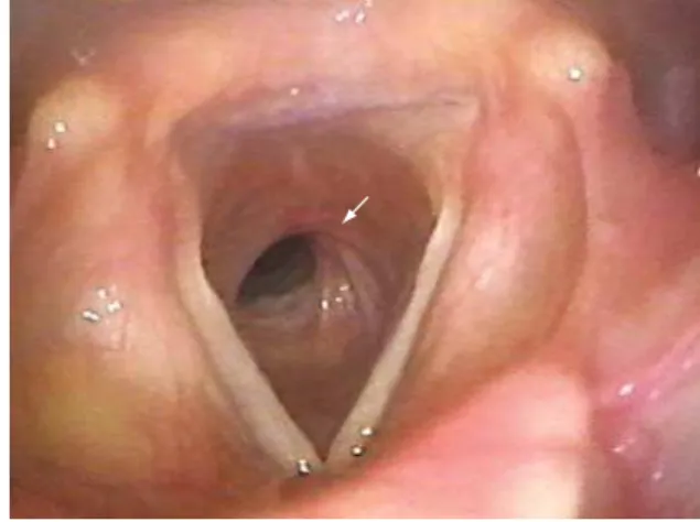
A 56-year-old, white female patient, who was a nonsmoker, sought treatment after experiencing progressive dyspnea and cough for six years. Her condition had deteriorated in the last six months. The patient had been treated unsuccessfully for ‘bronchitis’. Bronchoscopy revealed laryngotracheal stenosis (3-4 mm thick) with partial (50%) obstruc-tion of the tracheal lumen (Figure 3). Dilataobstruc-tion was performed using a rigid bronchoscope, and the biopsy revealed a nonspecific chronic inflamma-tory process. A three-week course of corticosteroid therapy was prescribed, and the patient remained asymptomatic during an eight-month outpatient follow-up period.
Discussion
Since first being reported by Brandenburg in 1972, idiopathic laryngotracheal stenosis has proven the have characteristics that are different from those of the other stenoses, notably the lack of a predis-posing factor and the almost exclusive prevalence in female patients.(1,2,10)
In the study of these four cases, all of which also involved female patients, we found no predisposing factors, either in the initial phase of the diagnosis or in the outpatient follow-up.
In the diagnosis of ITS, it is important to rule out the predisposing factors most often cited in the genesis of stenoses, such as prolonged intubation and tracheostomy, as well as tuberculosis, chem-ical burns resulting from aspiration, sarcoidosis, laryngotracheal fracture, and Wegener’s granuloma-tosis.(3,6,7) Other authors have proposed that estrogen
receptors are absent from the sites of stenosis, thereby allowing the increased release of fibroblast growth factor, which could also explain the almost exclusive incidence of ITS in women.(10) One study
even mentions the possibility that, in some cases, having occasionally been intubated for a short time, despite not causing post-intubation stenosis, as in the case of our second patient, who was intubated for a few hours, 24 years prior to the onset of ITS, can be a predisposing factor.(8)
It has been suggested that gastroesophageal reflux plays an important role in the genesis of laryn-gotracheal stenosis, although a definite conclusion regarding its role has not yet been reached, espe-cially since the lack of progression of the stenosis has also been reported.(2,7,11) None of our patients
complained of reflux or any other diseases of the digestive tract.
In addition to the above-mentioned treatment options, such as dilatation, laser treatment, and surgical resection, as well as systemic or topical corticosteroid therapy, topical administration of mitomycin C, an antineoplastic agent that inhibits the proliferation of fibroblasts, has also been used.(11)
The etiology of ITS, a rare entity, remains in dispute. This disease is absolutely predominant
Figure 2 - Cervical computed tomography scan: image of the laryngotracheal stenosis.
104 Rossi C, Colombari F, Guembarowsky AL, Ferreira Filho OF, Thomson JC
J Bras Pneumol. 2007;33(1):101-104
in women. In the differential diagnosis of ‘bron-chitis’, ITS should be contemplated when there is no response to the usual treatment. The management of ITS should range from dilatation (with or without the use of corticosteroids), as an initial treatment, to (in the event of the former being unsuccessful) surgical correction.
References
1. Brandenburg JH. Idiopathic subglottic stenosis. Trans Am Acad Ophthalmol Otolaryngol. 1972;76(5):1402-6.
2. Grillo HC. Idiopathic stenosis. In: Pearson FG, Cooper JD, Deslauries J, Ginsberg RJ, Hiebert CA, Patterson GA, et al. Thoracic surgery. 2nd ed. New York: Churchill Livingstone; 2002. p.314-20.
3. Lorenz RR. Adult laryngotracheal stenosis: etiology and surgical management. Curr Opin Otolaryngol Head Neck Surg. 2003;11(6):467-72.
4. Grillo HC, Mathisen DJ, Ashiku SK, Wright CD, Wain JC. Successful treatment of idiopathic laryngotracheal stenosis by resection and primary anastomosis. Ann Otol Rhinol Laryngol. 2003;112(9 Pt 1):798-800.
5. Ashiku SK, Kuzucu A, Grillo HC, Wright CD, Wain JC, Lo B, et al. Idiopathic laryngotracheal stenosis: Effective definitive treatment with laryngotracheal resection. J Thorac Cardiovasc Surg. 2004;127(1):99-107.
6. Grillo HC. Primary reconstruction of airway after resection of subglottic laryngeal and upper tracheal stenosis. Ann Thorac Surg. 1982;33(1):3-18.
7. Grillo HC, Mathisen DJ, Wain JC. Laryngotracheal resection and reconstruction for subglottic stenosis. Ann Thorac Surg. 1992;53(1):54-63.
8. Grillo HC. Management of idiopathic tracheal stenosis. Chest Surg Clin N Am. 1996;6(4):811-8.
9. Grillo HC. The history of tracheal surgery. Chest Surg Clin N Am. 2003;13(2):175-89.
10. Valdez TA, Shapsay SM. Idiopathic subglottic stenosis revisited. Ann Otol Rhinol Laryngol. 2002;111(8):690-5. 11. Dedo HH, Catien MD. Idiopathic progressive subglottic
stenosis: findings and treatment in 52 patients. Ann Otol Rhinol Laryngol. 2001;110(4):305-11.
12. Giudice M, Piazza C, Foccoli P, Toninelli C, Cavaliere S, Peretti G. Idiopathic subglottic stenosis: management by endoscopic and open-neck surgery in a serie of 30 patients. Eur Arch Otorhinolaryngol. 2003;260(5):235-8.
13. Ashiku SK, Mathisen DJ. Idiopathic laryngotracheal stenosis. Chest Surg Clin N Am. 2003;13(2):257-69.
14. Rea F, Callegaro D, Loy M, Zuin A, Narne S, Gobbi T, et al. Benign tracheal and laryngotracheal stenosis: surgical treatment and results. Eur J Cardithorac Surg. 2002;22(3):352-6.